Click image to see more details
-

-
-
-
-
+3
Product Info Summary
| SKU: | PA1971 |
|---|---|
| Size: | 100 μg/vial |
| Reactive Species: | Human, Mouse, Rat |
| Host: | Rabbit |
| Application: | IF, IHC, ICC, WB |
Customers Who Bought This Also Bought

Product info
Product Name
Anti-Tight junction protein ZO-2 TJP2 Antibody Picoband®
SKU/Catalog Number
PA1971
Size
100 μg/vial
Form
Lyophilized
Description
Boster Bio Anti-Tight junction protein ZO-2 TJP2 Antibody catalog # PA1971. Tested in IF, IHC, ICC, WB applications. This antibody reacts with Human, Mouse, Rat. The brand Picoband indicates this is a premium antibody that guarantees superior quality, high affinity, and strong signals with minimal background in Western blot applications. Only our best-performing antibodies are designated as Picoband, ensuring unmatched performance.
Storage & Handling
Store at -20˚C for one year from date of receipt. After reconstitution, at 4˚C for one month. It can also be aliquotted and stored frozen at -20˚C for six months. Avoid repeated freeze-thaw cycles.
Cite This Product
Anti-Tight junction protein ZO-2 TJP2 Antibody Picoband® (Boster Biological Technology, Pleasanton CA, USA, Catalog # PA1971)
Host
Rabbit
Contents
Each vial contains antibody formulated with stabilizing components, 0.9mg NaCl, 0.2mg Na2HPO4, 0.05mg Thimerosal, 0.01mg NaN3.
*This antibody is supplied in a stabilized formulation.
Compatibility with conjugation reactions depends on the chemistry of the conjugation method used.
For conjugation methods that are not compatible with the stabilizing components present in this formulation, a carrier-free antibody format is required.
Clonality
Polyclonal
Isotype
Rabbit IgG
Immunogen
A synthetic peptide corresponding to a sequence at the C-terminus of mouse TJP2, identical to the related rat sequence.
Cross-reactivity
No cross-reactivity with other proteins
Reactive Species
PA1971 is reactive to Tjp2 in Human, Mouse, Rat
Observed Molecular Weight
150 kDa
Calculated molecular weight
131.3 kDa
Background of Tjp2
TJP2 (Tight Junction Protein 2), also known as Zona Occludens 2 or ZO2, is a protein that in humans is encoded by the TJP2 gene. Tight junction proteins (TJPs) belong to a family of membrane-associated guanylate kinase (MAGUK) homologs that are involved in the organization of epithelial and endothelial intercellular junctions. Duclos et al. (1994) mapped the TJP2 gene telomeric to the Friedreich ataxia critical region on chromosome 9q13-q21. TJP2 lies about 70 kb centromeric to the X123 gene and is transcribed in the centromere-to-telomere direction. Using in vitro assays and immunoprecipitation studies, Itoh et al. (1999) showed that the mouse Tjp1, Tjp2, and Tjp3 PDZ1 domains interacted with the C-terminal cytoplasmic domains of Cldn1 through Cldn8. In the mouse inner ear, Walsh et al. (2010) found that Tjp2 expression decreased rapidly between E16.5 and age 1 week to a level in adult mice that was approximately 50% of the level at birth (P0).
Antibody Validation
Boster validates all antibodies on WB, IHC, ICC, Immunofluorescence, and ELISA with known positive control and negative samples to ensure specificity and high affinity, including thorough antibody incubations.
Application & Images
Applications
PA1971 is guaranteed for IF, IHC, ICC, WB Boster Guarantee
Recommend Dilution
| Application | Dilution | Species |
|---|---|---|
| Western blot | 0.1-0.5μg/ml | Human, Mouse, Rat |
| Immunohistochemistry (Paraffin-embedded Section) | 2-5μg/ml | Human, Mouse, Rat |
| Immunocytochemistry/Immunofluorescence | 5 μg/ml | Human |
Tested application
Suggested blocking solution with 5% non-fat milk or BSA; (*)Recommended protein loading: 20-40 µg per lane
Use TE buffer pH 9.0 for antigen retrieval; (*) citrate buffer pH 6.0 is an alternative.
Validation Images & Assay Conditions
Click image to see more details
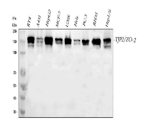
Western blot analysis of TJP2 using anti-TJP2 antibody (PA1971).
Electrophoresis was performed on a 5-20% SDS-PAGE gel at 70V (Stacking gel) / 90V (Resolving gel) for 2-3 hours. The sample well of each lane was loaded with 30 ug of sample under reducing conditions.
Lane 1: human RT4 whole cell lysates,
Lane 2: human A431 whole cell lysates,
Lane 3: human HepG2 whole cell lysates,
Lane 4: human MCF-7 whole cell lysates,
Lane 5: human U2OS whole cell lysates,
Lane 6: human Hela whole cell lysates,
Lane 7: human PC-3 whole cell lysates,
Lane 8: rat RH35 whole cell lysates,
Lane 9: rat HEPA1-6 whole cell lysates.
After electrophoresis, proteins were transferred to a nitrocellulose membrane at 150 mA for 50-90 minutes. Blocked the membrane with 5% non-fat milk/TBS for 1.5 hour at RT. The membrane was incubated with rabbit anti-TJP2 antigen affinity purified polyclonal antibody (Catalog # PA1971) at 0.5 μg/mL overnight at 4°C, then washed with TBS-0.1%Tween 3 times with 5 minutes each and probed with a goat anti-rabbit IgG-HRP secondary antibody at a dilution of 1:5000 for 1.5 hour at RT. The signal is developed using an Enhanced Chemiluminescent detection (ECL) kit (Catalog # EK1002) with Tanon 5200 system. A specific band was detected for TJP2 at approximately 150 kDa. The expected band size for TJP2 is at 131 kDa.

Click image to see more details
IHC analysis of ZO-2/TJP2 using anti-ZO-2/TJP2 antibody (PA1971).
ZO-2/TJP2 was detected in a paraffin-embedded section of human stoamch tissue. Heat mediated antigen retrieval was performed in EDTA buffer (pH 8.0, epitope retrieval solution). The tissue section was blocked with 10% goat serum. The tissue section was then incubated with 2 μg/ml rabbit anti-ZO-2/TJP2 Antibody (PA1971) overnight at 4°C. Peroxidase Conjugated Goat Anti-rabbit IgG was used as secondary antibody and incubated for 30 minutes at 37°C. The tissue section was developed using HRP Conjugated Rabbit IgG Super Vision Assay Kit (Catalog # SV0002) with DAB as the chromogen.

Click image to see more details
IHC analysis of ZO-2/TJP2 using anti-ZO-2/TJP2 antibody (PA1971).
ZO-2/TJP2 was detected in a paraffin-embedded section of human stomach tissue. Heat mediated antigen retrieval was performed in EDTA buffer (pH 8.0, epitope retrieval solution). The tissue section was blocked with 10% goat serum. The tissue section was then incubated with 2 μg/ml rabbit anti-ZO-2/TJP2 Antibody (PA1971) overnight at 4°C. Peroxidase Conjugated Goat Anti-rabbit IgG was used as secondary antibody and incubated for 30 minutes at 37°C. The tissue section was developed using HRP Conjugated Rabbit IgG Super Vision Assay Kit (Catalog # SV0002) with DAB as the chromogen.

Click image to see more details
IHC analysis of TJP2 using anti-TJP2 antibody (PA1971).
TJP2 was detected in a paraffin-embedded section of human stomach tissue. Heat mediated antigen retrieval was performed in EDTA buffer (pH 8.0, epitope retrieval solution). The tissue section was blocked with 10% goat serum. The tissue section was then incubated with 2 μg/ml rabbit anti-TJP2 Antibody (PA1971) overnight at 4°C. Peroxidase Conjugated Goat Anti-rabbit IgG was used as secondary antibody and incubated for 30 minutes at 37°C. The tissue section was developed using HRP Conjugated Rabbit IgG Super Vision Assay Kit (Catalog # SV0002) with DAB as the chromogen.

Click image to see more details
IHC analysis of TJP2 using anti-TJP2 antibody (PA1971).
TJP2 was detected in a paraffin-embedded section of mouse liver tissue. Heat mediated antigen retrieval was performed in EDTA buffer (pH 8.0, epitope retrieval solution). The tissue section was blocked with 10% goat serum. The tissue section was then incubated with 2 μg/ml rabbit anti-TJP2 Antibody (PA1971) overnight at 4°C. Peroxidase Conjugated Goat Anti-rabbit IgG was used as secondary antibody and incubated for 30 minutes at 37°C. The tissue section was developed using HRP Conjugated Rabbit IgG Super Vision Assay Kit (Catalog # SV0002) with DAB as the chromogen.

Click image to see more details
IHC analysis of TJP2 using anti-TJP2 antibody (PA1971).
TJP2 was detected in a paraffin-embedded section of rat kidney tissue. Heat mediated antigen retrieval was performed in EDTA buffer (pH 8.0, epitope retrieval solution). The tissue section was blocked with 10% goat serum. The tissue section was then incubated with 2 μg/ml rabbit anti-TJP2 Antibody (PA1971) overnight at 4°C. Peroxidase Conjugated Goat Anti-rabbit IgG was used as secondary antibody and incubated for 30 minutes at 37°C. The tissue section was developed using HRP Conjugated Rabbit IgG Super Vision Assay Kit (Catalog # SV0002) with DAB as the chromogen.

Click image to see more details
IF analysis of TJP2 using anti-TJP2 antibody (PA1971).
TJP2 was detected in an immunocytochemical section of U2OS cells. Enzyme antigen retrieval was performed using IHC enzyme antigen retrieval reagent (AR0022) for 15 mins. The cells were blocked with 10% goat serum. And then incubated with 5 μg/mL rabbit anti-TJP2 Antibody (PA1971) overnight at 4°C. DyLight®488 Conjugated Goat Anti-Rabbit IgG (BA1127) was used as secondary antibody at 1:500 dilution and incubated for 30 minutes at 37°C. The section was counterstained with DAPI. Visualize using a fluorescence microscope and filter sets appropriate for the label used.
Specific Publications For Anti-Tight junction protein ZO-2 TJP2 Antibody Picoband® (PA1971)
Loading publications
Recommended Resources
Here are featured tools and databases that you might find useful.
- Boster's Pathways Library
- Protein Databases
- Bioscience Research Protocol Resources
- Data Processing & Analysis Software
- Photo Editing Software
- Scientific Literature Resources
- Research Paper Management Tools
- Molecular Biology Software
- Primer Design Tools
- Bioinformatics Tools
- Phylogenetic Tree Analysis
Customer Reviews
Have you used Anti-Tight junction protein ZO-2 TJP2 Antibody Picoband®?
Share your experimental results or join a short interview to earn up to $1,000 in product credits or other rewards.
0 Reviews For Anti-Tight junction protein ZO-2 TJP2 Antibody Picoband®
Customer Q&As
Have a question?
Find answers in Q&As, reviews.
Can't find your answer?
Submit your question
1 Customer Q&As for Anti-Tight junction protein ZO-2 TJP2 Antibody Picoband®
Question
We are currently using anti-TJP2 antibody PA1971 for rat tissue, and we are happy with the WB results. The species of reactivity given in the datasheet says mouse, rat. Is it possible that the antibody can work on pig tissues as well?
H. Kulkarni
Verified customer
Asked: 2016-10-27
Answer
The anti-TJP2 antibody (PA1971) has not been validated for cross reactivity specifically with pig tissues, though there is a good chance of cross reactivity. We have an innovator award program that if you test this antibody and show it works in pig you can get your next antibody for free. Please contact me if I can help you with anything.
Boster Scientific Support
Answered: 2016-10-27
