Click image to see more details
-

-
-
-
-
+3
Product Info Summary
| SKU: | M00001-2 |
|---|---|
| Size: | 100 μl |
| Reactive Species: | Human |
| Host: | Rabbit |
| Application: | IF, ICC, WB |
Customers Who Bought This Also Bought

Product info
Product Name
Anti-p53 TP53 Rabbit Monoclonal Antibody
SKU/Catalog Number
M00001-2
BM4304 is an alternative SKU for this antibody, used in previous lots.
Size
100 μl
Form
Liquid
Description
Boster Bio Anti-p53 TP53 Rabbit Monoclonal Antibody catalog # M00001-2. Tested in WB, ICC/IF applications. This antibody reacts with Human.
Storage & Handling
Store at -20°C for one year. For short term storage and frequent use, store at 4°C for up to one month. Avoid repeated freeze-thaw cycles.
Cite This Product
Anti-p53 TP53 Rabbit Monoclonal Antibody (Boster Biological Technology, Pleasanton CA, USA, Catalog # M00001-2)
Host
Rabbit
Contents
Rabbit IgG in stabilizing components, phosphate buffered saline, pH 7.4, 150mM NaCl, 0.02% sodium azide and 50% glycerol.
*This antibody is supplied in a stabilized formulation.
Compatibility with conjugation reactions depends on the chemistry of the conjugation method used.
For conjugation methods that are not compatible with the stabilizing components present in this formulation, a carrier-free antibody format is required.
Clonality
Monoclonal
Clone Number
DDE-20
Isotype
Rabbit IgG
Immunogen
A synthesized peptide derived from human p53
Reactive Species
M00001-2 is reactive to TP53 in Human
Observed Molecular Weight
55 kDa
Calculated molecular weight
43.7 kDa
Antibody Validation
Boster validates all antibodies on WB, IHC, ICC, Immunofluorescence, and ELISA with known positive control and negative samples to ensure specificity and high affinity, including thorough antibody incubations.
Application & Images
Applications
M00001-2 is guaranteed for IF, ICC, WB Boster Guarantee
Recommend Dilution
WB 1:500-1:2000
ICC/IF 1:50-1:200
Validation Images & Assay Conditions
Click image to see more details
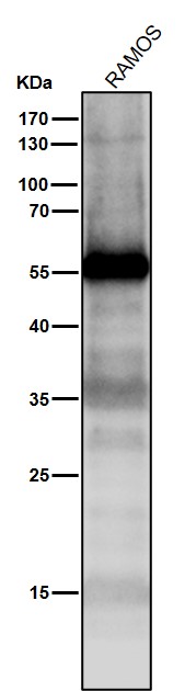
All lanes use the Antibody at 1:1K dilution for 1 hour at room temperature.

Click image to see more details
All lanes use the Antibody at 1:1K dilution for 1 hour at room temperature.

Click image to see more details
Western blot analysis of p53 expression in (1) Raji cell lysate; (2) HepG2 cell lysate.

Click image to see more details
All lanes use the Antibody at 1:1K dilution for 1 hour at room temperature.

Click image to see more details
Immunofluorescent analysis of Hela cells, using p53 Antibody .

Click image to see more details
Immunofluorescent analysis using the Antibody at 1:50 dilution.

Click image to see more details
Immunofluorescent analysis using the Antibody at 1:50 dilution.
Specific Publications For Anti-p53 TP53 Rabbit Monoclonal Antibody (M00001-2)
Loading publications
Recommended Resources
Here are featured tools and databases that you might find useful.
- Boster's Pathways Library
- Protein Databases
- Bioscience Research Protocol Resources
- Data Processing & Analysis Software
- Photo Editing Software
- Scientific Literature Resources
- Research Paper Management Tools
- Molecular Biology Software
- Primer Design Tools
- Bioinformatics Tools
- Phylogenetic Tree Analysis
Customer Reviews
Have you used Anti-p53 TP53 Rabbit Monoclonal Antibody?
Share your experimental results or join a short interview to earn up to $1,000 in product credits or other rewards.
0 Reviews For Anti-p53 TP53 Rabbit Monoclonal Antibody
Customer Q&As
Have a question?
Find answers in Q&As, reviews.
Can't find your answer?
Submit your question
5 Customer Q&As for Anti-p53 TP53 Rabbit Monoclonal Antibody
Question
We are currently using anti-p53 Rabbit Monoclonal antibody M00001-2 for human tissue, and we are content with the IF results. The species of reactivity given in the datasheet says human. Is it true that the antibody can work on pig tissues as well?
Verified Customer
Verified customer
Asked: 2020-03-04
Answer
The anti-p53 Rabbit Monoclonal antibody (M00001-2) has not been tested for cross reactivity specifically with pig tissues, though there is a good chance of cross reactivity. We have an innovator award program that if you test this antibody and show it works in pig you can get your next antibody for free. Please contact me if I can help you with anything.
Boster Scientific Support
Answered: 2020-03-04
Question
We ordered your anti-p53 Rabbit Monoclonal antibody for IF on colon in a previous experiment. I am using human, and We intend to use the antibody for WB next. We need examining colon as well as ovarian adenocarcinoma in our next experiment. Could give a recommendation on which antibody would work the best for WB?
Verified Customer
Verified customer
Asked: 2019-12-11
Answer
I viewed the website and datasheets of our anti-p53 Rabbit Monoclonal antibody and I see that M00001-2 has been validated on human in both IF and WB. Thus M00001-2 should work for your application. Our Boster satisfaction guarantee will cover this product for WB in human even if the specific tissue type has not been validated. We do have a comprehensive range of products for WB detection and you can check out our website bosterbio.com to find out more information about them.
Boster Scientific Support
Answered: 2019-12-11
Question
My lab would like using your anti-p53 Rabbit Monoclonal antibody for cellular response to glucose starvation studies. Has this antibody been tested with western blotting on raji cell lysate? We would like to see some validation images before ordering.
S. Huang
Verified customer
Asked: 2017-10-20
Answer
Thanks for your inquiry. This M00001-2 anti-p53 Rabbit Monoclonal antibody is tested on raji cell lysate, hepg2 cell lysate. It is guaranteed to work for IF, WB in human. Our Boster guarantee will cover your intended experiment even if the sample type has not been be directly tested.
Boster Scientific Support
Answered: 2017-10-20
Question
Our team were happy with the WB result of your anti-p53 Rabbit Monoclonal antibody. However we have observed positive staining in blood cytoplasm using this antibody. Is that expected? Could you tell me where is TP53 supposed to be expressed?
J. Zhao
Verified customer
Asked: 2015-07-23
Answer
From literature, blood does express TP53. Generally TP53 expresses in cytoplasm. Regarding which tissues have TP53 expression, here are a few articles citing expression in various tissues:
Colon, Pubmed ID: 16131611
Embryonic kidney, Pubmed ID: 17525332
Glial cell, and Glial tumor, Pubmed ID: 19011621
Kidney, Pubmed ID: 15489334
Lung carcinoma, Pubmed ID: 14660794
Boster Scientific Support
Answered: 2015-07-23
Question
We have been able to see staining in human colon. Do you have any suggestions? Is anti-p53 Rabbit Monoclonal antibody supposed to stain colon positively?
O. Carter
Verified customer
Asked: 2014-01-07
Answer
From literature colon does express TP53. From Uniprot.org, TP53 is expressed in epithelium of mammary gland, colon, kidney, lung carcinoma, blood, ovarian adenocarcinoma, glial cell glial tumor, embryonic kidney, among other tissues. Regarding which tissues have TP53 expression, here are a few articles citing expression in various tissues:
Colon, Pubmed ID: 16131611
Embryonic kidney, Pubmed ID: 17525332
Glial cell, and Glial tumor, Pubmed ID: 19011621
Kidney, Pubmed ID: 15489334
Lung carcinoma, Pubmed ID: 14660794
Boster Scientific Support
Answered: 2014-01-07
