Click image to see more details
Product Info Summary
| SKU: | PB9155 |
|---|---|
| Size: | 100 μg/vial |
| Reactive Species: | Human |
| Host: | Rabbit |
| Application: | WB |
Customers Who Bought This Also Bought

Product info
Product Name
Anti-Perforin/PRF1 Antibody Picoband®
SKU/Catalog Number
PB9155
PB0167 is an alternative SKU for this antibody, used in previous lots.
Size
100 μg/vial
Form
Lyophilized
Description
Boster Bio Anti-Perforin/PRF1 Antibody Picoband® catalog # PB9155. Tested in WB applications. This antibody reacts with Human. The brand Picoband indicates this is a premium antibody that guarantees superior quality, high affinity, and strong signals with minimal background in Western blot applications. Only our best-performing antibodies are designated as Picoband, ensuring unmatched performance.
Storage & Handling
Store at -20˚C for one year from date of receipt. After reconstitution, at 4˚C for one month. It can also be aliquotted and stored frozen at -20˚C for six months. Avoid repeated freeze-thaw cycles.
Cite This Product
Anti-Perforin/PRF1 Antibody Picoband® (Boster Biological Technology, Pleasanton CA, USA, Catalog # PB9155)
Host
Rabbit
Contents
Each vial contains 4 mg Trehalose, 0.9 mg NaCl and 0.2 mg Na2HPO4.
Clonality
Polyclonal
Isotype
Rabbit IgG
Immunogen
E.coli-derived human Perforin recombinant protein (Position: E175-W555). Human Perforin shares 68% amino acid (aa) sequence identity with both mouse and rat Perforin.
Cross-reactivity
No cross-reactivity with other proteins
Reactive Species
PB9155 is reactive to PRF1 in Human
Observed Molecular Weight
61 kDa
Calculated molecular weight
61.4 kDa
Background of PRF1
PRF1, also known as Perforin-1, is a protein that in humans is encoded by the PRF1 gene. It is mapped to 10q22.1. PRF1 is a cytolytic protein found in the granules of Cytotoxic T lymphocytes (CTLs) and NK cells. Upon degranulation, PRF1 inserts itself into the target cell's plasma membrane, forming a pore. The lytic membrane-inserting part of perforin is the MACPF domain. This region shares homology with cholesterol-dependent cytolysins from Gram-positive bacteria. PRF1 has structural and functional similarities to complement component 9 (C9). Like C9, this protein creates transmembrane tubules and is capable of lysing non-specifically a variety of target cells. This protein is one of the main cytolytic proteins of cytolytic granules, and it is known to be a key effector molecule for T-cell- and natural killer-cell-mediated cytolysis. PRF1 is thought to act by creating holes in the plasma membrane which triggers an influx of calcium and initiates membrane repair mechanisms. These repair mechanisms bring perforin and granzymes into early endosomes.
Antibody Validation
Boster validates all antibodies on WB, IHC, ICC, Immunofluorescence, and ELISA with known positive control and negative samples to ensure specificity and high affinity, including thorough antibody incubations.
Application & Images
Applications
PB9155 is guaranteed for WB Boster Guarantee
Recommend Dilution
| Application | Dilution | Species |
|---|---|---|
| Western blot | 0.1-0.5μg/ml | Human |
Tested application
Suggested blocking solution with 5% non-fat milk or BSA; (*)Recommended protein loading: 20-40 µg per lane
Validation Images & Assay Conditions
Click image to see more details
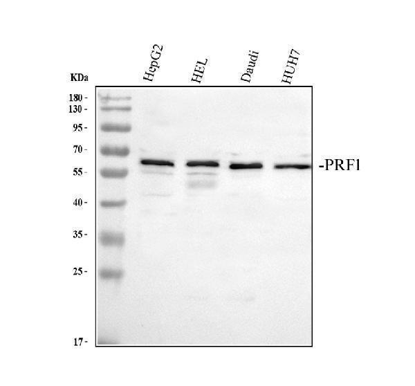
Western blot analysis of ADFP/Perforin using anti-ADFP/Perforin antibody (PB9155).
Electrophoresis was performed on a 10% SDS-PAGE gel at 80V (Stacking gel) / 120V (Resolving gel) for 2 hours. The sample well of each lane was loaded with 30 ug of sample under reducing conditions.
Lane 1: human HepG2 whole cell lysates,
Lane 2: human HEL whole cell lysates,
Lane 3: human Daudi whole cell lysates,
Lane 4: human HUH7 whole cell lysates.
After electrophoresis, proteins were transferred to a nitrocellulose membrane at 150 mA for 50-90 minutes. Blocked the membrane with 5% non-fat milk/TBS for 1.5 hour at RT. The membrane was incubated with rabbit anti-ADFP/Perforin antigen affinity purified polyclonal antibody (Catalog # PB9155) at 0.5 μg/mL overnight at 4°C, then washed with TBS-0.1%Tween 3 times with 5 minutes each and probed with a goat anti-rabbit IgG-HRP secondary antibody at a dilution of 1:5000 for 1.5 hour at RT. The signal is developed using an ECL Plus Western Blotting Substrate (Catalog # AR1196-200) with Tanon 5200 system. A specific band was detected for ADFP/Perforin at approximately 61 kDa. The expected band size for ADFP/Perforin is at 61 kDa.
Specific Publications For Anti-Perforin/PRF1 Antibody Picoband® (PB9155)
Loading publications
Recommended Resources
Here are featured tools and databases that you might find useful.
- Boster's Pathways Library
- Protein Databases
- Bioscience Research Protocol Resources
- Data Processing & Analysis Software
- Photo Editing Software
- Scientific Literature Resources
- Research Paper Management Tools
- Molecular Biology Software
- Primer Design Tools
- Bioinformatics Tools
- Phylogenetic Tree Analysis
Customer Reviews
Have you used Anti-Perforin/PRF1 Antibody Picoband®?
Share your experimental results or join a short interview to earn up to $1,000 in product credits or other rewards.
0 Reviews For Anti-Perforin/PRF1 Antibody Picoband®
Customer Q&As
Have a question?
Find answers in Q&As, reviews.
Can't find your answer?
Submit your question
4 Customer Q&As for Anti-Perforin/PRF1 Antibody Picoband®
Question
My boss were satisfied with the WB result of your anti-Perforin/PRF1 antibody. However we have seen positive staining in natural killer cell cytoplasmic granule lumen. secreted. cell using this antibody. Is that expected? Could you tell me where is PRF1 supposed to be expressed?
Verified Customer
Verified customer
Asked: 2019-10-15
Answer
According to literature, natural killer cell does express PRF1. Generally PRF1 expresses in cytoplasmic granule lumen. secreted. cell. Regarding which tissues have PRF1 expression, here are a few articles citing expression in various tissues:
Liver, Pubmed ID: 19159218
Natural killer cell, Pubmed ID: 3419519, 8676885
Spleen, Pubmed ID: 14702039
Boster Scientific Support
Answered: 2019-10-15
Question
We are currently using anti-Perforin/PRF1 antibody PB9155 for human tissue, and we are satisfied with the WB results. The species of reactivity given in the datasheet says human. Is it likely that the antibody can work on zebrafish tissues as well?
Verified Customer
Verified customer
Asked: 2018-06-15
Answer
The anti-Perforin/PRF1 antibody (PB9155) has not been validated for cross reactivity specifically with zebrafish tissues, but there is a good chance of cross reactivity. We have an innovator award program that if you test this antibody and show it works in zebrafish you can get your next antibody for free. Please contact me if I can help you with anything.
Boster Scientific Support
Answered: 2018-06-15
Question
We have seen staining in human spleen. Any tips? Is anti-Perforin/PRF1 antibody supposed to stain spleen positively?
Verified Customer
Verified customer
Asked: 2018-05-17
Answer
From literature spleen does express PRF1. From Uniprot.org, PRF1 is expressed in mononuclear cell, natural killer cell, spleen, liver, among other tissues. Regarding which tissues have PRF1 expression, here are a few articles citing expression in various tissues:
Liver, Pubmed ID: 19159218
Natural killer cell, Pubmed ID: 3419519, 8676885
Spleen, Pubmed ID: 14702039
Boster Scientific Support
Answered: 2018-05-17
Question
you antibody using your anti-Perforin/PRF1 antibody for t cell mediated cytotoxicity studies. Has this antibody been tested with western blotting on colo320 whole cell lysate? We would like to see some validation images before ordering.
N. Krishna
Verified customer
Asked: 2015-08-14
Answer
I appreciate your inquiry. This PB9155 anti-Perforin/PRF1 antibody is validated on hela whole cell lysate, colo320 whole cell lysate, hepg2 whole cell lysate. It is guaranteed to work for WB in human. Our Boster guarantee will cover your intended experiment even if the sample type has not been be directly tested.
Boster Scientific Support
Answered: 2015-08-14
