Click image to see more details
Product Info Summary
| SKU: | M00104-3 |
|---|---|
| Size: | 50 µl |
| Reactive Species: | Human, Mouse |
| Host: | Rabbit |
| Application: | WB |
Customers Who Bought This Also Bought

Product info
Product Name
Anti-Phospho-Erk1/2(Thr202/Tyr204) Antibody
SKU/Catalog Number
M00104-3
Size
50 µl
Description
Boster Bio Anti-Phospho-Erk1/2(Thr202/Tyr204) Antibody (Catalog # M00104-3). Tested in WB application(s). This antibody reacts with Human, Mouse.
Storage & Handling
Maintain refrigerated at 2-8°C for up to 2 weeks. For long-term storage, store at -20°C in small aliquots to prevent freeze-thaw cycles.
Cite This Product
Anti-Phospho-Erk1/2(Thr202/Tyr204) Antibody (Boster Biological Technology, Pleasanton CA, USA, Catalog # M00104-3)
Host
Rabbit
Contents
Purified polyclonal antibody supplied in PBS with 0.09% (W/V) sodium azide.
Clonality
Polyclonal
Isotype
Rabbit IgG
Immunogen
This Phospho-Erk1/2 (Thr202/Tyr204) antibody is generated from a rabbit immunized with a KLH conjugated synthetic peptide between 176-208 amino acids from human Phospho-Erk1/2 (Thr202/Tyr204).
Reactive Species
M00104-3 is reactive to MAPK3 in Human, Mouse
Calculated molecular weight
43.1 kDa
Background of MAPK3
Serine/threonine kinase which acts as an essential component of the MAP kinase signal transduction pathway. MAPK1/ERK2 and MAPK3/ERK1 are the 2 MAPKs which play an important role in the MAPK/ERK cascade. They participate also in a signaling cascade initiated by activated KIT and KITLG/SCF. Depending on the cellular context, the MAPK/ERK cascade mediates diverse biological functions such as cell growth, adhesion, survival and differentiation through the regulation of transcription, translation, cytoskeletal rearrangements. The MAPK/ERK cascade plays also a role in initiation and regulation of meiosis, mitosis, and postmitotic functions in differentiated cells by phosphorylating a number of transcription factors. About 160 substrates have already been discovered for ERKs. Many of these substrates are localized in the nucleus, and seem to participate in the regulation of transcription upon stimulation. However, other substrates are found in the cytosol as well as in other cellular organelles, and those are responsible for processes such as translation, mitosis and apoptosis. Moreover, the MAPK/ERK cascade is also involved in the regulation of the endosomal dynamics, including lysosome processing and endosome cycling through the perinuclear recycling compartment (PNRC); as well as in the fragmentation of the Golgi apparatus during mitosis. The substrates include transcription factors (such as ATF2, BCL6, ELK1, ERF, FOS, HSF4 or SPZ1), cytoskeletal elements (such as CANX, CTTN, GJA1, MAP2, MAPT, PXN, SORBS3 or STMN1), regulators of apoptosis (such as BAD, BTG2, CASP9, DAPK1, IER3, MCL1 or PPARG), regulators of translation (such as EIF4EBP1) and a variety of other signaling-related molecules (like ARHGEF2, FRS2 or GRB10). Protein kinases (such as RAF1, RPS6KA1/RSK1, RPS6KA3/RSK2, RPS6KA2/RSK3, RPS6KA6/RSK4, SYK, MKNK1/MNK1, MKNK2/MNK2, RPS6KA5/MSK1, RPS6KA4/MSK2, MAPKAPK3 or MAPKAPK5) and phosphatases (such as DUSP1, DUSP4, DUSP6 or DUSP16) are other substrates which enable the propagation the MAPK/ERK signal to additional cytosolic and nuclear targets, thereby extending the specificity of the cascade.
Antibody Validation
Boster validates all antibodies on WB, IHC, ICC, Immunofluorescence, and ELISA with known positive control and negative samples to ensure specificity and high affinity, including thorough antibody incubations.
Application & Images
Applications
M00104-3 is guaranteed for WB Boster Guarantee
Recommend Dilution
WB: 1:1000
Validation Images & Assay Conditions
Click image to see more details
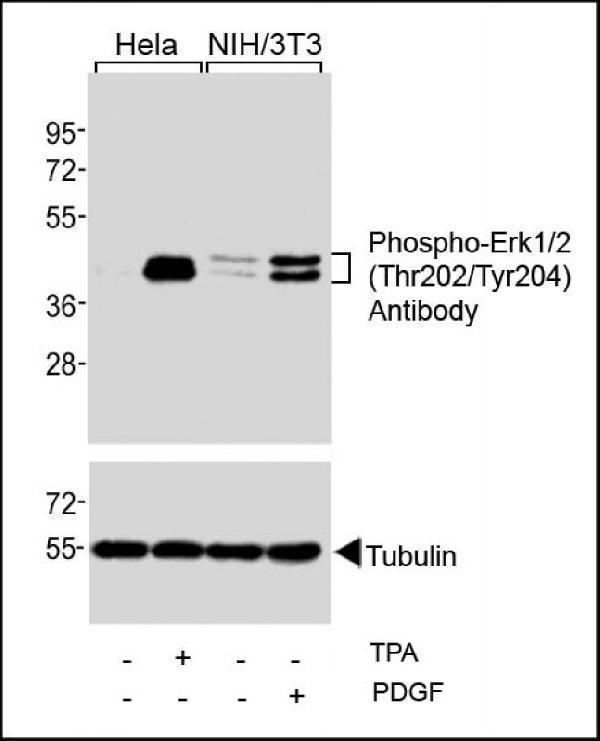
Western blot analysis of extracts from Hela cells, untreated or treated with TPA (200nM), and NIH/3T3 cells, untreated or treated with PDGF (100ng/ml), using Phospho-Erk1/2 (Thr202/Tyr204) Antibody (upper) or Tubulin (lower).

Click image to see more details
Western blot analysis of extracts from Hela cells, untreated or treated with TPA (200nM), and NIH/3T3 cells, untreated or treated with PDGF (100ng/ml), using Phospho-Erk1/2(Thr202/Tyr204) Antibody (upper) or Tubulin (lower).
Specific Publications For Anti-Phospho-Erk1/2(Thr202/Tyr204) Antibody (M00104-3)
Loading publications
Recommended Resources
Here are featured tools and databases that you might find useful.
- Boster's Pathways Library
- Protein Databases
- Bioscience Research Protocol Resources
- Data Processing & Analysis Software
- Photo Editing Software
- Scientific Literature Resources
- Research Paper Management Tools
- Molecular Biology Software
- Primer Design Tools
- Bioinformatics Tools
- Phylogenetic Tree Analysis
Customer Reviews
Have you used Anti-Phospho-Erk1/2(Thr202/Tyr204) Antibody?
Share your experimental results or join a short interview to earn up to $1,000 in product credits or other rewards.
0 Reviews For Anti-Phospho-Erk1/2(Thr202/Tyr204) Antibody
Customer Q&As
Have a question?
Find answers in Q&As, reviews.
Can't find your answer?
Submit your question
