
Click image to see more details
-

-
-
-
-
+1
Product Info Summary
| SKU: | PA2087 |
|---|---|
| Size: | 100 μg/vial |
| Reactive Species: | Human, Mouse, Rat |
| Host: | Rabbit |
| Application: | IHC, WB |
Customers Who Bought This Also Bought

Product info
Product Name
Anti-Prolactin Receptor/PRLR Antibody Picoband®
SKU/Catalog Number
PA2087
BA3818 is an alternative SKU for this antibody, used in previous lots.
Size
100 μg/vial
Form
Lyophilized
Description
Boster Bio Anti-Prolactin Receptor/PRLR Antibody catalog # PA2087. Tested in IHC, WB applications. This antibody reacts with Human, Mouse, Rat. The brand Picoband indicates this is a premium antibody that guarantees superior quality, high affinity, and strong signals with minimal background in Western blot applications. Only our best-performing antibodies are designated as Picoband, ensuring unmatched performance.
Storage & Handling
Store at -20˚C for one year from date of receipt. After reconstitution, at 4˚C for one month. It can also be aliquotted and stored frozen at -20˚C for six months. Avoid repeated freeze-thaw cycles.
Cite This Product
Anti-Prolactin Receptor/PRLR Antibody Picoband® (Boster Biological Technology, Pleasanton CA, USA, Catalog # PA2087)
Host
Rabbit
Contents
Each vial contains antibody formulated with stabilizing components, 0.9mg NaCl, 0.2mg Na2HPO4, 0.05mg Thimerosal, 0.05mg NaN3.
*This antibody is supplied in a stabilized formulation.
Compatibility with conjugation reactions depends on the chemistry of the conjugation method used.
For conjugation methods that are not compatible with the stabilizing components present in this formulation, a carrier-free antibody format is required.
Clonality
Polyclonal
Isotype
Rabbit IgG
Immunogen
A synthetic peptide corresponding to a sequence at the C-terminus of mouse PRLR, identical to the related rat sequence, and different from the related human sequence by three amino acids.
Cross-reactivity
No cross-reactivity with other proteins
Reactive Species
PA2087 is reactive to Prlr in Human, Mouse, Rat
Observed Molecular Weight
95 kDa
Calculated molecular weight
68.2 kDa
Background of Prlr
PRLR (Prolactin Receptor), is a cytokine receptor. By somatic cell hybrid analysis and by in situ hybridization, Arden et al. (1989, 1990) demonstrated that the prolactin receptor gene resides in the same chromosomal region as the growth hormone receptor gene, which has been mapped to 5p13-p12. Cunningham et al. (1990) demonstrated that zinc greatly increases the affinity of GH for the extracellular binding domain of PRLR, although it is not required for binding of GH to the growth hormone receptor or for binding of prolactin to the prolactin receptor. By mutational analysis, they showed that a cluster of 3 residues (histidine-18, histidine-21, and glutamic acid-174) in GH and histidine-188 in PRLR (conserved in all PRL receptors but not GH receptors) are likely zinc-ion ligands.
Antibody Validation
Boster validates all antibodies on WB, IHC, ICC, Immunofluorescence, and ELISA with known positive control and negative samples to ensure specificity and high affinity, including thorough antibody incubations.
Application & Images
Applications
PA2087 is guaranteed for IHC, WB Boster Guarantee
Recommend Dilution
| Application | Dilution | Species |
|---|---|---|
| Immunohistochemistry (Paraffin-embedded Section) | 0.5-1μg/ml | Human, Rat, Mouse |
| Western blot | 0.1-0.5μg/ml | Human, Mouse, Rat |
Tested application
Suggested blocking solution with 5% non-fat milk or BSA; (*)Recommended protein loading: 20-40 µg per lane
Use TE buffer pH 9.0 for antigen retrieval; (*) citrate buffer pH 6.0 is an alternative.
Validation Images & Assay Conditions
Click image to see more details
Anti-PRLR antibody, PA2087, IHC(P)
IHC(P): Human Mammary Cancer Tissue

Click image to see more details
Anti-PRLR antibody, PA2087, IHC(P)
IHC(P): Rat Testis Tissue

Click image to see more details
Western blot analysis of PRLR using anti-PRLR antibody (PA2087).
Electrophoresis was performed on a 5-20% SDS-PAGE gel at 70V (Stacking gel) / 90V (Resolving gel) for 2-3 hours. The sample well of each lane was loaded with 50ug of sample under reducing conditions.
Lane 1: rat PC-12 whole cell lysates.
After Electrophoresis, proteins were transferred to a Nitrocellulose membrane at 150mA for 50-90 minutes. Blocked the membrane with 5% Non-fat Milk/ TBS for 1.5 hour at RT. The membrane was incubated with rabbit anti-PRLR antigen affinity purified polyclonal antibody (Catalog # PA2087) at 0.5 μg/mL overnight at 4°C, then washed with TBS-0.1%Tween 3 times with 5 minutes each and probed with a goat anti-rabbit IgG-HRP secondary antibody at a dilution of 1:10000 for 1.5 hour at RT. The signal is developed using an Enhanced Chemiluminescent detection (ECL) kit (Catalog # EK1002) with Tanon 5200 system. A specific band was detected for PRLR at approximately 90KD. The expected band size for PRLR is at 70KD.

Click image to see more details
Western blot analysis of PRLR using anti-PRLR antibody (PA2087).
Electrophoresis was performed on a 5-20% SDS-PAGE gel at 70V (Stacking gel) / 90V (Resolving gel) for 2-3 hours. The sample well of each lane was loaded with 50ug of sample under reducing conditions.
Lane 1: human Hela whole cell lysates,
Lane 2: human MCF-7 whole cell lysates.
After Electrophoresis, proteins were transferred to a Nitrocellulose membrane at 150mA for 50-90 minutes. Blocked the membrane with 5% Non-fat Milk/ TBS for 1.5 hour at RT. The membrane was incubated with rabbit anti-PRLR antigen affinity purified polyclonal antibody (Catalog # PA2087) at 0.5 μg/mL overnight at 4°C, then washed with TBS-0.1%Tween 3 times with 5 minutes each and probed with a goat anti-rabbit IgG-HRP secondary antibody at a dilution of 1:10000 for 1.5 hour at RT. The signal is developed using an Enhanced Chemiluminescent detection (ECL) kit (Catalog # EK1002) with Tanon 5200 system. A specific band was detected for PRLR at approximately 95KD. The expected band size for PRLR is at 70KD.

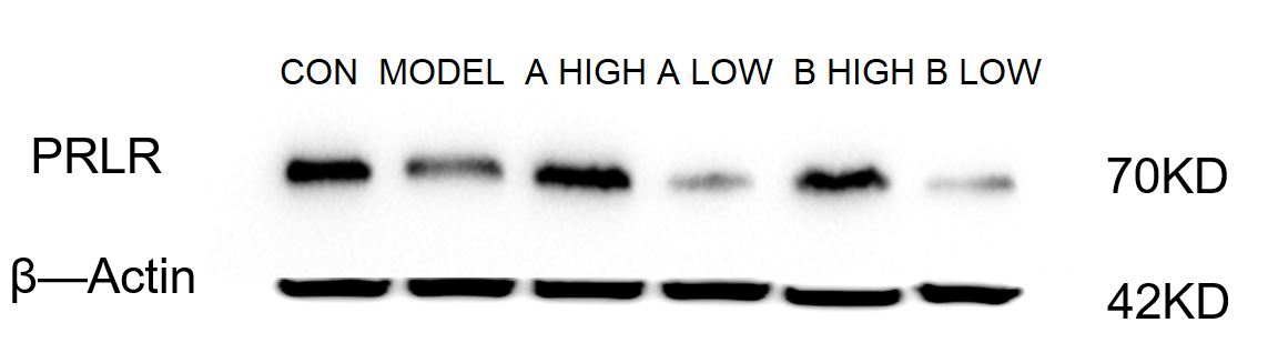
Click image to see more details
Western blot analysis of PRLR using anti-PRLR antibody (PA2087).
Electrophoresis was performed on a 5-20% SDS-PAGE gel at 70V (Stacking gel) / 90V (Resolving gel) for 2-3 hours. The sample well of each lane was loaded with 30 ug of sample under reducing conditions.
Lane 1: control group-mouse brain tissue lysates,
Lane 2: model group-mouse brain tissue lysates,
Lane 3: high-dose A group-mouse brain tissue lysates,
Lane 4: low-dose A group-mouse brain tissue lysates,
Lane 5: high-dose B group-mouse brain tissue lysates,
Lane 6: low-dose B group-mouse brain tissue lysates.
After electrophoresis, proteins were transferred to a nitrocellulose membrane at 150 mA for 50-90 minutes. Blocked the membrane with 5% non-fat milk/TBS for 1.5 hour at RT. The membrane was incubated with rabbit anti-PRLR antigen affinity purified monoclonal antibody (Catalog # PA2087) at 1:1000 overnight at 4°C, then washed with TBS-0.1%Tween 3 times with 5 minutes each and probed with a HRP Conjugated AffiniPure Goat Anti-Rabbit IgG (H+L) (BA1054) at a dilution of 1:2000 for 1 hour at RT. The signal is developed using an Enhanced Chemiluminescent detection (ECL) kit (Catalog # EK1002) with ChemiDoc MP system. A specific band was detected for PRLR at approximately 70 kDa. The expected band size for PRLR is at 68 kDa.
Specific Publications For Anti-Prolactin Receptor/PRLR Antibody Picoband® (PA2087)
Loading publications
Recommended Resources
Here are featured tools and databases that you might find useful.
- Boster's Pathways Library
- Protein Databases
- Bioscience Research Protocol Resources
- Data Processing & Analysis Software
- Photo Editing Software
- Scientific Literature Resources
- Research Paper Management Tools
- Molecular Biology Software
- Primer Design Tools
- Bioinformatics Tools
- Phylogenetic Tree Analysis
Customer Reviews
Have you used Anti-Prolactin Receptor/PRLR Antibody Picoband®?
Share your experimental results or join a short interview to earn up to $1,000 in product credits or other rewards.
1 Reviews For Anti-Prolactin Receptor/PRLR Antibody Picoband®
Anti-PRLR Antibody (PA2087) demonstrated clear and specific detection of PRLR in mouse brain tissue by Western blot, with distinct differences observed among the control, model, and AB treatment groups.
Excellent
| SKU | PA2087 |
|---|---|
| Application | Western Blot |
| Sample | mouse brain tissue |
| Sample Processing Description | Mouse brain tissues were lysed in RIPA buffer containing a protease inhibitor cocktail at 4 °C for 2 hours. After centrifugation, the supernatant was collected for protein quantification. The protein concentration was adjusted accordingly, mixed with 5× protein loading buffer, and denatured by heating for 10 minutes. Then, 15 μl of protein sample was loaded per lane for electrophoresis. |
| Other Reagents | blocking buffer |
| Primary Antibody | Prolactin Receptor/PRLR Antibody Picoband® |
| Primary Incubation | 1:1000, overnight at 4 ℃ |
| Secondary Antibody | HRP Conjugated AffiniPure Goat Anti-Rabbit IgG (H+L) (BA1054) |
| Secondary Incubation | 1:2000, 1 h in RT |
| Detection | Substrate: ECL substrate, Image system:ChemiDoc MP |
| Results Summary | The figure shows representative Western blot results of PRLR and the internal control β-actin in brain tissues from normal mice, the model group, and mice treated with low and high doses of AB. The antibody produced clear bands, and distinct differences among the experimental groups were clearly observed. |
Yetao Ju, Liaoning University of Traditional Chinese Medicine
Verified customer
Submitted 2026-02-28
Customer Q&As
Have a question?
Find answers in Q&As, reviews.
Can't find your answer?
Submit your question
4 Customer Q&As for Anti-Prolactin Receptor/PRLR Antibody Picoband®
Question
I was wanting to use your anti-Prolactin Receptor/PRLR antibody for IHC-P for human mammary carcinoma on frozen tissues, but I want to know if it has been validated for this particular application. Has this antibody been validated and is this antibody a good choice for human mammary carcinoma identification?
Verified Customer
Verified customer
Asked: 2020-02-19
Answer
You can see on the product datasheet, PA2087 anti-Prolactin Receptor/PRLR antibody has been tested for IHC-P, WB on human, mouse, rat tissues. We have an innovator award program that if you test this antibody and show it works in human mammary carcinoma in IHC-frozen, you can get your next antibody for free.
Boster Scientific Support
Answered: 2020-02-19
Question
Is a blocking peptide available for product anti-Prolactin Receptor/PRLR antibody (PA2087)?
Verified Customer
Verified customer
Asked: 2019-10-31
Answer
We do provide the blocking peptide for product anti-Prolactin Receptor/PRLR antibody (PA2087). If you would like to place an order for it please contact support@bosterbio.com and make a special request.
Boster Scientific Support
Answered: 2019-10-31
Question
Please see the WB image, lot number and protocol we used for mammary carcinoma using anti-Prolactin Receptor/PRLR antibody PA2087. Please let me know if you require anything else.
Verified Customer
Verified customer
Asked: 2018-06-18
Answer
Thank you very much for the data. Our lab team are working to resolve this as quickly as possible, and we appreciate your patience and understanding! You have provided everything we needed. Please let me know if there is anything you need in the meantime.
Boster Scientific Support
Answered: 2018-06-18
Question
Is there a BSA free version of anti-Prolactin Receptor/PRLR antibody PA2087 available?
J. Roberts
Verified customer
Asked: 2018-05-01
Answer
We appreciate your recent telephone inquiry. I can confirm that some lots of this anti-Prolactin Receptor/PRLR antibody PA2087 are BSA free. For now, these lots are available and we can make a BSA free formula for you free of charge. It will take 3 extra days to prepare. If you require this antibody BSA free again in future, please do not hesitate to contact me and I will be pleased to check which lots we have in stock that are BSA free.
Boster Scientific Support
Answered: 2018-05-01
