Click image to see more details
Product Info Summary
| SKU: | A00131 |
|---|---|
| Size: | 100 μg/vial |
| Reactive Species: | Human |
| Host: | Rabbit |
| Application: | WB |
Customers Who Bought This Also Bought

Product info
Product Name
Anti-TLR2 Antibody Picoband®
SKU/Catalog Number
A00131
Size
100 μg/vial
Form
Lyophilized
Description
Boster Bio Anti-TLR2 Antibody Picoband® catalog # A00131. Tested in WB applications. This antibody reacts with Human. The brand Picoband indicates this is a premium antibody that guarantees superior quality, high affinity, and strong signals with minimal background in Western blot applications. Only our best-performing antibodies are designated as Picoband, ensuring unmatched performance. TLR2 signals via MYD88 and TRAF6 leading to NF-κB activation, cytokine secretion, and inflammatory responses (well-established). Assay context: primary antibody reactive with human, validated for WB on the product page. TLR2 pathway activation is often evaluated by WB of downstream nodes (e.g., phospho-signaling or adaptor abundance; study-dependent), aligning with methodological considerations in Western blotting or higher-throughput in-cell western blot; innate immune interpretation can be complemented by myeloid activation markers such as TREM2 (putative).
Storage & Handling
Store at -20˚C for one year from date of receipt. After reconstitution, at 4˚C for one month. It can also be aliquotted and stored frozen at -20˚C for six months. Avoid repeated freeze-thaw cycles.
Cite This Product
Anti-TLR2 Antibody Picoband® (Boster Biological Technology, Pleasanton CA, USA, Catalog # A00131)
Host
Rabbit
Contents
Each vial contains 4mg Trehalose, 0.9mg NaCl, 0.2mg Na2HPO4, 0.05mg NaN3.
Clonality
Polyclonal
Isotype
Rabbit IgG
Immunogen
E.coli-derived human TLR2 recombinant protein (Position: H610-S784). Human TLR2 shares 87.4% amino acid (aa) sequence identity with mouse TLR2.
Cross-reactivity
No cross-reactivity with other proteins.
Reactive Species
A00131 is reactive to TLR2 in Human
Observed Molecular Weight
105 kDa
Calculated molecular weight
89.8 kDa
Background of TLR2
Toll-like receptor 2, also known as TLR-2, is a protein that in humans is encoded by the TLR2 gene. It has also been designated as CD282 (cluster of differentiation 282). TLR-2 plays a role in the immune system. And TLR-2 is a membrane protein, a receptor, which is expressed on the surface of certain cells and recognizes foreign substances and passes on appropriate signals to the cells of the immune system. TLR2 expresses as 4- and 4.4-kb mRNAs in heart, brain, and muscle.
Antibody Validation
Boster validates all antibodies on WB, IHC, ICC, Immunofluorescence, and ELISA with known positive control and negative samples to ensure specificity and high affinity, including thorough antibody incubations.
Application & Images
Applications
A00131 is guaranteed for WB Boster Guarantee
Recommend Dilution
| Application | Dilution | Species |
|---|---|---|
| Western blot | 0.1-0.5μg/ml | Human |
Tested application
Suggested blocking solution with 5% non-fat milk or BSA; (*)Recommended protein loading: 20-40 µg per lane
Validation Images & Assay Conditions
Click image to see more details
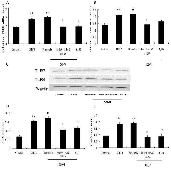
Effect of Prdx6-iPLA2 on Toll-like receptor 2 (TLR2) and TLR4 expression in microglia in response to OGD/R. TLR2 and TLR4 were activated during OGD/R exposure. Prdx6-iPLA2 siRNA or MJ33 treatment could inhibit TLR2 and TLR4 mRNA (A,B) and protein expression (C–E) . Results are expressed as the mean ± SEM of three independent experiments. ** p < 0.01 vs. Control; # p < 0.05 vs. Scramble; ## p < 0.01 vs. Scramble.
Index in PubMed under a CC BY license. PMID: 28424593

Click image to see more details
Expression of TLR2 and TLR4 in response to Prdx6 siRNA and MJ33. Real-time PCR showed that TLR2 (A) and TLR4 (B) expression increased after MCAO or giving Prdx6-siRNA. Combined treatment with Prdx6-siRNA and MJ33 downregulated TLR2 and TLR4 levels. Representative Western blot (C) and semi-quantitative analyses of the levels of TLR2 (D) and TLR4 (E) in the cortex after MCAO. Results are expressed as the mean ± SEM of three independent experiments. & p < 0.01 vs. Sham; ** p < 0.01 vs. Scramble; # p < 0.05, vs. Prdx6 siRNA; ## p < 0.01 vs. Prdx6 siRNA.
Index in PubMed under a CC BY license. PMID: 28424593

Click image to see more details
Expression of TLR2 in splenocytes of different vaccinated groups on day 2 after infection with S . aureus . (A) Western blot detection of TLR2 in splenocytes. Lane A: PBS group; lane B pEGFP-C2 group; lane C pEGFP /HA2 group; lane D pEGFP/Ag85A group; lane E pEGFP/Ag85A-HA2 group; lane F iPR group. (B) RT-PCR detection of TLR2 in splenocytes. Lanes 1, 8: pEGFP/Ag85A-HA2 group; lanes 2, 9: pEGFP-C2 group; lanes 3, 10: pEGFP /HA2 group; lanes 4, 11: pEGFP/Ag85A group; lanes 5, 12: PBS group; lanes 7, 13: iPR group. Lanes 1–5, 7: TLR2 (410 bp); lanes 8–13: GADPH (531 bp); lanes 6, 14: marker.
Index in PubMed under a CC BY license. PMID: 23369570

Click image to see more details
Western blot analysis of TLR2 using anti-TLR2 antibody (A00131).
Electrophoresis was performed on a 5-20% SDS-PAGE gel at 70V (Stacking gel) / 90V (Resolving gel) for 2-3 hours. The sample well of each lane was loaded with 30 ug of sample under reducing conditions.
Lane 1: human THP-1 (-LPS) whole cell lysates,
Lane 2: human THP-1 (+LPS) whole cell lysates,
Lane 3: human Hacat whole cell lysates,
Lane 4: human PC-3 whole cell lysates,
Lane 5: human Caco-2 whole cell lysates,
Lane 6: human HepG2 whole cell lysates.
After electrophoresis, proteins were transferred to a nitrocellulose membrane at 150 mA for 50-90 minutes. Blocked the membrane with 5% non-fat milk/TBS for 1.5 hour at RT. The membrane was incubated with rabbit anti-TLR2 antigen affinity purified polyclonal antibody (A00131) at 0.5 μg/mL overnight at 4°C, then washed with TBS-0.1%Tween 3 times with 5 minutes each and probed with a goat anti-rabbit IgG-HRP secondary antibody at a dilution of 1:5000 for 1.5 hour at RT. The signal is developed using an Enhanced Chemiluminescent detection (ECL) kit (Catalog # EK1002) with Tanon 5200 system. A specific band was detected for TLR2 at approximately 105 kDa. The expected band size for TLR2 is at 90 kDa.
Specific Publications For Anti-TLR2 Antibody Picoband® (A00131)
Loading publications
Recommended Resources
Here are featured tools and databases that you might find useful.
- Boster's Pathways Library
- Protein Databases
- Bioscience Research Protocol Resources
- Data Processing & Analysis Software
- Photo Editing Software
- Scientific Literature Resources
- Research Paper Management Tools
- Molecular Biology Software
- Primer Design Tools
- Bioinformatics Tools
- Phylogenetic Tree Analysis
Customer Reviews
Have you used Anti-TLR2 Antibody Picoband®?
Share your experimental results or join a short interview to earn up to $1,000 in product credits or other rewards.
0 Reviews For Anti-TLR2 Antibody Picoband®
Customer Q&As
Have a question?
Find answers in Q&As, reviews.
Can't find your answer?
Submit your question
15 Customer Q&As for Anti-TLR2 Antibody Picoband®
Question
We appreciate helping with my inquiry over the phone. Here are the WB image, lot number and protocol we used for leukocyte prostate using anti-TLR2 antibody A00131. Let me know if you need anything else.
Verified Customer
Verified customer
Asked: 2020-04-23
Answer
Thank you for the data. You have provided everything we needed. Our lab team are working to resolve your inquiry as quickly as possible, and we appreciate your patience and understanding! Please let me know if there is anything you need in the meantime.
Boster Scientific Support
Answered: 2020-04-23
Question
We have seen staining in rat leukocyte prostate. What should we do? Is anti-TLR2 antibody supposed to stain leukocyte prostate positively?
Verified Customer
Verified customer
Asked: 2020-02-10
Answer
From literature leukocyte prostate does express TLR2. From Uniprot.org, TLR2 is expressed in blood, leukocyte prostate, fetal lung, t-cell, monocyte, among other tissues. Regarding which tissues have TLR2 expression, here are a few articles citing expression in various tissues:
Blood, Pubmed ID: 15489334
Fetal lung, Pubmed ID: 9751057
Leukocyte, and Prostate, Pubmed ID: 9596645, 9435236
Monocyte, Pubmed ID: 16622205
T-cell, Pubmed ID: 10426995, 21078852
Boster Scientific Support
Answered: 2020-02-10
Question
Does A00131 anti-TLR2 antibody work on parafin embedded sections? If so, which fixation method do you recommend we use (PFA, paraformaldehyde, other)?
Verified Customer
Verified customer
Asked: 2019-10-16
Answer
As indicated on the product datasheet, A00131 anti-TLR2 antibody as been validated on WB. It is best to use PFA for fixation because it has better tissue penetration ability. PFA needs to be prepared fresh before use. Long term stored PFA turns into formalin, as the PFA molecules congregate and become formalin.
Boster Scientific Support
Answered: 2019-10-16
Question
My lab would like to test anti-TLR2 antibody A00131 on rat leukocyte prostate for research purposes, then I may be interested in using anti-TLR2 antibody A00131 for diagnostic purposes as well. Is the antibody suitable for diagnostic purposes?
Verified Customer
Verified customer
Asked: 2019-10-09
Answer
The products we sell, including anti-TLR2 antibody A00131, are only intended for research use. They would not be suitable for use in diagnostic work. If you have the means to develop a product into diagnostic use, and are interested in collaborating with us and develop our product into an IVD product, please contact us for more discussions.
Boster Scientific Support
Answered: 2019-10-09
Question
We are currently using anti-TLR2 antibody A00131 for human tissue, and we are content with the WB results. The species of reactivity given in the datasheet says human, mouse, rat. Is it likely that the antibody can work on canine tissues as well?
Verified Customer
Verified customer
Asked: 2019-08-20
Answer
The anti-TLR2 antibody (A00131) has not been validated for cross reactivity specifically with canine tissues, but there is a good chance of cross reactivity. We have an innovator award program that if you test this antibody and show it works in canine you can get your next antibody for free. Please contact me if I can help you with anything.
Boster Scientific Support
Answered: 2019-08-20
Question
Is this A00131 anti-TLR2 antibody reactive to the isotypes of TLR2?
Verified Customer
Verified customer
Asked: 2019-08-13
Answer
The immunogen of A00131 anti-TLR2 antibody is E.coli-derived human TLR2 recombinant protein (Position: H610-S784). Human TLR2 shares 87.4% amino acid (aa) sequence identity with mouse TLR2. Could you tell me which isotype you are interested in so I can help see if the immunogen is part of this isotype?
Boster Scientific Support
Answered: 2019-08-13
Question
Does anti-TLR2 antibody A00131 work for WB with leukocyte prostate?
Verified Customer
Verified customer
Asked: 2019-05-20
Answer
According to the expression profile of leukocyte prostate, TLR2 is highly expressed in leukocyte prostate. So, it is likely that anti-TLR2 antibody A00131 will work for WB with leukocyte prostate.
Boster Scientific Support
Answered: 2019-05-20
Question
I have a question about product A00131, anti-TLR2 antibody. I was wondering if it would be possible to conjugate this antibody with biotin. I would need it to be without BSA or sodium azide. I am planning on using a buffer exchange of sodium azide with PBS only. Would there be problems for me to conjugate the antibody and store it in -20 degrees in small aliquots?
Verified Customer
Verified customer
Asked: 2019-04-23
Answer
It is not recommended storing this antibody with PBS buffer only in -20 degrees. If you want to store it in -20 degrees it is best to add some cryoprotectant like glycerol. If you want carrier free A00131 anti-TLR2 antibody, we can provide it to you in a special formula with trehalose and/or glycerol. These molecules will not interfere with conjugation chemistry and provide a good level of protection for the antibody from degradation. Please be sure to specify this in your purchase order.
Boster Scientific Support
Answered: 2019-04-23
Question
We are interested in using your anti-TLR2 antibody for cellular response to bacterial lipopeptide studies. Has this antibody been tested with western blotting on mouse spleen tissue? We would like to see some validation images before ordering.
Verified Customer
Verified customer
Asked: 2018-09-24
Answer
I appreciate your inquiry. This A00131 anti-TLR2 antibody is validated on rat spleen tissue, mouse spleen tissue. It is guaranteed to work for WB in human, mouse, rat. Our Boster guarantee will cover your intended experiment even if the sample type has not been be directly tested.
Boster Scientific Support
Answered: 2018-09-24
Question
We were well pleased with the WB result of your anti-TLR2 antibody. However we have been able to see positive staining in t-cell membrane using this antibody. Is that expected? Could you tell me where is TLR2 supposed to be expressed?
Verified Customer
Verified customer
Asked: 2018-03-12
Answer
From literature, t-cell does express TLR2. Generally TLR2 expresses in membrane. Regarding which tissues have TLR2 expression, here are a few articles citing expression in various tissues:
Blood, Pubmed ID: 15489334
Fetal lung, Pubmed ID: 9751057
Leukocyte, and Prostate, Pubmed ID: 9596645, 9435236
Monocyte, Pubmed ID: 16622205
T-cell, Pubmed ID: 10426995, 21078852
Boster Scientific Support
Answered: 2018-03-12
Question
I was wanting to use your anti-TLR2 antibody for WB for rat leukocyte prostate on frozen tissues, but I want to know if it has been tested for this particular application. Has this antibody been tested and is this antibody a good choice for rat leukocyte prostate identification?
H. Rodriguez
Verified customer
Asked: 2018-02-15
Answer
As indicated on the product datasheet, A00131 anti-TLR2 antibody has been validated for WB on human, mouse, rat tissues. We have an innovator award program that if you test this antibody and show it works in rat leukocyte prostate in IHC-frozen, you can get your next antibody for free.
Boster Scientific Support
Answered: 2018-02-15
Question
Is a blocking peptide available for product anti-TLR2 antibody (A00131)?
Verified Customer
Verified customer
Asked: 2017-12-18
Answer
We do provide the blocking peptide for product anti-TLR2 antibody (A00131). If you would like to place an order for it please contact support@bosterbio.com and make a special request.
Boster Scientific Support
Answered: 2017-12-18
Question
Do you have a BSA free version of anti-TLR2 antibody A00131 available?
B. Rodriguez
Verified customer
Asked: 2017-07-25
Answer
Thank you for your recent telephone inquiry. I can confirm that some lots of this anti-TLR2 antibody A00131 are BSA free. For now, these lots are available and we can make a BSA free formula for you free of charge. It will take 3 extra days to prepare. If you require this antibody BSA free again in future, please do not hesitate to contact me and I will be pleased to check which lots we have in stock that are BSA free.
Boster Scientific Support
Answered: 2017-07-25
Question
See attached the WB image, lot number and protocol we used for leukocyte prostate using anti-TLR2 antibody A00131. Please let me know if you require anything else.
K. Yang
Verified customer
Asked: 2014-08-11
Answer
Thank you very much for the data. Our lab team are working to resolve this as quickly as possible, and we appreciate your patience and understanding! You have provided everything we needed. Please let me know if there is anything you need in the meantime.
Boster Scientific Support
Answered: 2014-08-11
Question
I see that the anti-TLR2 antibody A00131 works with WB, what is the protocol used to produce the result images on the product page?
W. Li
Verified customer
Asked: 2013-05-22
Answer
You can find protocols for WB on the "support/technical resources" section of our navigation menu. If you have any further questions, please send an email to support@bosterbio.com
Boster Scientific Support
Answered: 2013-05-22
