


Click image to see more details
-
-
-
-
-
+2
Product Info Summary
| SKU: | PB9071 |
|---|---|
| Size: | 100 μg/vial |
| Reactive Species: | Human, Mouse, Rat |
| Host: | Rabbit |
| Application: | Flow Cytometry, IHC, WB |
Customers Who Bought This Also Bought

Product info
Product Name
Anti-VEGF/VEGFA Antibody Picoband®
SKU/Catalog Number
PB9071
Size
100 μg/vial
Form
Lyophilized
Description
Boster Bio Anti-VEGF/VEGFA Antibody Picoband® catalog # PB9071. Tested in Flow Cytometry, IHC, WB applications. This antibody reacts with Human, Mouse, Rat. The brand Picoband indicates this is a premium antibody that guarantees superior quality, high affinity, and strong signals with minimal background in Western blot applications. Only our best-performing antibodies are designated as Picoband, ensuring unmatched performance.
Storage & Handling
Store at -20˚C for one year from date of receipt. After reconstitution, at 4˚C for one month. It can also be aliquotted and stored frozen at -20˚C for six months. Avoid repeated freeze-thaw cycles.
Cite This Product
Anti-VEGF/VEGFA Antibody Picoband® (Boster Biological Technology, Pleasanton CA, USA, Catalog # PB9071)
Host
Rabbit
Contents
Each vial contains 4 mg Trehalose, 0.9 mg NaCl and 0.2 mg Na2HPO4.
Clonality
Polyclonal
Isotype
Rabbit IgG
Immunogen
E.coli-derived human VEGF recombinant protein (Position: A27-R191). Human VEGF shares 78% amino acid (aa) sequence identity with both mouse and rat VEGF.
Cross-reactivity
No cross-reactivity with other proteins
Reactive Species
PB9071 is reactive to VEGFA in Human, Mouse, Rat
Observed Molecular Weight
18, 27 kDa
Calculated molecular weight
27.0 kDa
Background of VEGFA
VEGF, a homodimeric glycoprotein of relative molecular mass 45,000, is the only mitogen that specifically acts on endothelial cells. It may be a major regulator of tumor angiogenesis in vivo. It is, however, structurally related to platelet-derived growth factor. VEGF shares homology with the PDGF A chain and B chain, including conservation of all 8 cysteines found in PDGFA and PDGFB. VEGF gene contains 8 exons. VEGF induces remodeling and enhances TH2-mediated sensitization and inflammation in the lung. And this gene also can regulate haematopoietic stem cell survival by an internal autocrine loop mechanism. What’s more, it also stimulates neurogenesis in vitro and in vivo.
Antibody Validation
Boster validates all antibodies on WB, IHC, ICC, Immunofluorescence, and ELISA with known positive control and negative samples to ensure specificity and high affinity, including thorough antibody incubations.
Application & Images
Applications
PB9071 is guaranteed for Flow Cytometry, IHC, WB Boster Guarantee
Recommend Dilution
| Application | Dilution | Species |
|---|---|---|
| Western blot | 0.1-0.5μg/ml | Human, Mouse, Rat |
| Immunohistochemistry (Paraffin-embedded Section) | 2-5μg/ml | Human |
| Flow Cytometry (Fixed) | 1-3μg/1x106 cells | Human |
Note: For flow cytometry (FCM), validated for fixed cells only. Not validated for FACS or surface flow cytometry using live cells.
Tested application
Suggested blocking solution with 5% non-fat milk or BSA; (*)Recommended protein loading: 20-40 µg per lane
Use TE buffer pH 9.0 for antigen retrieval; (*) citrate buffer pH 6.0 is an alternative.
Validation Images & Assay Conditions
Click image to see more details
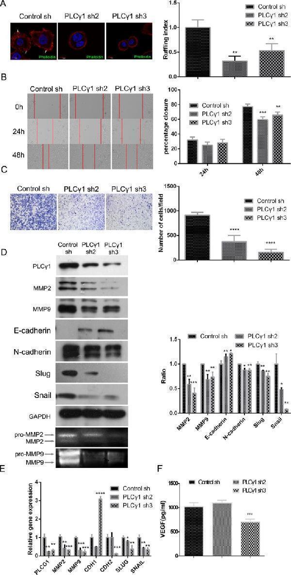
Lentivirus-mediated PLCγ1 shRNA could suppress migration in human gastric adenocarcinoma BGC-823 cells. Cells were transduced with lentivirus-mediated PLCγ1 shRNA2/3 vectors. ( A ) The formation of membrane ruffles was detected using Ruffling assay as described in Materials and Methods. The cell nuclei were stained DAPI (blue) and the membrane ruffles were stained rhodamine-conjugated phalloidin (red). Scale bar = 10 μm. ( B and C ) The migration ability was measured using Transwell assay (B, magnification × 100) and Scratch assay (C, magnification × 400) as described in Materials and Methods. ( D ) The protein levels of MMP2, MMP9, E-cadherin, N-cadherin, snail, slug, and GAPDH were detected with Western blotting analysis, and the pro and active forms of MMP2/9 were observed using gelatin zymography assay as described in Materials and Methods. ( E ) The mRNA levels of PLCG1, MMP2, MMP9, CDHI, CDH2, SNAIL, SLUG, and GAPDH were detected using Real-time PCR analysis as described in Materials and Methods. ( F ) The level of VEGF in extracellular matrix was detected using ELISA as described in Materials and Methods. Data are reported as means ± S.D. of three independent experiments (* P < 0.05, ** P < 0.01, *** P < 0.001, **** P < 0.0001, vs respective control).
Index in PubMed under a CC BY license. PMID: 26811493
Click image to see more details
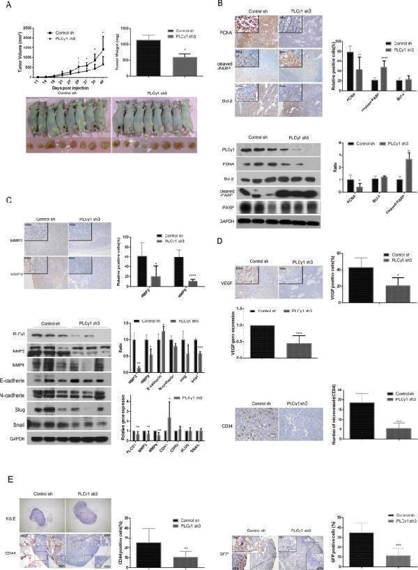
Depletion of PLCγ1 suppresses growth and metastasis of gastric adenocarcinoma in a nude mouse tumor xenograft model. ( A ) Volume and weight of tumor samples from nude mice. ( B ) The protein levels of PCNA, cleaved-PARP, PARP, and Bcl-2 in the tumor samples were detected by Immunohistochemistry (Magnificationx100, x400) and Western blotting analyses as described in Materials and Methods. ( C ) The levels of MMP2 and MMP9 in the tumor samples were detected by Immunohistochemistry analysis as described in Materials and Methods (Magnificationx100, x400). The protein and mRNA levels of MMP2, MMP9, E-cadherin(CDH1), N-cadherin(CDH2), snail(SNAIL), and slug(SLUG) in the tumor samples were detected by Western Blotting and Real-time PCR analyses as described in Materials and Methods. ( D ) The protein levels of VEGF and CD34 and the mRNA level of VEGF in tumor samples were detected by Immunohistochemistry and Real-time PCR analysis as described in Materials and Methods. The number of microvessels was accounted under OLYPUS x41 microscope (Magnification x100, x400). ( E ) The lymphoid follicles in inguinal lymph nodes of nude mice were observed under OLYPUS x41microscope, and the protein levels of CD44 and GFP in inguinal lymph nodes of nude mice was detected by Immunohistochemistry analysis as described in Materials and Methods (Magnificationx40, x400). Data are reported as means ± S.D. of three independent experiments (* P < 0.05, ** P < 0.01, *** P < 0.001, **** P < 0.0001, vs respective control).
Index in PubMed under a CC BY license. PMID: 26811493
Click image to see more details
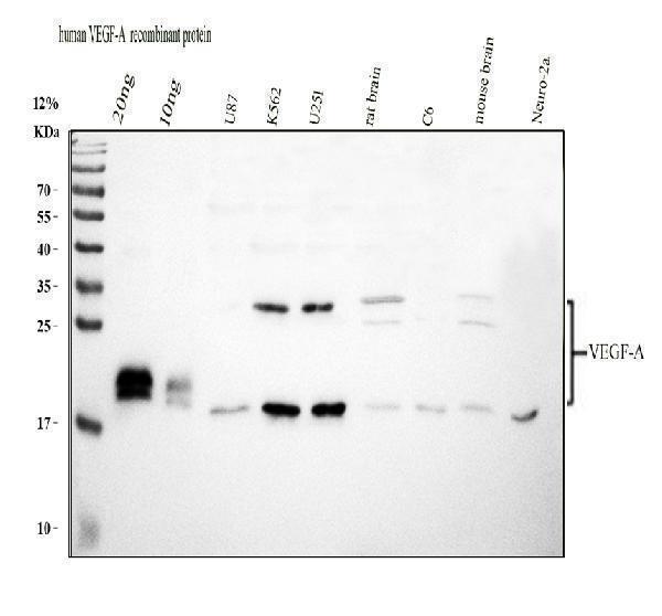
Western blot analysis of VEGF/VEGFA using anti-VEGF/VEGFA antibody (PB9071).
Electrophoresis was performed on a 12% SDS-PAGE gel at 80V (Stacking gel) / 120V (Resolving gel) for 2 hours. The sample well of each lane was loaded with 30 ug of sample under reducing conditions.
Lane 1: recombinant human VEGF/VEGFA protein 20ng,
Lane 2: recombinant human VEGF/VEGFA protein 10ng,
Lane 3: human U87 whole cell lysates,
Lane 4: human K562 whole cell lysates,
Lane 5: rat brain tissue lysates,
Lane 6: rat C6 whole cell lysates,
Lane 7: mouse brain tissue lysates,
Lane 8: mouse Neuro-2a whole cell lysates.
After electrophoresis, proteins were transferred to a nitrocellulose membrane at 150 mA for 50-90 minutes. Blocked the membrane with 5% non-fat milk/TBS for 1.5 hour at RT. The membrane was incubated with rabbit anti-VEGF/VEGFA antigen affinity purified polyclonal antibody (PB9071) at 0.5 μg/mL overnight at 4°C, then washed with TBS-0.1%Tween 3 times with 5 minutes each and probed with a goat anti-rabbit IgG-HRP secondary antibody (Catalog # BA1054) at a dilution of 1:5000 for 1.5 hour at RT. The signal is developed using an ECL Plus Western Blotting Substrate (Catalog # AR1196-200) with Tanon 5200 system. A specific band was detected for VEGF/VEGFA at approximately 18, 27 kDa. The expected band size for VEGF/VEGFA is at 27 kDa.
Click image to see more details
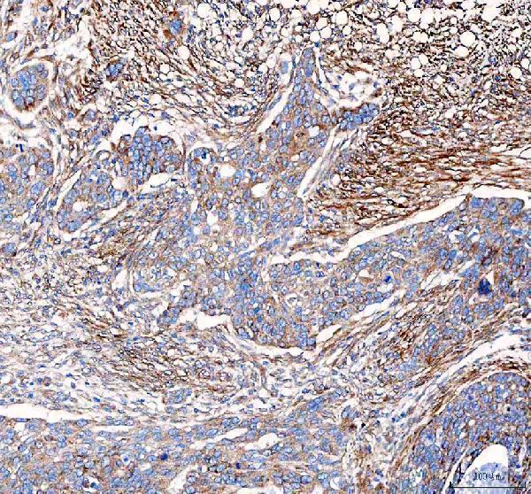
IHC analysis of VEGF/VEGFA using anti-VEGF/VEGFA antibody (PB9071).
VEGF/VEGFA was detected in a paraffin-embedded section of human ovarian serous adenocarcinoma tissue. Heat mediated antigen retrieval was performed in EDTA buffer (pH 8.0, epitope retrieval solution). The tissue section was blocked with 10% goat serum. The tissue section was then incubated with 2 μg/ml rabbit anti-VEGF/VEGFA Antibody (PB9071) overnight at 4°C. Peroxidase Conjugated Goat Anti-rabbit IgG was used as secondary antibody and incubated for 30 minutes at 37°C. The tissue section was developed using HRP Conjugated Rabbit IgG Super Vision Assay Kit (Catalog # SV0002) with DAB as the chromogen.
Click image to see more details
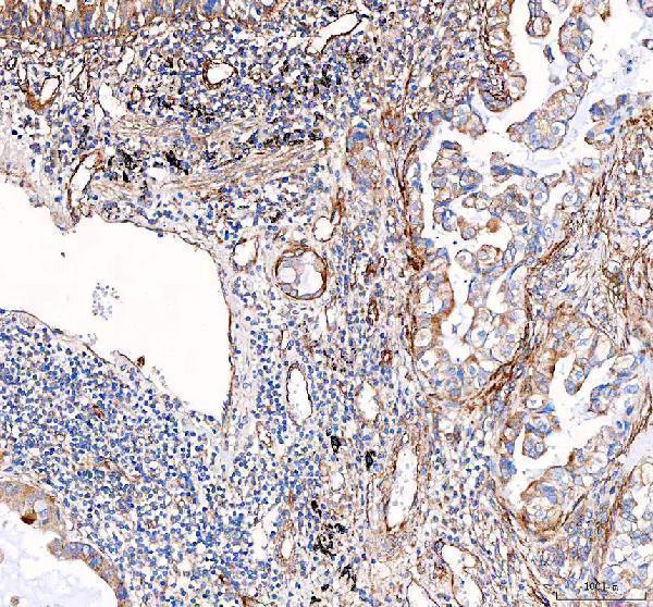
IHC analysis of VEGF/VEGFA using anti-VEGF/VEGFA antibody (PB9071).
VEGF/VEGFA was detected in a paraffin-embedded section of human lung cancer tissue. Heat mediated antigen retrieval was performed in EDTA buffer (pH 8.0, epitope retrieval solution). The tissue section was blocked with 10% goat serum. The tissue section was then incubated with 2 μg/ml rabbit anti-VEGF/VEGFA Antibody (PB9071) overnight at 4°C. Peroxidase Conjugated Goat Anti-rabbit IgG was used as secondary antibody and incubated for 30 minutes at 37°C. The tissue section was developed using HRP Conjugated Rabbit IgG Super Vision Assay Kit (Catalog # SV0002) with DAB as the chromogen.
Click image to see more details
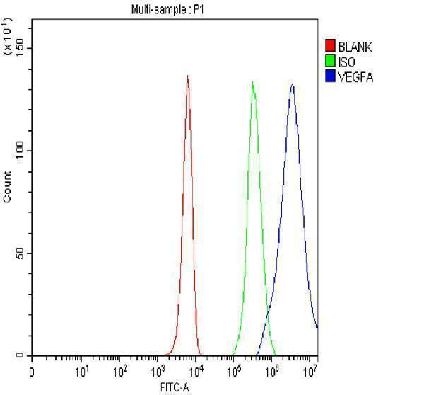
Flow Cytometry analysis of K562 cells using anti-VEGF/VEGFA antibody (PB9071).
Overlay histogram showing K562 cells stained with PB9071 (Blue line). The cells were fixed with 4% paraformaldehyde and blocked with 10% normal goat serum. And then incubated with rabbit anti-VEGF/VEGFA Antibody (PB9071, 1 μg/1x106 cells) for 30 min at 20°C. DyLight®488 conjugated goat anti-rabbit IgG (BA1127, 5-10 μg/1x106 cells) was used as secondary antibody for 30 minutes at 20°C. Isotype control antibody (Green line) was rabbit IgG (1 μg/1x106) used under the same conditions. Unlabelled sample without incubation with primary antibody and secondary antibody (Red line) was used as a blank control.
Specific Publications For Anti-VEGF/VEGFA Antibody Picoband® (PB9071)
Loading publications
Recommended Resources
Here are featured tools and databases that you might find useful.
- Boster's Pathways Library
- Protein Databases
- Bioscience Research Protocol Resources
- Data Processing & Analysis Software
- Photo Editing Software
- Scientific Literature Resources
- Research Paper Management Tools
- Molecular Biology Software
- Primer Design Tools
- Bioinformatics Tools
- Phylogenetic Tree Analysis
Customer Reviews
Have you used Anti-VEGF/VEGFA Antibody Picoband®?
Share your experimental results or join a short interview to earn up to $1,000 in product credits or other rewards.
0 Reviews For Anti-VEGF/VEGFA Antibody Picoband®
Customer Q&As
Have a question?
Find answers in Q&As, reviews.
Can't find your answer?
Submit your question
5 Customer Q&As for Anti-VEGF/VEGFA Antibody Picoband®
Question
We need using your anti-VEGF/VEGFA antibody for positive regulation of cell proliferation by vegf-activated platelet derived growth factor receptor signaling pathway studies. Has this antibody been tested with western blotting on tissue lysate? We would like to see some validation images before ordering.
Verified Customer
Verified customer
Asked: 2020-03-13
Answer
I appreciate your inquiry. This PB9071 anti-VEGF/VEGFA antibody is tested on human placenta tissue, lung cancer tissue, tissue lysate, rat thymus tissue, brain tissue. It is guaranteed to work for IHC, WB in human, rat. Our Boster guarantee will cover your intended experiment even if the sample type has not been be directly tested.
Boster Scientific Support
Answered: 2020-03-13
Question
Our lab were satisfied with the WB result of your anti-VEGF/VEGFA antibody. However we have observed positive staining in left lobe of thyroid gland secreted using this antibody. Is that expected? Could you tell me where is VEGFA supposed to be expressed?
Verified Customer
Verified customer
Asked: 2020-01-17
Answer
According to literature, left lobe of thyroid gland does express VEGFA. Generally VEGFA expresses in secreted. Regarding which tissues have VEGFA expression, here are a few articles citing expression in various tissues:
Heart, Pubmed ID: 19054851
Kidney, Pubmed ID: 9878851, 12124351
Lung, Pubmed ID: 15489334
Mammary gland, Pubmed ID: 9450968
Renal glomerulus, Pubmed ID: 10464055
Retina, Pubmed ID: 10067980, 11563986
Boster Scientific Support
Answered: 2020-01-17
Question
We are currently using anti-VEGF/VEGFA antibody PB9071 for rat tissue, and we are satisfied with the WB results. The species of reactivity given in the datasheet says human, rat. Is it true that the antibody can work on dog tissues as well?
Verified Customer
Verified customer
Asked: 2019-11-13
Answer
The anti-VEGF/VEGFA antibody (PB9071) has not been validated for cross reactivity specifically with dog tissues, though there is a good chance of cross reactivity. We have an innovator award program that if you test this antibody and show it works in dog you can get your next antibody for free. Please contact me if I can help you with anything.
Boster Scientific Support
Answered: 2019-11-13
Question
We have been able to see staining in human retina. Do you have any suggestions? Is anti-VEGF/VEGFA antibody supposed to stain retina positively?
R. Jha
Verified customer
Asked: 2019-11-11
Answer
According to literature retina does express VEGFA. According to Uniprot.org, VEGFA is expressed in left lobe of thyroid gland, kidney, mammary gland, renal glomerulus, hemangioendothelioma, heart, lung, retina, among other tissues. Regarding which tissues have VEGFA expression, here are a few articles citing expression in various tissues:
Heart, Pubmed ID: 19054851
Kidney, Pubmed ID: 9878851, 12124351
Lung, Pubmed ID: 15489334
Mammary gland, Pubmed ID: 9450968
Renal glomerulus, Pubmed ID: 10464055
Retina, Pubmed ID: 10067980, 11563986
Boster Scientific Support
Answered: 2019-11-11
Question
We purchased anti-VEGF/VEGFA antibody for IHC on mammary gland in the past. I am using human, and We want to use the antibody for WB next. you antibody examining mammary gland as well as retina in our next experiment. Could you please give me some suggestion on which antibody would work the best for WB?
R. Anderson
Verified customer
Asked: 2018-03-08
Answer
I looked at the website and datasheets of our anti-VEGF/VEGFA antibody and it appears that PB9071 has been validated on human in both IHC and WB. Thus PB9071 should work for your application. Our Boster satisfaction guarantee will cover this product for WB in human even if the specific tissue type has not been validated. We do have a comprehensive range of products for WB detection and you can check out our website bosterbio.com to find out more information about them.
Boster Scientific Support
Answered: 2018-03-08

