This website uses cookies to ensure you get the best experience on our website.
- Table of Contents
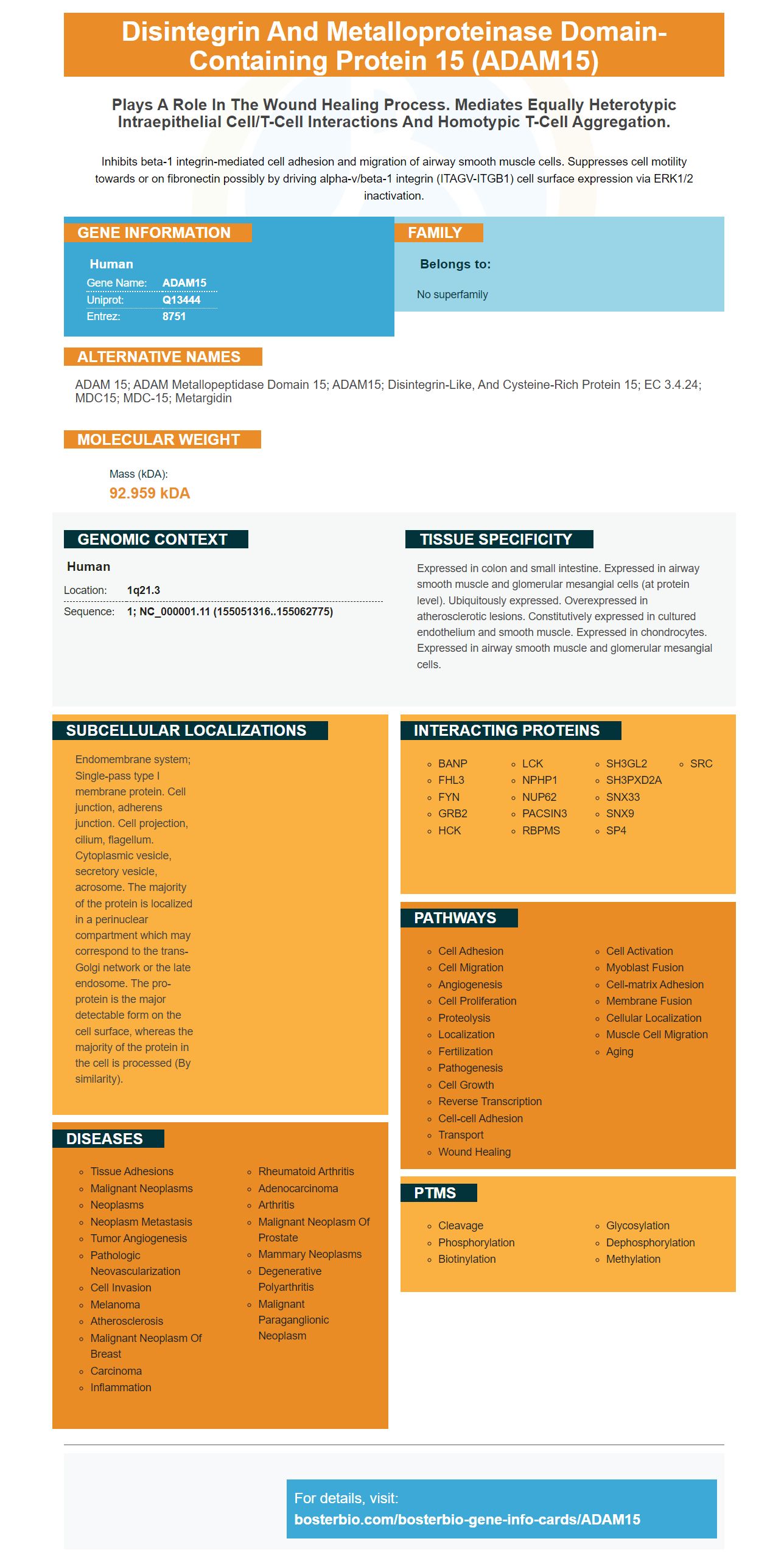
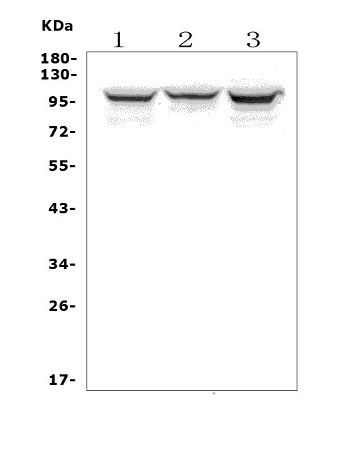
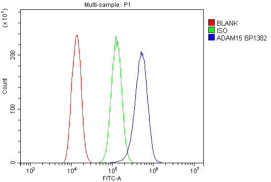
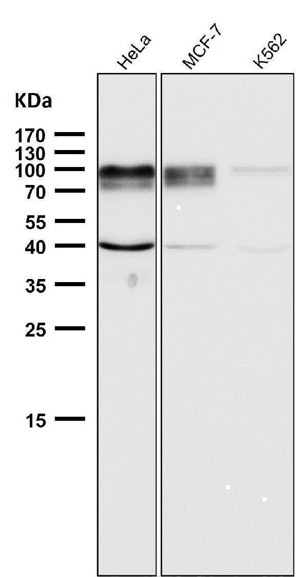
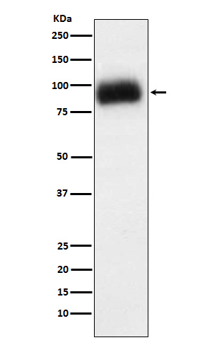
Facts about Disintegrin and metalloproteinase domain-containing protein 15.
Inhibits beta-1 integrin-mediated cell adhesion and migration of airway smooth muscle cells. Suppresses cell motility towards or on fibronectin possibly by driving alpha-v/beta-1 integrin (ITAGV-ITGB1) cell surface expression via ERK1/2 inactivation.
Human | |
---|---|
Gene Name: | ADAM15 |
Uniprot: | Q13444 |
Entrez: | 8751 |
Belongs to: |
---|
No superfamily |
ADAM 15; ADAM metallopeptidase domain 15; ADAM15; disintegrin-like, and cysteine-rich protein 15; EC 3.4.24; MDC15; MDC-15; Metargidin
Mass (kDA):
92.959 kDA
Human | |
---|---|
Location: | 1q21.3 |
Sequence: | 1; NC_000001.11 (155051316..155062775) |
Expressed in colon and small intestine. Expressed in airway smooth muscle and glomerular mesangial cells (at protein level). Ubiquitously expressed. Overexpressed in atherosclerotic lesions. Constitutively expressed in cultured endothelium and smooth muscle. Expressed in chondrocytes. Expressed in airway smooth muscle and glomerular mesangial cells.
Endomembrane system; Single-pass type I membrane protein. Cell junction, adherens junction. Cell projection, cilium, flagellum. Cytoplasmic vesicle, secretory vesicle, acrosome. The majority of the protein is localized in a perinuclear compartment which may correspond to the trans-Golgi network or the late endosome. The pro-protein is the major detectable form on the cell surface, whereas the majority of the protein in the cell is processed (By similarity).
In this article, you will learn about the best uses of the ADAM15 Marker. This marker is used to determine the transfer efficiency of proteins within cells. It has multiple applications and is also useful for research. It is a highly recommended marker for many research projects. Here are some examples of how to use it. Also, you will learn about its benefits. Read on to learn more!
This monoclonal antibody recognizes the disintegrin/cysteine-rich/EGF repeat domain in mouse ADAM15. ADAM15 is a member of the family of proteins called ADAMs, which are involved in a variety of cell-cell interactions and physiological processes. ADAM15 is a membrane-anchored glycoprotein. Recent research indicates that ADAM15 may be involved in prostate cancer metastasis and pathological neovascularization.
Several suppliers offer anti-ADA15 ADAM15 antibodies. This antibody reacts with human, mouse, and rat. It is not recommended for use in clinical applications. However, it is recommended for research purposes, and may be used to enhance a clinical study. The Boster Bio ADAM15 Antibody is available as a recombinant protein or as an ELISA Kit. The ADAM15 antibody is not intended for resale or commercial use. It can serve as a great boost for your research.
The expression of ADAM15 has been found to be correlated with immune cell infiltration in different types of cancer. Its knockdown has been associated with reduced expression of Bcl-2, N-Cadherin, Snail, and Bax. Conversely, ADAM15 overexpression has the opposite effect. These findings suggest that ADAM15 may be a potential biomarker for HCC.
In HCC, ADAM15 overexpression is associated with decreased survival and reduced survival. In the same way, ADAM15 expression in HCC tissues was higher than in noncancerous tissues. Moreover, TCGA and GEO databases validated that ADAM15 is overexpressed in HCC. Further studies are needed to understand the role of ADAM15 in these processes. So, buy Anti-ADA15 ADAM15 Antibody from Boster Bio
The ADA15 marker is the protein that encodes the antisense recombinant human cytokine p53, which is found in the cytosol of cells. The ADAM15 gene encodes the cellular response to hormones, cytokines, and other signaling proteins. This protein is expressed by fibroblasts and plays a major role in aging. Boster Bio's ADAM15 antibody is an ideal solution for various biological applications. The antibodies are available in a variety of formats including Western Blotting, Immunohistochemistry, and ELISA.
Improved membrane staining can dramatically increase the speed and sensitivity of Western blotting. Western blotting uses two types of membranes: PVDF and nitrocellulose. The difference between these two types of membranes depends on their binding abilities. PVDF is stronger, and NC is weaker. The NC membrane was also tested for its re-probing ability. Both membranes improved protein transfer efficiency.
The first membrane staining method involves preparing colloidal gold in TBST, a solution that binds to the protein bands. The gold binds strongly to proteins that have a concentration of two nanograms per band. The gold binds strongly to membranes, so it is possible to use commercial ready-to-use stains for the nitrocellulose membrane. The nitrocellulose membranes are stained in 50% methanol/water solution for 10 min.
For PVDF membranes, the amount of serum proteins needed for IgG staining was four-fold less than for NC membranes. A2M staining required 1.5 mg of serum proteins. Those proteins that are over 20 KD are best suited for NC membranes with their higher sensitivity, resolution, and affinity. For small molecular weight proteins, a PVDF membrane is the best choice.
In WB, the molecular weight of proteins transferred can greatly affect the efficiency of the protein transfer. A thicker gel will result in incomplete transfer since protein has to travel farther to reach the membrane. Thicker gels are also better for low-molecular-weight proteins, but the resulting transfer efficiency will be lowered by the reduced removal of protein from the gel matrix. This improved membrane staining method improves protein transfer efficiency by membrane staining in Western blotting applications.
While both semi-dry and wet transfer methods are effective, the wet method is generally more time-consuming. The efficiency of semi-dry transfer is significantly better than that of wet transfer. Both methods require the sandwich to be suspended in a transfer buffer. The sandwich is placed in the transfer buffer vertically, allowing the protein to pass through the membrane under an electric field. The duration of time required for both methods depends on the molecular weight of the target protein.
The ADAM15 Marker has many benefits in the field of cancer immunotherapy. This marker is associated with diverse biological functions in cancer, including reducing cell-cell adhesions, releasing growth factors, and increasing the expression of genes involved in apoptosis. In prostate cancer, ADAM15 targeting with siRNA inhibits prostate cancer cell metastasis. Prostate cancer cells have two distinct mechanisms for dissemination, vascular intravasation and local invasion.
ADAM15 protein expression has been studied in various clinical specimens and has been found to be a specific focal overexpression in advanced bladder cancer. All low grade bladder cancer samples showed low or moderate staining indexes, whereas 48% of invasive bladder cancer and 72% of metastatic disease specimens exhibited moderate to high staining levels of the ADAM15 marker. ADAM15 is expressed in normal urothelium and umbrella cells, a structure that is associated with tumor metastasis.
ADAM15 has been found to mediate the shedding of ectodomains. In addition, it is associated with integrin binding. Its substrates include epidermal growth factor family growth factors, which may be associated with atherosclerosis. The human ADAM15 protein contains an arginine-glycine-aspartate (RGD) sequence, while rodent ADAM-15 has avb3 and a5b1 integrins.
The ADAM15 Marker has also shown great potential in cancer immunotherapy. In studies, the protein is detected in urine from cancer patients. Interestingly, this marker has also been detected in breast cancer patients' urine. In one study, patients with breast cancer showed significantly higher levels of ADAM12 than healthy controls. Further, the percentage of patients with high levels of ADAM12 was higher in patients with the disease than in healthy controls.
PMID: 8617717 by Kraetzschmar J., et al. Metargidin, a membrane-anchored metalloprotease-disintegrin protein with an RGD integrin binding sequence.
PMID: 9039960 by Herren B., et al. Expression of a disintegrin-like protein in cultured human vascular cells and in vivo.