This website uses cookies to ensure you get the best experience on our website.
- Table of Contents
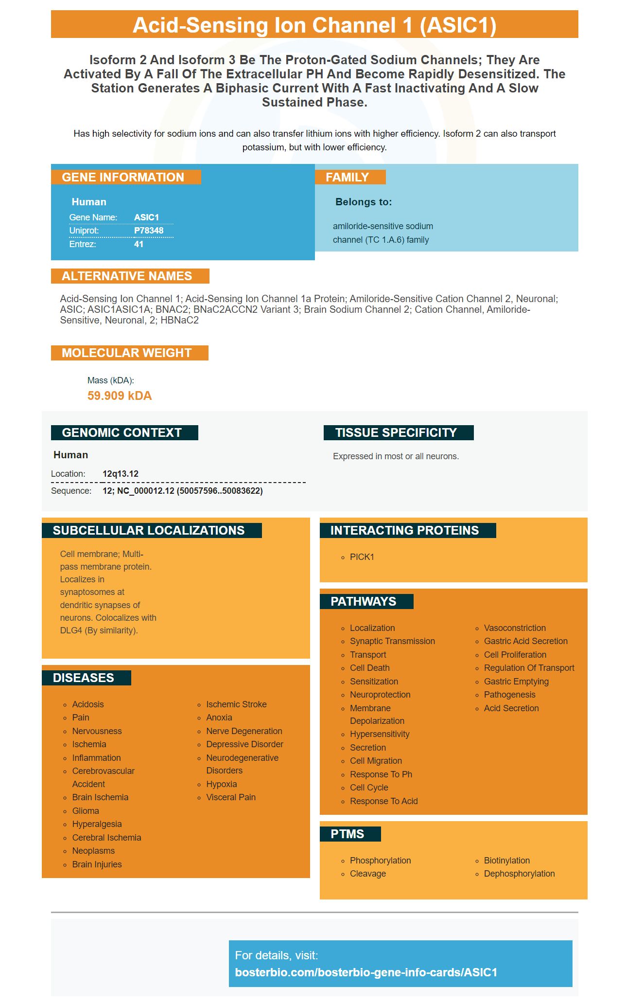
Facts about Acid-sensing ion channel 1.
Has high selectivity for sodium ions and can also transfer lithium ions with higher efficiency. Isoform 2 can also transport potassium, but with lower efficiency.
Human | |
---|---|
Gene Name: | ASIC1 |
Uniprot: | P78348 |
Entrez: | 41 |
Belongs to: |
---|
amiloride-sensitive sodium channel (TC 1.A.6) family |
Acid-sensing ion channel 1; acid-sensing ion channel 1a protein; amiloride-sensitive cation channel 2, neuronal; ASIC; ASIC1ASIC1A; BNAC2; BNaC2ACCN2 variant 3; Brain sodium channel 2; Cation channel, amiloride-sensitive, neuronal, 2; hBNaC2
Mass (kDA):
59.909 kDA
Human | |
---|---|
Location: | 12q13.12 |
Sequence: | 12; NC_000012.12 (50057596..50083622) |
Expressed in most or all neurons.
Cell membrane; Multi-pass membrane protein. Localizes in synaptosomes at dendritic synapses of neurons. Colocalizes with DLG4 (By similarity).
Boster has many resources for information about the ASIC1 gene. Boster's gene infographics provide basic information about each gene and cover all genes found in mice and humans. It also has a search bar that allows you to search for specific genes. It is a relatively recent gene so it takes some research to understand its role.
It is fascinating to study the regulatory relationship between miR-485 (ASIC1) and acid-sensitive Ion channel 1. Recent studies have shown that miR-485 suppresses ASIC1, an enterosensory gene. Enterodynia occurs when ASIC1 is reduced. ASIC1 may also be linked to the development a functional gastrointestinal disorder such as IBS. The environment can influence its expression.
MiR-485-5p was expressed at low levels within HCC tissues. Bioinformatics analyses and mechanism experiments confirmed that miR-485-5p, ASIC1, and ASIC1 work in tandem to suppress their expression. FAM83H-AS1 is the predicted negative regulator of miR-485 expression. MiR-485 regulates the expression of a subset, including FAM83H-AS1, MEF2D and ASIC1.
The Regulatory relationship between miR-485 and the ASIC1 gene has been demonstrated in experimental models of enterodynia. Agomir's upregulation of miR-485 results in enterodynia. It also suppresses ASIC1 expression at the spinal dorsalhorn and inhibits synaptic transmitting. Furthermore, miR-485 was linked to several types of carcinoma and has been associated in various ways with migration, proliferation, invasion and growth. Whether miR-485 regulates the expression of ASIC1 and thereby prevents enterodynia is unknown.
The Regulatory relationship between miR-485 and the ASIC1 gene has been identified in different mouse models. ASIC1 can be found in neurons, but is expressed in the spine dorsal. Although ASIC1 expression was limited to the spinal dorsalhorn, it was difficult not to exclude primary sensory nerve ends from it. ASIC1 could also be a presynaptic component of spinal synaptic transmission, as it was found to be.
Several mice transfected with miR-485-3p were found to have significantly reduced levels of this molecule. Infection of these mice by the miR-485-5p replica resulted in inhibitions of cell growth and migration, invasion, and death. This study confirms the importance and role of miR-485 when it comes to controlling cell growth, migration, and invasion. This gene was also shown to play an important role during the differentiation of different types of cancer cells.
MiRNAs regulate gene expression by targeting the 3'UTR region of a target mRNA. ASIC1 is responsible for controlling the expression of a wide array of genes in human spinal dorsalhorn. This evidence suggests that ASIC1 and miR-485 have a targeted regulatory relationship. They are both expressed in the same neurons of the spinal dorsal horn, which is important for understanding the role of miRs.
The current study shows that the expression of ASIC1 is increased in the dorsal horn of the spinal cord. The presence or absence of neurons and/or glial cells is what determines the expression of ASIC1 in this area. ASIC1 was significantly elevated in the NMD spinal chord model than it was in CON rats. This protein could be a therapeutic target. Further studies are needed in order to determine if ASIC1 can be found in other structures of spinal cord.
ASIC1 expression was compared to the expression of CGRP+ or IB4+ neuronal markers. This study revealed that ASIC1 expression was strongly correlated with neuronal marker expressions in the L4 DRG. However, ASIC1b was found to be higher in L5 DRGs than in L5 DRGs.
ASIC1 expression is high within dendritic spines. It shows a higher relative density than LckGFP in spines and membranes. This suggests that ASIC1a preferentially visits dendritic spines. ASIC1a also responds better to acid-induced changes of Ca2+ concentrations within ASIC1a// slice neuron.
It is possible that the two proteins are interconnected by co-expression in spinal dorsal horn neuron cells. ASIC1 has been shown in the reduction of inhibition of BMT by blocking NKCC1, a NKCC1 protein that is implicated in chronic visceral discomfort. It also decreases inhibitory and synaptic transmission in spinal dorsalhorn neuron cells. This suggests that both proteins could interact to reduce pain.
ASIC1 is important in the regulation and prevention of acidosis-induced death neuronal. The effect of acidosis on ASICs has been attributed to forward trafficking of ASICs, but the role of dendritic targeting is unclear. ASIC1 mutants whose dendritic levels are reduced have the opposite effect upon acidosis-induced vertebral loss.
The maturation or maturation of ASICs is what regulates their function in dorsal hair. In response to serum deprivation, most ASIC1a is trapped in the endoplasmic reticulum and undergoes rapid membrane insertion. ASIC2 expression is found in smooth muscle cells of the vascular system when glycerol has been present. MCAO reduces ASIC1a's expression by approximately 30 minutes following ischemia. Alternate biogenesis of ASICs could also be responsible for ischemic-induced neuronal damage.
Immunofluorescence staining revealed ASIC1 is primarily co-localized alongside NeuN. In contrast, ASIC1 is little or not co-localized with GFAP-positive astrocytes or with the microglial marker CD11b. The co-staining rates for ASIC1 from NeuN-positive cells were 8.5% and 83.5%, respectively. These results support the hypothesis that ASIC1 plays a role in activating the innate immune system.
NeuN antibody is immunoreactive in most neurons of the mammalian Central Nervous System (CNS). It is not detected in tissues outside the nervous system, such as glial cells and immature neural progenitor cells. The cell nucleus and the perinuclear cells mediate NeuN antibody binding. However, both NeuN protein forms are not equally prevalent in each location.
In one study, miR-485 was found to negatively regulate ASIC1 transcription. This compound was administered to PMS offspring rats for 7 days and the ASIC1 protein expression was determined by Western blotting and qPCR. MiR-485 treatment decreased ASIC1 expression but did not alter mRNA expression in comparison to the NC agomir groups.
ASIC1 costuining is low in neurons that have not been NeuN-positive. It is not known how NeuN immunoreactivity can be found in non-neuronal cells. Some methods could influence the results. ASIC1 is also highly co-localized within NeuN-positive neurons, according to the authors. But this is not conclusive.
The pathogenesis of inflammatory conditions such as arthritis has been linked to antibodies to NeuN protein. Although NeuN is found in many forms and intracellular locations, its presence has been limited to nervous tissue, where it is only detected in small amounts. ASIC1 can only bind to NeuN if it contains at least one phosphate group.
The antigenic determinaant is another factor. The molecule could also be fixed in formaldehyde or paraffin may be poured over it. This can reduce the anti-NeuN antibody affinity, but the high concentrations of synapsin 1 compensate for the low affinity. ASIC1 is not the answer as it is not a reliable way to identify antigens.
Application of ASIC1 markers in cell biology has made it possible to measure the cellular response to various stimuli. ASICs can be used for calcium imaging and molecular biology research. ASIC1 is also a useful diagnostic tool to assess the OLC's function. It can also be used to research human ovarian tumors. While further research is needed to determine the relative contributions of the ASIC1 variants to human cells, these results suggest that the ASIC1 marker contributes towards the formation of OP.
ASIC transcripts have been detected by RTPCR using total RNA templates isolated from adult rat cortical gray matter, corpus callosum, and optic nerve. Table 1 shows the primers for each sample. After 35 cycles (PCR), the conventional products are visualized via gel electrophoresis. The figure below shows a white arrowhead that marks the expected product dimensions in base pairs.
The ASIC1 gene is found in the oligodendrocyte cell lineage. It performs multiple functions. A number of DNA sequences regulate the expression level of this gene. The gene is expressed in oligodendrocytes during different stages of development, including the embryonic, adult, and follicle-like phases. ASIC1 is also expressed in all three stages, including the embryonic stage, adult stage, and the follicle-like phase of cultured OLC. However, the ASIC3 protein template was only weakly detected by the sensitive dye SYBR Gold.
The nervous system has many acid-sensing, ion channel expressions. They are involved in sensory processes and regulate acid and pain levels. ASIC1a channel regulates pH levels in CNS. They are also involved as regulators of learning and memory. ASIC1a also provides neuroprotection in stroke models. It may also be useful for detecting ischemic injury.
The ASIC1Marker protein is an essential component for studying ASIC1's functions in the heart as well as the esophagus. Their roles in mechanotransduction or esophageal pH sensing are also implicated. However, the exact role of ASIC in these processes is not fully understood due to insufficient knowledge of the ASIC subunit expression profiles. It is important that ASIC Subunits Form Heteromultimeric Channels, Each with a Different Functional Property.
PMID: 9037075 by Garcia-Anoveros J., et al. BNaC1 and BNaC2 constitute a new family of human neuronal sodium channels related to degenerins and epithelial sodium channels.
PMID: 21036899 by Hoagland E.N., et al. Identification of a calcium permeable human acid-sensing ion channel 1 transcript variant.