This website uses cookies to ensure you get the best experience on our website.
- Table of Contents
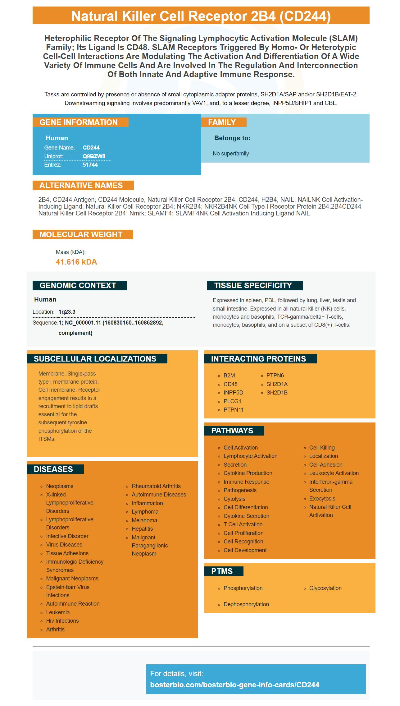
Facts about Natural killer cell receptor 2B4.

Tasks are controlled by presence or absence of small cytoplasmic adapter proteins, SH2D1A/SAP and/or SH2D1B/EAT-2. Downstreaming signaling involves predominantly VAV1, and, to a lesser degree, INPP5D/SHIP1 and CBL.
| Human | |
|---|---|
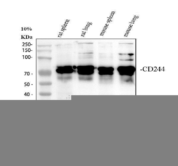
| Gene Name: | CD244 |
| Uniprot: | Q9BZW8 |
| Entrez: | 51744 |

| Belongs to: |
|---|
| No superfamily |

2B4; CD244 antigen; CD244 molecule, natural killer cell receptor 2B4; CD244; h2B4; NAIL; NAILNK cell activation-inducing ligand; natural killer cell receptor 2B4; NKR2B4; NKR2B4NK cell type I receptor protein 2B4,2B4CD244 natural killer cell receptor 2B4; Nmrk; SLAMF4; SLAMF4NK cell activation inducing ligand NAIL
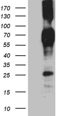
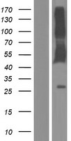
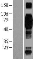
Mass (kDA):
41.616 kDA

| Human | |
|---|---|
| Location: | 1q23.3 |
| Sequence: | 1; NC_000001.11 (160830160..160862892, complement) |
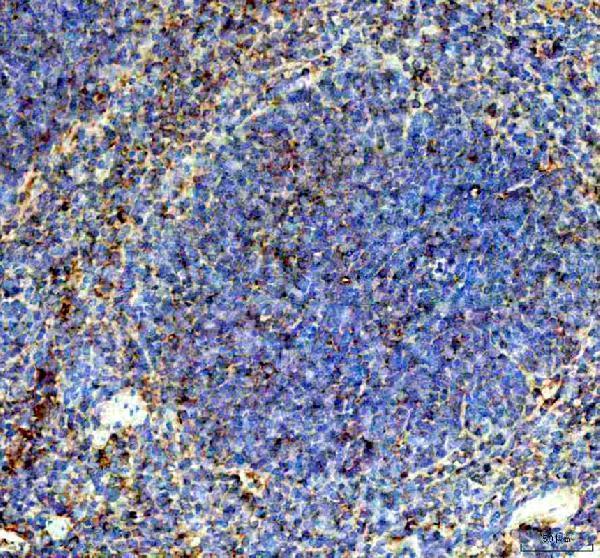
Expressed in spleen, PBL, followed by lung, liver, testis and small intestine. Expressed in all natural killer (NK) cells, monocytes and basophils, TCR-gamma/delta+ T-cells, monocytes, basophils, and on a subset of CD8(+) T-cells.
Membrane; Single-pass type I membrane protein. Cell membrane. Receptor engagement results in a recruitment to lipid drafts essential for the subsequent tyrosine phosphorylation of the ITSMs.


PMID: 10359122 by Nakajima H., et al. Activating interactions in human NK cell recognition: the role of 2B4-CD48.
PMID: 10556801 by Kubin M.Z., et al. Molecular cloning and biological characterization of NK cell activation-inducing ligand, a counterstructure for CD48.