This website uses cookies to ensure you get the best experience on our website.
- Table of Contents
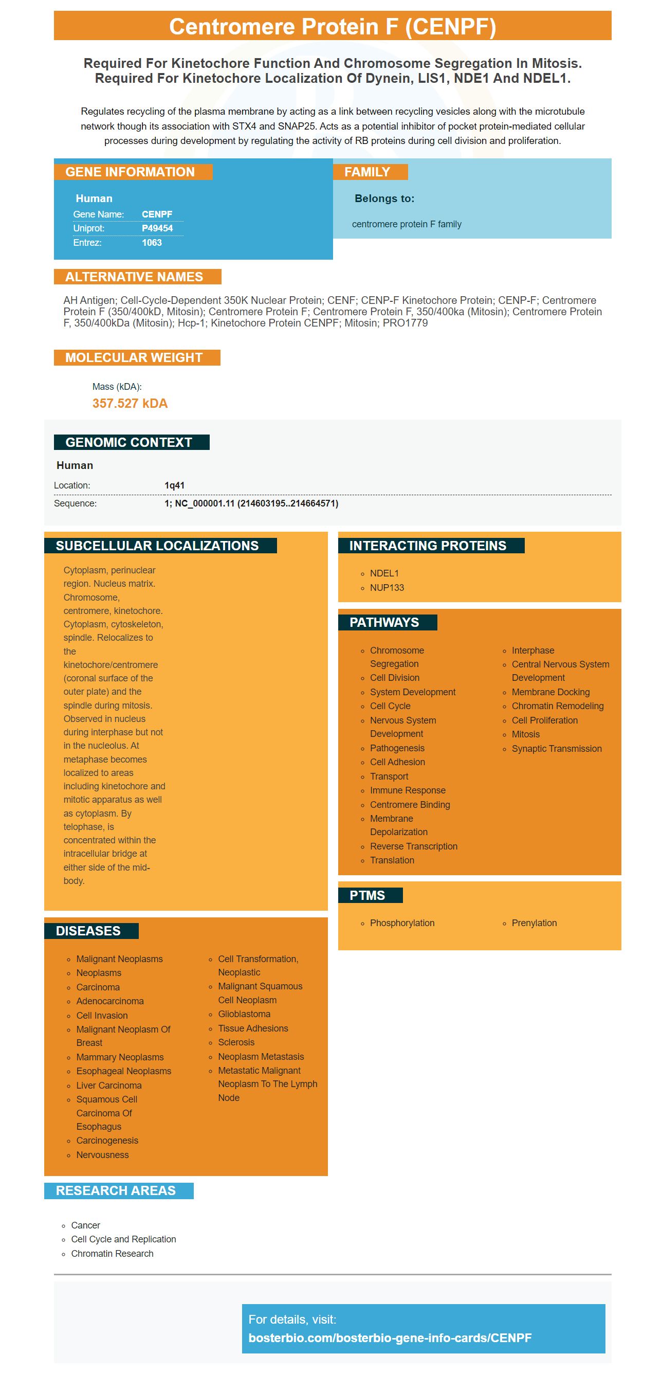
Facts about Centromere protein F.
Regulates recycling of the plasma membrane by acting as a link between recycling vesicles along with the microtubule network though its association with STX4 and SNAP25. Acts as a potential inhibitor of pocket protein-mediated cellular processes during development by regulating the activity of RB proteins during cell division and proliferation.
Human | |
---|---|
Gene Name: | CENPF |
Uniprot: | P49454 |
Entrez: | 1063 |
Belongs to: |
---|
centromere protein F family |
AH antigen; cell-cycle-dependent 350K nuclear protein; CENF; CENP-F kinetochore protein; CENP-F; centromere protein F (350/400kD, mitosin); centromere protein F; centromere protein F, 350/400ka (mitosin); centromere protein F, 350/400kDa (mitosin); hcp-1; Kinetochore protein CENPF; mitosin; PRO1779
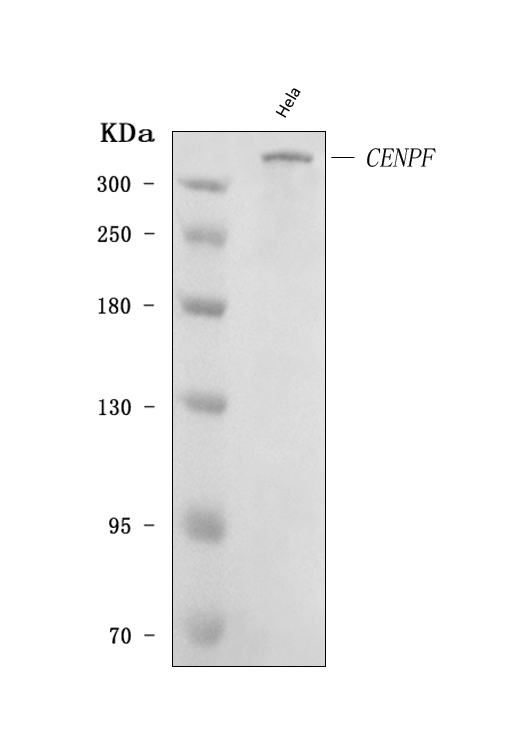
Mass (kDA):
357.527 kDA
Human | |
---|---|
Location: | 1q41 |
Sequence: | 1; NC_000001.11 (214603195..214664571) |
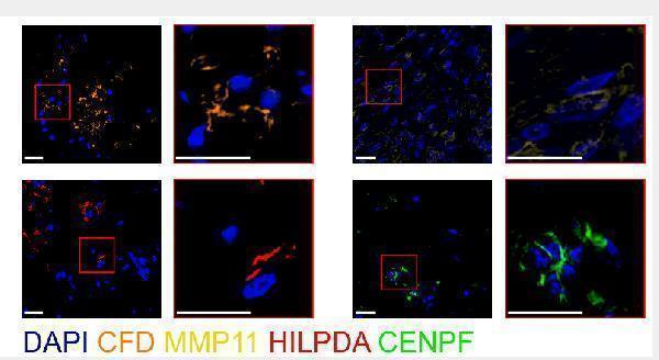
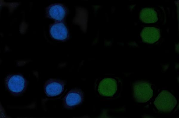
Cytoplasm, perinuclear region. Nucleus matrix. Chromosome, centromere, kinetochore. Cytoplasm, cytoskeleton, spindle. Relocalizes to the kinetochore/centromere (coronal surface of the outer plate) and the spindle during mitosis. Observed in nucleus during interphase but not in the nucleolus. At metaphase becomes localized to areas including kinetochore and mitotic apparatus as well as cytoplasm. By telophase, is concentrated within the intracellular bridge at either side of the mid-body.
CENPF is an epigenetic regulator that regulates lipid and histone metabolism. This article will discuss how this marker regulates cell proliferation and migration as well as colony formation. Continue reading to learn more about this gene marker and its uses. You'll also learn how it can aid in cancer research. It's an epigenetic regulator that regulates cell proliferation, migration, colony formation, and other aspects of cell growth.
Scientists have been focusing on CENPF's role in histone methylation over the past decade. However, several reports have shown that CENPF also regulates non-histone proteins like SIRT1 or PRMT. These proteins play an important role in the development and progression of many types of cancers. This gene may also play a role in hepatocellular cancer.
Researchers have many advantages from studying this gene. It can help prevent cancer in the developing cells, which is an important step towards treating diabetes. It is also beneficial for improving cardiovascular health as it is associated with a lower risk of stroke and heart disease. Scientists working on diseases of the heart and other major parts of the body will find this research a valuable resource in the future.
The epigenetic regulation plays a key role in metabolic regulation as well as tumorigenesis. To harmonize their genes, cells must respond dynamically to changes in their environment. Drugs that target one or both of epigenetics or metabolism may have a greater therapeutic effect than drugs that crosstalk. The interplay between epigenetic and metabolic regulation could help identify the underlying mechanism for carcinogenesis.
In addition, tumor cells require massive amounts of energy and nutrients. Drugs that alter tumor cells' metabolism might be of benefit. By altering the activity of EZH2 inhibitors, tumor cells may become resistant to drugs that affect the tumor's metabolism and therefore its prognosis. The development of targeted therapies may be possible.
The CENPF marker for PC is a novel biomarker. It serves clinical and functional purposes. CENPF plays a role in the progression of PC. Additionally, it decreases the activity a key enzyme involved in glycolysis, the Pyruvate Kinase. We also tested the CENPF indicator in a cell-culture model. We found an increase in the phosphorylation of PKM2 in CENPFKO-cells but no change to total PKM2.
Researchers used CRISPR/Cas9 to knock out CENPF in PC3 cell lines. The CENPFKO cell lines were then carefully characterized. They found morphological changes in the cells and decreased cell proliferation. They also found significantly decreased expression of mesenchymal markers. This result suggests that CENPF regulates the proliferation of cells in tissue engineering.
The phenotypes in the depleted cells were divergent, which suggests a common role of CENP-F for kinetochore assembly. Hypomorphic cells displayed a milder, more severe phenotype. Cells that were completely deficient showed a more severe form of the disease. The divergent cell phenotypes of CENPF-deficient cells prove that CENPF plays a crucial role in the kinetics kinetochore formation.
Moreover, we observed that CENP-F-depleted cells had three kinetochores that were positively labeled by Mad2; whereas, the intensity of Mad2 was absent in 60% of the control cells. Quantification of Mad2 in individual kinetochores confirmed Mad2 recruitment. Therefore, the CENPF marker regulates cell proliferation by controlling the cell cycle.
Recent research revealed that the CENPF Marker (a cell cycle factor) was overexpressed in human ACC tissues. This was associated with poor prognosis. Our research also indicated that CENPF overexpression may increase the progression of ACC. This resemblance with a tumor's Hallmark description suggests that CENPF may serve as a prognostic biomarker or therapeutic target for ACC. We also found that CENPF overexpression affected the expression of CDK1 which regulated cell proliferation and invasion.
Our results showed that CENPF is highly expressed in human ACC tissue and is correlated with the cell proliferation index Ki67. Therefore, the hyper-expression of CENPF may play a pivotal role in cell proliferation. We also conducted a thorough analysis of multiple arrays. This is a method that is both feasible and practical to test for CENPF Marker. We also considered immunotherapy, chemotherapy, and other therapeutic options for CENPF.
CENPF expression in breast cancer tissues can also be associated with poor prognosis. It is possible that this marker could also influence bone metastasis. According to a study published in Cancer Cell Int, tumors that have high levels of CENPF could be at risk for poor prognosis. These findings suggest that CENPF might play a key role in the cell-cycle mediated by G2/M phase transition.
The CENPF Marker regulates cells' proliferation and migration. CENPF overexpression is closely linked to the development of various neoplasms. Although CENPF has a complex biological function, its overexpression has been associated with the development many types of carcinomas. The gene could also be a factor in ACC prognosis, although its exact function is still unknown.
The role of the CENPF marker in osteosarcoma remains largely elusive. We examined the relationship of CENPF expression to prognosis among patients with osteosarcoma. This study revealed that CENPF expression was associated with poor prognosis among patients with osteosarcoma. Although CENPF's role in osteosarcoma is not yet clear, it could be a biomarker of poor prognosis.
We first performed a cell-cycle analysis using a flowcytometer to determine whether CENPF regulates TNBC cell colony formation. We first digested CENPF knockdown cells with 0.25% trypsin for 3 min at 37degC. After digestion, cells are washed in PBS buffer. They were then fixed in 70% ethanol for 30min at room temperature. The cells were then stained with propidium isodide, which was added to the solution in the presence RNaseA. Annexin V–FITC, and PI were used to double-stain the living cells. Then, we counted every sample to determine how many colonies were present.
To determine whether CENPF has an effect on colony formation, cells were transduced with lentivirus using myeloid and other erythroid media. Five days after transduction, cells could be sorted for GFP+ cells. They were then plated onto methylcellulose media for CFU-E and CFUGM counts. We used GFP and mCherry-sorted hematopoietic cells suspensions to determine if CENPF was involved.
Anoikis is the activation of apoptosis by cells. Cells lose their attachment to the extracellular matrix (ECM) or to neighboring cells. Steven M. Frisch, Hunter Francis, and Steven M. Frisch discovered the link between cell adhesion & apoptosis in 1994. They observed that cell death that is triggered via detachment occurs within fibroblasts as well as epithelial lines. The term anoikis is derived from Greek words meaning "homelessness," and its function is to prevent cells from adhering to inappropriate locations and translocating to ectopic environments.
The attachment of cells to the extracellular matrix is essential for cell physiology. This attachment is lost and induces anoikis in normal cell cells. Cancer cells can become resistant to anoikis. This allows them to survive, disperse, and fight off metastasis. This resistance is caused a Yes-associated protein (YAP), a Human Oncogene that regulates body size. Hippo tumor suppressor pathway regulates YAP. This pathway phosphorylates/inhibits/activates kinases that regulate YAP.
Archazolid inhibits V-ATPase, BIM, and other proteins involved in anoikis. T24 cells are susceptible to archazolid, which increases cell death when it is combined with NAC. Research on archazolid has provided important insights into the regulation of anoikis resistance in cancer. The physiologic mechanisms of anoikis are crucial to tissue homeostasis. Most cell types require interplay between cells, ECM, and other factors to maintain tissue homeostasis. Prosurvival signals are also triggered by cell interaction with the ECM.
Anoikis is a characteristic of metastatic cancer cells. While it is not possible to fully understand the mechanisms behind anoikis, there are a variety of drugs that can be used to improve anoikis. Early diagnosis is critical to identify anoikis resistance genes. Patients can develop anoikis resistant tumors by targeting these markers.
PMID: 7542657 by Liao H., et al. CENP-F is a protein of the nuclear matrix that assembles onto kinetochores at late G2 and is rapidly degraded after mitosis.
PMID: 7651420 by Zhu X., et al. Characterization of a novel 350-kilodalton nuclear phosphoprotein that is specifically involved in mitotic-phase progression.