This website uses cookies to ensure you get the best experience on our website.
- Table of Contents
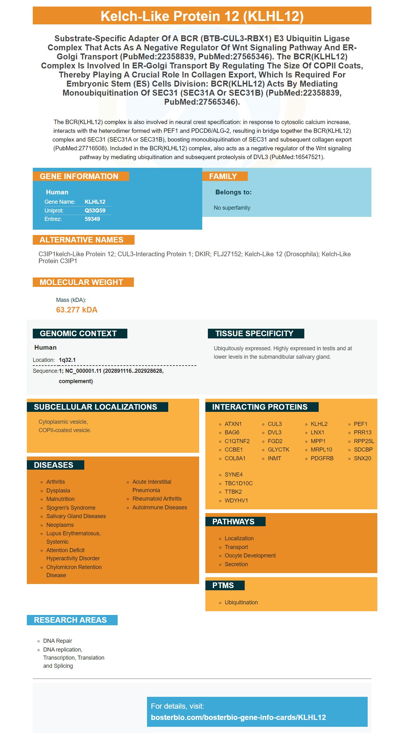
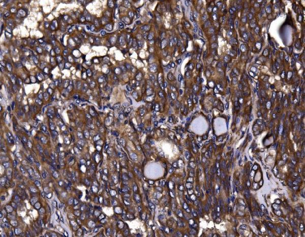
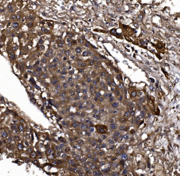
Facts about Kelch-like protein 12.

The BCR(KLHL12) complex is also involved in neural crest specification: in response to cytosolic calcium increase, interacts with the heterodimer formed with PEF1 and PDCD6/ALG-2, resulting in bridge together the BCR(KLHL12) complex and SEC31 (SEC31A or SEC31B), boosting monoubiquitination of SEC31 and subsequent collagen export (PubMed:27716508). Included in the BCR(KLHL12) complex, also acts as a negative regulator of the Wnt signaling pathway by mediating ubiquitination and subsequent proteolysis of DVL3 (PubMed:16547521).
| Human | |
|---|---|
| Gene Name: | KLHL12 |
| Uniprot: | Q53G59 |
| Entrez: | 59349 |

| Belongs to: |
|---|
| No superfamily |

C3IP1kelch-like protein 12; CUL3-interacting protein 1; DKIR; FLJ27152; kelch-like 12 (Drosophila); kelch-like protein C3IP1
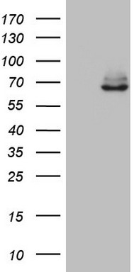
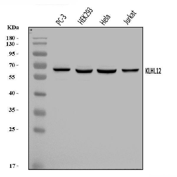
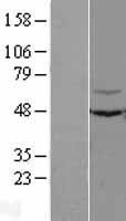
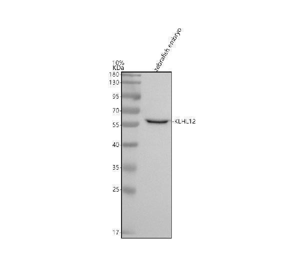
Mass (kDA):
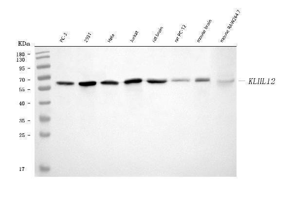
63.277 kDA

| Human | |
|---|---|
| Location: | 1q32.1 |
| Sequence: | 1; NC_000001.11 (202891116..202928628, complement) |
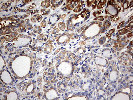
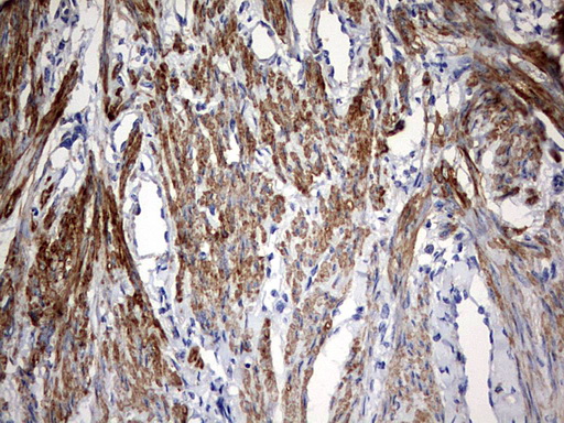
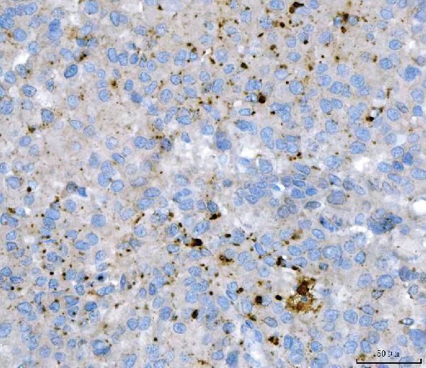
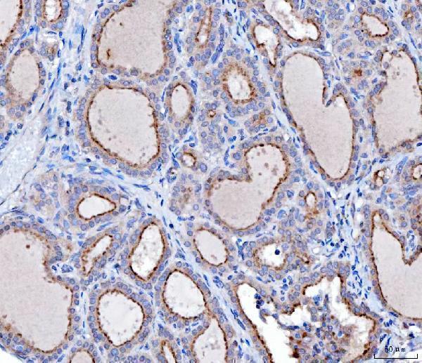
Ubiquitously expressed. Highly expressed in testis and at lower levels in the submandibular salivary gland.
Cytoplasmic vesicle, COPII-coated vesicle.


PMID: 15383316 by Mai A., et al. hDKIR, a human homologue of the Drosophila kelch protein, involved in a ring-like structure.
PMID: 16108817 by Uchida K., et al. Identification of specific autoantigens in Sjoegren's syndrome by SEREX.