This website uses cookies to ensure you get the best experience on our website.
- Table of Contents
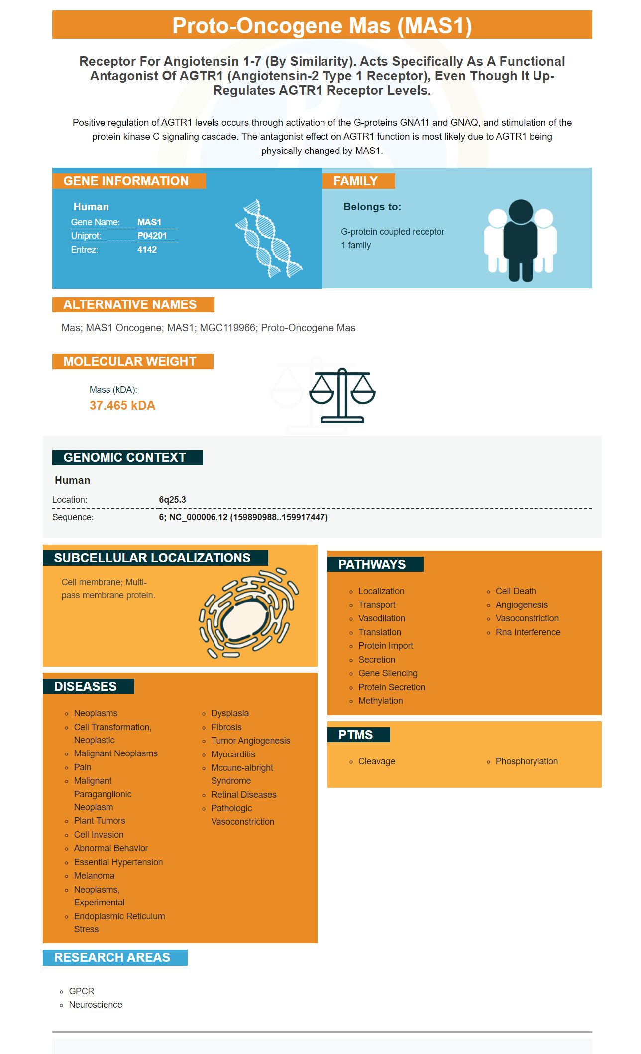
Facts about Proto-oncogene Mas.
Positive regulation of AGTR1 levels occurs through activation of the G-proteins GNA11 and GNAQ, and stimulation of the protein kinase C signaling cascade. The antagonist effect on AGTR1 function is most likely due to AGTR1 being physically changed by MAS1.
Human | |
---|---|
Gene Name: | MAS1 |
Uniprot: | P04201 |
Entrez: | 4142 |
Belongs to: |
---|
G-protein coupled receptor 1 family |
Mas; MAS1 oncogene; MAS1; MGC119966; proto-oncogene Mas
Mass (kDA):
37.465 kDA
Human | |
---|---|
Location: | 6q25.3 |
Sequence: | 6; NC_000006.12 (159890988..159917447) |
Cell membrane; Multi-pass membrane protein.
Boster Bio makes it easy for you to use the MAS1 mark in your experiments. There are many options for setting up your flow procedures. Refer to our flow procedures guide for more information. Follow these steps to optimize your experiments:
The MAS1 receptor marker is a protein found on the cell surface that has been shown activity in angiotensin. It is also believed to play a role in the development and maintenance of the hippocampus. The MAS1 protein is orthologous in function to the human MAS1 prototype-oncogene. It has been shown interaction with 17-beta estradiol as well as the renin–angiotensin cacade.
Boster Bio's MAS1Marker is an antigen based on peptides that has been shown significant immunomodulatory activity in various types of inflammatory diseases. This antibody is used to detect Mas1 expression in the bronchial epithelium, and airway smooth cell. Using Mas1 in the diagnosis of asthma and allergies is critical. Its expression decreases as a result of acute allergic asthma inflammation.
The primary-secondary-ABC system is a reliable and highly sensitive method of detecting proteins. The primary antibody binds to a protein of interest and the secondary antibody recognizes it. This combination has a low background and high specificity making it a great choice for researchers. Other detection systems are based on polysaccharides or organic polymers. These kits are highly useful in immunohistochemistry, ELISA, and Western Blotting.
MAS1, a protein from the s1s family that is expressed by insect cells, is known as MAS1. It can be expressed as a fusion protein with MBP in E. coli. MBP1s-expressing cells from mice were immunized. The mice were then monitored using an ELISA Kit that used FLAG s1s for the antigen. The experiment revealed two IgG2a MAbs, which were highly specific for s1s using immunoblotting and the ELISA assay.
A secondary antigen is an antigen that a human or a mouse has specific affinity for. Secondary antibodies are produced in response to a pool IgG of a specific species. This pool allows secondary antibodies from a specific species to bind all Ig subclasses, light chain and conserved domains. In some cases secondary antibodies can be produced against specific antigens by immunizing a host animals with an antigen from another species.
One immunofluorescent staining was done to determine whether the secondary antibody could bind a specific antigen. We used 0.3% tritonX-100 to permeate the slices and then blocked them with either 1% bovine serum albin or 5% normal goat serum to determine if the secondary antibody was cross-species. To prevent nonspecific staining, we also used normal donkey serum.
The APC/laminB1 couple had a stronger correlation coefficient than the corresponding antirat and antimouse secondary antibody pairs. This correlation was attributed to the fact that the APC and keratin signals overlapped to a significant extent. Although we could not prove causality, these results suggest that secondary antibodies may compete for binding to a specific antigen. This technique will be used to determine the best secondary antibody for a specific application.
To identify specific neuronal population in the brain, a secondary antimouse antibody is used to stain a rat’s hippocampus. The anti-mouse antibodies would prevent rat-anti-GFAP antibodies from binding. This strategy would also help determine if the secondary is cross-species. While there is some degree of cross-species antibody binding, this is not always possible.
A primary antibody binds a protein of interest and can be used to identify a protein. Secondary antibodies, such those offered by Boster Bio can be used for immunohistochemistry, ICC/IF, and immunohistochemistry. They can also be used for Western Blotting. This article explains how to perform the procedure. It is also easy to follow. Follow the instructions for your application.
The primary antibody used was a mouse antiCRM1 antibody. It was used at 1 mg/106 cellular cells. The secondary antibody was goat anti-mouse IgG conjugated with DyLight(r)488. The cells were then incubated for 30 minutes with a secondary antibody using MAS1 marker. They were counterstained with DAPI and hematoxylin.
The s1s protein was expressed as a fusion protein in insect cells and in E. coli. Then, mice were immunized with MBP-s1s. The mice's response to the antigen was determined using ELISA with FLAG-s1s antigen. Two IgG2a MAbs (2F4 and 3E2) were obtained. Both were capable immunoblotting, immunostaining and PCR.
In addition to detecting MAS1 in human cells a primary antibody that targets the reovirus s1s proteins, called s1s receptor inhibits MAPK pathwaying pathways. This antibody decreases production of CCL2, an important molecule in airway remodeling, and inhibits p38 MAPK/ERK1/2. These antibodies are useful for determining whether the Mas receptor is present in the airway epithelium and bronchial smooth muscles.
PMID: 3708691 by Young D., et al. Isolation and characterization of a new cellular oncogene encoding a protein with multiple potential transmembrane domains.
PMID: 3419518 by Jackson T.R., et al. The mas oncogene encodes an angiotensin receptor.