This website uses cookies to ensure you get the best experience on our website.
- Table of Contents
5 Q&As
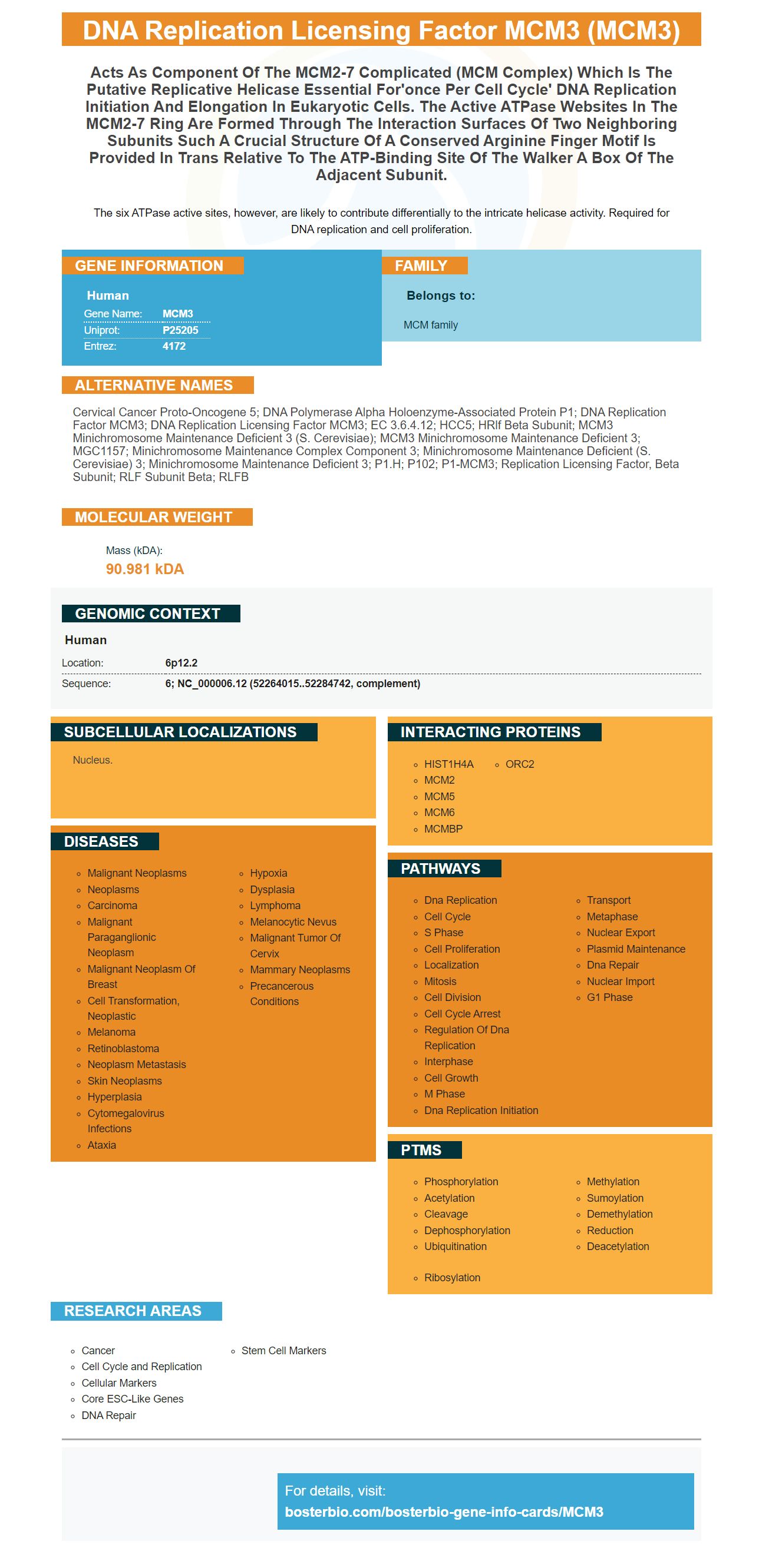
Facts about DNA replication licensing factor MCM3.
The six ATPase active sites, however, are likely to contribute differentially to the intricate helicase activity. Required for DNA replication and cell proliferation.
Human | |
---|---|
Gene Name: | MCM3 |
Uniprot: | P25205 |
Entrez: | 4172 |
Belongs to: |
---|
MCM family |
cervical cancer proto-oncogene 5; DNA polymerase alpha holoenzyme-associated protein P1; DNA replication factor MCM3; DNA replication licensing factor MCM3; EC 3.6.4.12; HCC5; hRlf beta subunit; MCM3 minichromosome maintenance deficient 3 (S. cerevisiae); MCM3 minichromosome maintenance deficient 3; MGC1157; minichromosome maintenance complex component 3; minichromosome maintenance deficient (S. cerevisiae) 3; minichromosome maintenance deficient 3; P1.h; p102; P1-MCM3; replication licensing factor, beta subunit; RLF subunit beta; RLFB
Mass (kDA):
90.981 kDA
Human | |
---|---|
Location: | 6p12.2 |
Sequence: | 6; NC_000006.12 (52264015..52284742, complement) |
Nucleus.
You are interested in learning more information about BosterBio and the MCM3 Marker. Read on to find out how this high-affinity primary antibody can help you with your research. This marker can be used for species-specific samples, applications, and research papers. Boster Bio can provide product credits for scientists from all over the world. Learn more about this innovative product by reading this Boster Bio review.
Scientists will have probably heard of MCM3 Marker. The MCM3 is found in the cell membrane and is one of the most common markers for tumors. Because it is so common in tumors, it is a valuable target for many research studies. Boster antibodies are highly specific and can be used to detect this protein in a variety of settings, including cancer cell lines, tumors, and other types of tissues.
High MCM3 expression has been shown to be associated with poor prognosis among IDC patients. Moreover, gene microarray data analysis confirmed that MCM3 is a more accurate proliferative marker than Ki-67. MCM3 expression is independent of HER2 and ER status. These results support its use in proliferative markers. Further research is needed to confirm the proliferative potential of this marker.
The MCM3 complex members are expressed by a wide range of cancers. MCM3 is expressed in many types of cancers and is controlled by many factors. Mcm3 is subjected to dynamic posttranslational changes, including phosphorylation. Mcm3 undergoes cell-cycle-specific modifications. In the present study, Mcm3 expression levels are shown for 20 types of tumors.
The MCM3 genes may be a biomarker for cervical cancer. High levels of MCM3 gene expression are associated with a longer life expectancy, but shorter relapse-free mortality. MCM3 expression is positively correlated to PRIM2 & MCM6, although the role MCM3 in cervical carcinoma is not fully understood. More clinical tissue specimens and cell experiments are needed to confirm this gene's function.
Researchers have discovered that MCM3 expression limits the effectiveness of endocrine therapy for ER+ breast cancer cells. By detecting tumor-specific MCM3 expression, they can tailor a patient's treatment. MCM3 is one of the most important biomarkers in clinical practice. It can be used to help doctors determine whether a particular treatment is appropriate for a patient.
B cells must express either the IgD receptor or IgM receptor in order to create high-affinity primary antibody. The IgD or IgM receptor inhibits the production of autoreactive IgM primary antibodies and speeds up generation of protective memory IgM. Monovalent antigens are unable to activate the IgD/BCR whereas polyvalent antigens stimulate IgG memory responses in activated B cells. This study demonstrates the importance of the IgD BCR in regulating the dynamics of the B cell activation and immune response.
The antibody was produced using affinity chromatography and an epitope-specific immunogen. Immunohistochemical reactions were performed on 4-mm paraffin sections or TMA blocks. After the antibody was removed, it was used to detect the antigen and purify it. The results were then analysed using immunofluorescence microscopy. This procedure can be repeated many times to identify specific molecules.
Indirect IF confirmed the existence of affinity maturation. Three independent experiments showed that PR-IgMs can be affinity matured using three different color curves. Abcam scientists reviewed the affinity maturation data. This allows researchers to identify the most powerful antibody for a given target. And, most importantly, the high-affinity antibodies are more potent than their non-affinity counterparts.
With the Duolink II near-ligation kit, it is possible to develop high-affinity secondary antibodies using MCM3, the enzyme that activates MCM3's protein. The kit was validated by demonstrating that it recognizes MCM3 in chromatin, and soluble fractions of cells. Open Biosystems can sell the corresponding antibodies.
The MCM protein is more effective as a proliferative marker that PCNA or Ki67, according to research. However, the MCM protein complex serves other functions than DNA replication. The MCM complex plays many different roles in cellular functions. A 90% decrease in MCM proteins did no harm to DNA replication or cell cycle progression. This paradox is still unknown, and will be further investigated.
PMID: 8265339 by Hu B., et al. The P1 family: a new class of nuclear mammalian proteins related to the yeast Mcm replication proteins.
PMID: 7758114 by Kubota Y., et al. Identification of the yeast MCM3-related protein as a component of Xenopus DNA replication licensing factor.