This website uses cookies to ensure you get the best experience on our website.
- Table of Contents
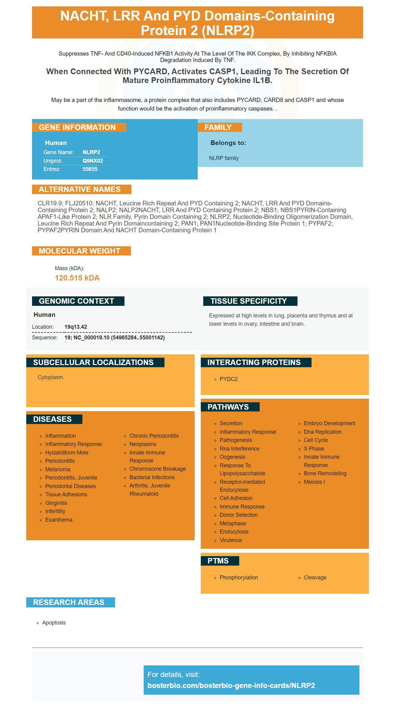
Facts about NACHT, LRR and PYD domains-containing protein 2.
Suppresses TNF- and CD40-induced NFKB1 activity at the level of the IKK complex, by inhibiting NFKBIA degradation induced by TNF.
When connected with PYCARD, activates CASP1, leading to the secretion of mature proinflammatory cytokine IL1B.May be a part of the inflammasome, a protein complex that also includes PYCARD, CARD8 and CASP1 and whose function would be the activation of proinflammatory caspases. .
Human | |
---|---|
Gene Name: | NLRP2 |
Uniprot: | Q9NX02 |
Entrez: | 55655 |
Belongs to: |
---|
NLRP family |
CLR19.9; FLJ20510; NACHT, leucine rich repeat and PYD containing 2; NACHT, LRR and PYD domains-containing protein 2; NALP2; NALP2NACHT, LRR and PYD containing protein 2; NBS1; NBS1PYRIN-containing APAF1-like protein 2; NLR family, pyrin domain containing 2; NLRP2; nucleotide-binding oligomerization domain, leucine rich repeat and pyrin domaincontaining 2; PAN1; PAN1Nucleotide-binding site protein 1; PYPAF2; PYPAF2PYRIN domain and NACHT domain-containing protein 1
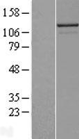
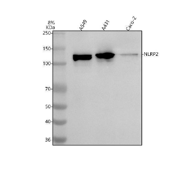
Mass (kDA):
120.515 kDA
Human | |
---|---|
Location: | 19q13.42 |
Sequence: | 19; NC_000019.10 (54965284..55001142) |
Expressed at high levels in lung, placenta and thymus and at lower levels in ovary, intestine and brain.
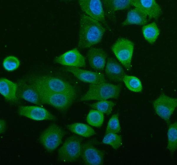
Cytoplasm.
If you are interested in pursuing the discovery of the NLRP2 gene, you've come to the right place. In this article, you'll learn how this pyrin-like protein functions as a nucleotide-binding site and promotes cell apoptosis during acute kidney injury. As well as these applications, you'll learn about its function in disease progression, drug discovery, and toxicity testing.
The NLRP2 gene is a member of the family of proteins called pyrin-like receptors. It is believed to play a role in regulation of immune responses. It is also important in ensuring the health of reproductive tissues, such as the ovaries and eggs. The NLRP2 gene produces the pyrin-like protein NALP2, whose PYD domain is responsible for its N-terminal pyrin characterization. In addition, NLRP2 is important in the activation of caspase-1.
Its function is not known, but it is thought to regulate the activity of the inflammatory response in the intestine. The gene encodes a protein that is localized in multiple organelles, where it participates in different intracellular functions. Researchers have used boster protein capture as a tool to identify proteins with different functions. But what makes this technique unique is its ease of use.
The NLRP2 gene has become an innate immune sensor for pathogens and a key player in the sterile stress signal. This finding has implications for how the gene functions in innate immune responses. The protein is also involved in the regulation of the inflammasome. The inflammasome triggers NLRP2 when it detects a pathogen.
This antibody is produced from rabbit anti-NLRP2 polyclonal antibody. It was used to probe sections of kidney tissues with a primary rabbit anti-NLRP2 antibody. The membranes were then blocked with 5% bovine serum albumin at room temperature for 1 h. After blocking, the samples were incubated with primary rabbit anti-NLRP2 antibody overnight at 4degC. The primary antibody was detected at a band size of 121kD, which is consistent with the expected band size.
The role of NLRP2 is unclear, but its function in regulating various stages of development is critical. It regulates various stages of development, including the oocyte, preimplantation embryo, and follicle. By blocking this protein, pregnant women can avoid potentially harmful pathogens. And the NLRP2 gene is associated with many different functions in the immune system.
Melatonin inhibits NLRP3-dependent inflammasome triggering. Melatonin inhibits this process, reducing ROS production. This drug has an anti-inflammatory effect in both humans and animals. The effects of melatonin have been demonstrated. By blocking this protein, melatonin inhibits the expression of IL-1b and other pro-inflammatory proteins.
The CCK-8 assay was used to determine the IC50 of LPS/ATP. The dose of LPS/ATP did not affect the viability of RLE-6TN cells. LPS/ATP co-stimulation increased NLRP3 and GSDMD-N proteomic content. The LPS/ATP treatment also enhanced LDH release. Further, LPS/ATP co-stimulation enhanced apoptosis-related proteomic proteins, such as NLRP3 and Caspase-1 P20.
To study the effects of Nlrp2 on oocyte maturation, NLRP2 siRNA was transfected into human ovarian cells. It is important to note that Nlrp2 siRNA is highly selective and does not affect the expression of other Nlrp genes, such as Nlrp4f, Nlrp9c and Nlrp14.
A unique NLRP protein interacts with TBK1 protein through its ATP-binding site and ubiquitin-like domain. The TBK1 protein is a key innate immune adaptor that mediates IFN production. By inhibiting NLRP2 activity, the proteins can regulate the expression of certain genes that are associated with immunity. This mechanism of NLRP inhibition is currently under investigation.
Expression of NLRP2 was investigated in different kidney derived cell lines. Tubule cells expressed NLRP2 at higher levels than non-tubule cells. Knockdown of NLRP2 reduced OGD-induced cell apoptosis, suggesting that NLRP2 plays an important role in acute kidney injury. Further, NLRP2 might play an important role in kidney regeneration.
Knockdown of NLRP2 may interfere with cell cycle progression. This is determined by alterations in DNA content using PI staining and flow cytometry. Transfection of siNLRP2 resulted in a greater proportion of S-phase cells and a decreased proportion of G1-phase cells. Knockdown of NLRP2 may stop cell cycle progression at the S phase.
NLRP2 is a cytoplasmic pattern-recognition receptor. It is activated in response to the presence of inflammatory microbes and other pathogens. It has been associated with inflammatory diseases. In addition, the NLRP2 protein has been shown to inhibit mitophagy. Inhibition of mitophagy results in a marked increase in NLRP3 inflammasome activity.
In the present study, NLRP2 was upregulated in a mouse model of ischemic stroke. Knockdown of the gene inhibits cell proliferation in HUVECs, which indicates a protective role for this protein. Furthermore, knockdown of NLRP2 suppresses cell migration and invasion. Consequently, NLRP2 may have a therapeutic role in these disease states.
The siNLRP2 transfection reduced HUVEC invasion in the Matrigel assay, which reflects the migration of vascular cells. Knockdown of NLRP2 markedly inhibits HUVEC migration and invasion. Therefore, the ability of this gene to prevent wound closure may improve the healing of damaged tissues. The findings support the notion that NLRP2 is a novel molecular scaffold that promotes wound healing.
The function of NLRP12 is defined by protein-protein interactions. Using human PYD + NBD as bait in yeast two-hybrid screen, we identified a variety of proteins that are specifically bound to NLRP12's PYD. The resulting results show that HCK is a multifunctional protein that likely depends on protein-protein interactions.
Recent studies have revealed that NLRP2 is expressed in both normal and kidney-injured cells, and that NLRP2 knockdown decreased apoptosis in renal I/R injured cells. Therefore, targeting NLRP2 may be a new target for acute kidney injury treatment. In addition, this protein is involved in inflammatory responses. This makes targeting NLRP2 a novel therapeutic option for patients with acute kidney injury.
We studied the role of NLRP2 in acute kidney injury by using mouse models, OGD treatment, and OGD. We used cell-based and in vitro models and analyzed protein levels and the proportion of apoptotic HK-2 cells. We then knocked down NLRP2 expression and analyzed apoptosis in HK-2 cells using flow cytometry.
NLRP2 is also involved in the activation of the NLRP3 inflammasome. In addition to NLRP2, this protein is also implicated in a number of diseases, including pneumonia and diabetic nephropathy. NOX activity in peritoneal macrophages of mice exposed to CLP is increased. Although its role in acute kidney injury is not fully understood, it is linked to RIPK3.
NLRs are intracellular multiprotein signalling complexes that assemble in the cytosol. Inflammasomes recruit native immune cells to infected sites and activate caspase-1 and NLRC. These molecules also stimulate the activity of natural killer cells and induce pyroptosis, an inflammatory form of programmed cell death. This mechanism is implicated in inflammatory diseases, including heart and kidney disease.
Necroptosis and regulated necrosis are both related to renal inflammation and cell death. However, these processes are regulated and influenced by specific molecules. These molecules may act as therapeutic targets to prevent and treat acute kidney injury. However, it is necessary to determine which pathway NLRP2 inhibits to gain more understanding of how these processes function in acute kidney injury.
To identify whether NLRP2 affects cellular apoptosis, we used fluorescent dyes to measure the levels of the protein in cell cultures. NLRP2-siRNA was designed to mimic the LPS-induced cell death marker. In the present study, LPS-induced cells were treated with LPS for 24 hours. Cell viability was measured using fluorescent dyes and a microplate reader.
Immunohistochemical staining of renal tissue was performed using rabbit anti-NLRP2 antibody (1:5000) and a goat anti-caspase-3 antibody (catalog no. SA00001-2, ProteinTech Group, Inc.). The primary antibodies were used as the primary antibody and the secondary antibody was added 1 hour later. The immunohistochemical staining was completed in three separate experiments.
PMID: 11270363 by Bertin J., et al. The PYRIN domain: a novel motif found in apoptosis and inflammation proteins.
PMID: 11250163 by Martinon F., et al. The pyrin domain: a possible member of the death domain-fold family implicated in apoptosis and inflammation.