This website uses cookies to ensure you get the best experience on our website.
- Table of Contents

1 Citations
Facts about Phosphate-regulating neutral endopeptidase PHEX.

Regulates osteogenic cell differentiation and bone mineralization through the cleavage of this MEPE-derived ASARM peptide (PubMed:18597632). Promotes dentin mineralization and renal phosphate reabsorption by cleaving DMP1- and MEPE-derived ASARM peptides (PubMed:18597632, PubMed:18162525).
| Human | |
|---|---|
| Gene Name: | PHEX |
| Uniprot: | P78562 |
| Entrez: | 5251 |

| Belongs to: |
|---|
| peptidase M13 family |

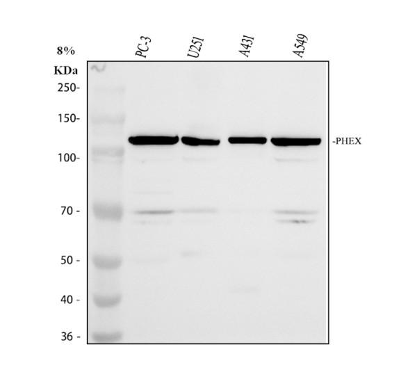
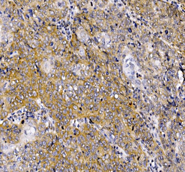
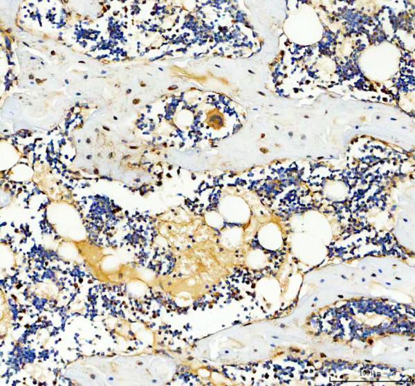
Phosphate-regulating neutral endopeptidase PHEX
Mass (kDA):
86.474 kDA

| Human | |
|---|---|
| Location: | Xp22.11 |
| Sequence: | X; NC_000023.11 (22032325..22251310) |
Specifically expressed in ovary (PubMed:9070861). Expressed at low levels in kidney (PubMed:9070861).
Cell membrane; Single-pass type II membrane protein.


PMID: 9199930 by Francis F., et al. Genomic organization of the human PEX gene mutated in X-linked dominant hypophosphatemic rickets.
PMID: 9077527 by Beck L., et al. Pex/PEX tissue distribution and evidence for a deletion in the 3' region of the Pex gene in X-linked hypophosphatemic mice.