This website uses cookies to ensure you get the best experience on our website.
- Table of Contents
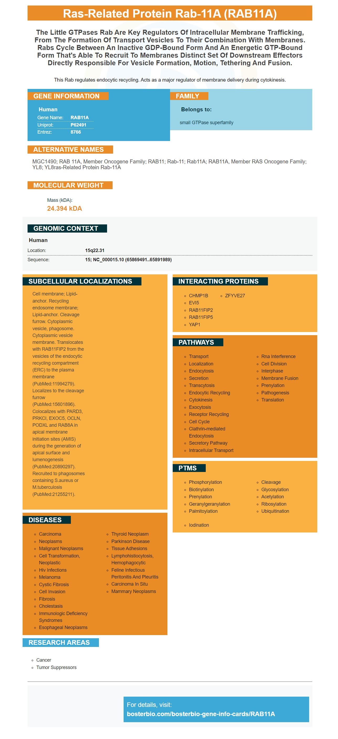
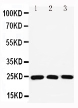
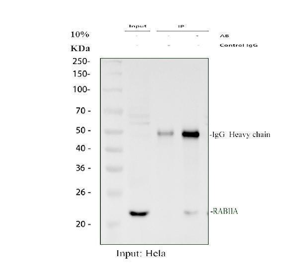
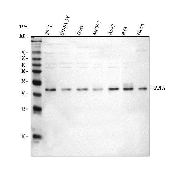
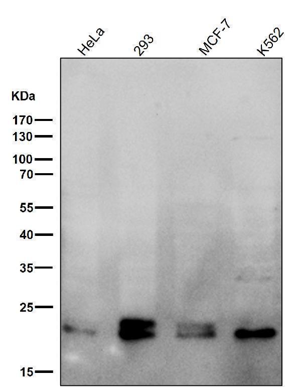
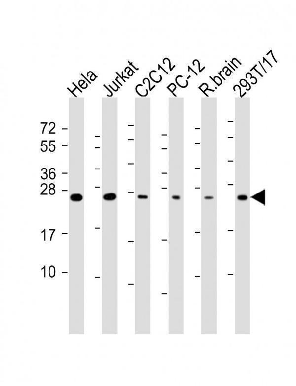
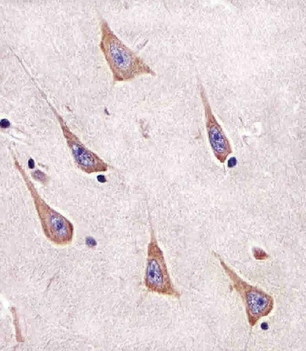
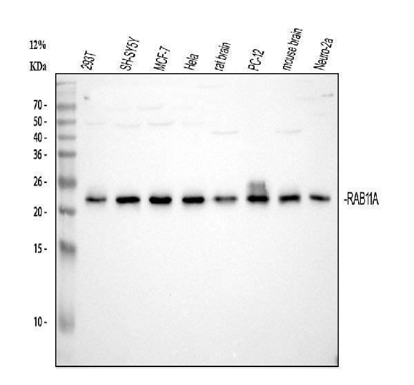
Facts about Ras-related protein Rab-11A.
This Rab regulates endocytic recycling. Acts as a major regulator of membrane delivery during cytokinesis.
Human | |
---|---|
Gene Name: | RAB11A |
Uniprot: | P62491 |
Entrez: | 8766 |
Belongs to: |
---|
small GTPase superfamily |
MGC1490; RAB 11A, member oncogene family; RAB11; Rab-11; Rab11A; RAB11A, member RAS oncogene family; YL8; YL8ras-related protein Rab-11A
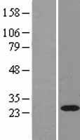
Mass (kDA):
24.394 kDA
Human | |
---|---|
Location: | 15q22.31 |
Sequence: | 15; NC_000015.10 (65869491..65891989) |
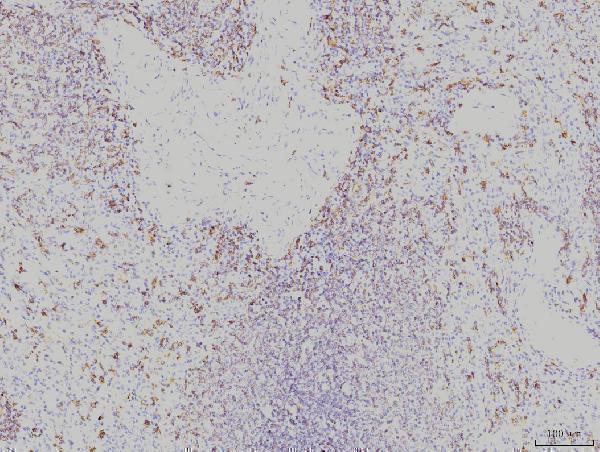
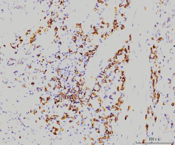
Cell membrane; Lipid-anchor. Recycling endosome membrane; Lipid-anchor. Cleavage furrow. Cytoplasmic vesicle, phagosome. Cytoplasmic vesicle membrane. Translocates with RAB11FIP2 from the vesicles of the endocytic recycling compartment (ERC) to the plasma membrane (PubMed:11994279). Localizes to the cleavage furrow (PubMed:15601896). Colocalizes with PARD3, PRKCI, EXOC5, OCLN, PODXL and RAB8A in apical membrane initiation sites (AMIS) during the generation of apical surface and lumenogenesis (PubMed:20890297). Recruited to phagosomes containing S.aureus or M.tuberculosis (PubMed:21255211).
The RAB11A marker is an internal marker for exosomes interacts with the proteins PSEN1, DLGAP5 & TBC1D2. The protein could be used as an exosome signature. These features make Rab11A an excellent biomarker for discerning between tumour EVs and normal ones. This article provides a brief description of the RAB11A marker.
We found that sEVs had many non-exosome particles using a single antibody to detect Rab11a. The number of exosomes is calculated as the percentage of CD63+ particles per milliliter. We also noticed that UC isolates had higher CD81 and CD9 levels. We also used the iFCM test to determine the accuracy of subsets of sEV.
Exosomes' composition is vital to determine the role of these cells in the process of aging. Recent studies suggest that the lipid content of exosomes could function as an molecular marker to detect the presence of diseases and their prognosis. It is a reliable internal exosome marker and will allow for targeted treatment. A group of researchers from the University of California San Francisco discovered Rab11a.
Utilizing Rab11a as a legitimate internal marker for exosomal function provides a more complete picture than microscopy by itself. In addition, BioID provides 16-fold higher confidence in the interaction of proteins than AP-MS likely representing cargo proteins that play a role in the process of endosomal transit. Furthermore, the ratio of new interactions vs. previously known interactions in BioID is two-fold higher than with AP-MS, suggesting that it is sensitive to transient interactions.
Using iFCM to identify the composition of exosomes in circulation can help you accurately and conveniently quantify their composition. It also quantifies the subtype-specific recovery skew in studies of exosomes. It is possible to improve our understanding of exosome function by using Rab11a as an internal marker of exosomes. With this level of accuracy, it is possible to identify which exosomes are responsible different disease-related diseases.
This luciferase test reveals the most numerous copies of a cellular protein called Ra11a. The KO cells are deficient in this protein, leading to defects in the assembly of DNA as well as an increased production of defective particles. Additionally the cells with KO are more vulnerable to DNA damage than the controls. This is why the RAB11A assay allows researchers to identify and characterize defective proteins and DNA.
The antibody reacts with Human, Rat and Mouse and has been tested for Western Blotting, immunohistochemistry, and applications using immunohistochemistry. This antibody is produced from a mouse that has been immunized with purified human recombinant RAB11A protein. If you prefer, you can buy a blocking peptide to use alongside the antibody. If you are using an anti-RAB11A MAb, ensure that the antibody is specifically designed for the target protein.
A KO cell doesn't have VRNP-mediated association with the ER. Additionally, it fails to form supramolecular vRNP bundles when Rab11a is absent. This indicates that the RAB11a KO cell lacks the protein, but shows similar levels of vRNP as control cells. The RAB11A mutation could play a role post-nuclear export of vRNA.
We observed a decrease in Rab11a in the post-replication stage of the IAV life cycle. These post-replication processes are affected by the loss of RAB11a. The loss of RAB11a in the NP-Tc virus alters the post-replication steps of IAV. This study will further determine whether this protein is essential to detect the virus or not.
Recent research has shown that Rab11a and the vRNPs communicate. Infectious viruses utilize Rab11a as an anchoring site for vRNPs. Rab11a+ vesicles facilitate interaction between vRNP segments , and they also form specific viruses. Rab11a deficiency in a cell also increases the number of non-infectious particles suggesting that there is a defect in the specific assembly of the viral genome.
Our results demonstrate that RAB5A, RAB11A, and T-DM1 sensitivity have strong correlations. We also confirmed that RAB11A is more sensitive T-DM1 in breast cancer cells than non-invasive breast cancer cell lines. The RAB11A test is compatible with various kinds of cells including breast cancer cells, fibroblasts and other types of cancer cells.
Exosomes are classified according to the function of a specific signaling pathway. Munc13-4 functions only when the RAB11A gene is active. Transient fusion of Rab11+ endsosomes produces exosomes. These exosomes have specific components that permit their Ca2+-dependent fusion to the plasma membrane. The RAB11A gene is a potential candidate for exosome subtype signature.
To study the exosomes, a number of biochemical methods have been employed. One of these is the immunoaffinity-based exosome separation. Researchers have developed a patentable method that was based on RAB11A which is an RNA protein. This technique is a promising method to identify exosomes in complex samples. This technique allows for the identification of antigens within exosomes.
Many scientists had previously considered exosomes "junk", but recent research has revealed fresh information regarding their functions. Although exosomes were initially thought to be junk, they do contain proteins, lipids and genetic material. However, exosomes play crucial roles in cell communication as well as epigenetic regulation. Because their contents are tightly controlled by parent cells, exosome-based diagnostics could potentially be extremely beneficial.
Nanowire-based exosome-trip system offers an easy method to isolate exosomes in small samples. Its unique features include porous silicon nanowires etched on the sidewalls of micropillars with evenly spaced pillars. This way, it creates exosomes of defined dimensions. Another benefit of this method is the capability to process hundreds of mL per minute.
The ISEV position paper declares that "exosome" should be used for both the exosome as a whole and its constituent components. However, there is currently no consensus on whether or not the term "exosome" is a single entity and it has been suggested that the term is a collective term , which includes all types of small extracellular vesicles.
RAB11A is a potential possibility to be an exosome subtype signature. However, there is still an absence of information about the microvesicles' function and properties which may make it difficult to establish the existence of exosome particularity. Further research is required to understand the function of RAB11A in the detection of exosomes within cells. This technology could revolutionize the diagnosis of exosomes treatment, monitoring, and treatment.
EVs are the most frequent kind of microvesicles that are found in the body. They facilitate various processes between cells, including intercellular communication. Exosomes are released by immune cells. They act as antigen-presenting vessels. They also stimulate the antitumoral immune system and reduce inflammation. These extracellular microvesicles are used by tumour cells to enhance the disease. These EVs are thought to also aid in neuronal survival as well as myelin formation as well as neurite outgrowth.
EVs are generally pooled and their composition can be analyzed by DNA or RNA sequencing. EVs are generally poor quality biomarkers and unreliable, but only a few EVs could be enough for a diagnostic analysis. The pooled EVs can also be studied with flow cytometry, a method that is more conventional than PCR.
Since the size range and the volume of EVs differ, the Boster Bio RAB11A marker is able to identify tumour EVs from non-tumour EVs. EVs can be stained or unstained either single-animal or multi-animal. EVs can be classified into CFSE+ and CD9+ instances, and the percentage of each is presented as a mean plus SEM.
ExoQuick(r) technology creates samples that have 80% of the structures of the vesicular. As shown in Figure 3B the majority of particles are CFSE+ EVs. ExoQuick(r) is a purified specimen, has lower levels of CD9+EVs compared to other samples. This suggests that ExoQuick(r) could be able to select one of the EV subpopulations that are different.
CFSE is widely used for FC applications. However, Jennifer Jones and colleagues have evaluated the suitability of CFSE-labelled EVs. CFSE-labeled EVs were found to be less likely to aggregate. These results are consistent in the data obtained with serum-free media as well as 10% EV depleted FBS. The results aren't conclusivehowever.
Boster Bio RAB11A marker, which can be used to detect tumour EVs using internal reflection fluorescence microscopes, is also useful. In contrast to clinical samples, murine biofluids show less variability which makes it easier to identify of biomarkers that are novel. However, the sample size is limited to how many EVs can be examined with one biofluid.
PMID: 1704119 by Drivas G.T., et al. Identification and characterization of a human homolog of the Schizosaccharomyces pombe ras-like gene YPT-3.
PMID: 9662449 by Gromov P.S., et al. Human rab11a: transcription, chromosome mapping and effect on the expression levels of host GTP-binding proteins.