This website uses cookies to ensure you get the best experience on our website.
- Table of Contents

1 Citations 17 Q&As


Facts about TP53-binding protein 1.

In response to DSBs, phosphorylation by ATM promotes interaction with RIF1 and dissociation from NUDT16L1/TIRR, leading to recruitment to DSBs websites (PubMed:28241136). Recruited to DSBs websites by recognizing and binding histone H2A monoubiquitinated at'Lys-15' (H2AK15Ub) and histone H4 dimethylated at'Lys-20' (H4K20me2), two histone marks that are found at DSBs websites (PubMed:23760478, PubMed:28241136, PubMed:17190600).
| Human | |
|---|---|
| Gene Name: | TP53BP1 |
| Uniprot: | Q12888 |
| Entrez: | 7158 |

| Belongs to: |
|---|
| No superfamily |

53BP1; FLJ41424; MGC138366; p202; p53-binding protein 1; p53bp1; TP53BP1; tumor protein 53-binding protein, 1; tumor protein p53 binding protein 1; tumor protein p53-binding protein, 1; tumor suppressor p53-binding protein 1
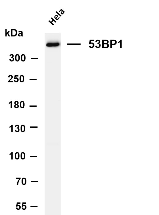
Mass (kDA):
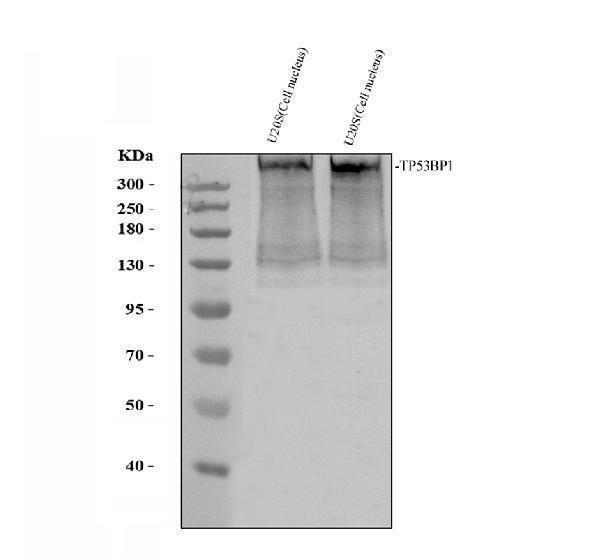
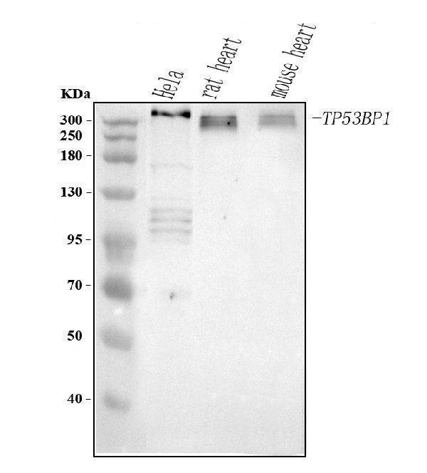
213.574 kDA

| Human | |
|---|---|
| Location: | 15q15.3 |
| Sequence: | 15; NC_000015.10 (43403061..43510640, complement) |
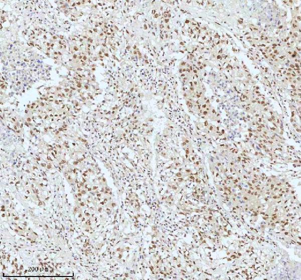
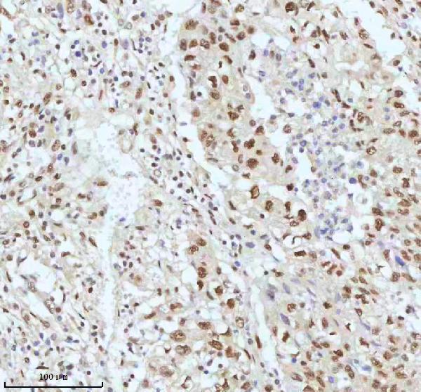
Nucleus. Chromosome. Chromosome, centromere, kinetochore. Localizes to the nucleus in absence of DNA damage (PubMed:28241136). Following DNA damage, recruited to sites of DNA damage, such as double stand breaks (DSBs): recognizes and binds histone H2A monoubiquitinated at 'Lys-15' (H2AK15Ub) and histone H4 dimethylated at 'Lys-20' (H4K20me2), two histone marks that are present at DSBs sites (PubMed:23333306, PubMed:23760478, PubMed:24703952, PubMed:28241136, PubMed:17190600). Associated with kinetochores during mitosis (By similarity).


PMID: 9748285 by Iwabuchi K., et al. Stimulation of p53-mediated transcriptional activation by the p53- binding proteins, 53BP1 and 53BP2.
PMID: 8016121 by Iwabuchi K., et al. Two cellular proteins that bind to wild-type but not mutant p53.