This website uses cookies to ensure you get the best experience on our website.
- Table of Contents
1 Citations 7 Q&As
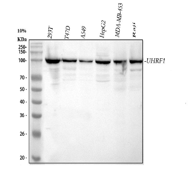
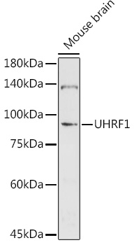
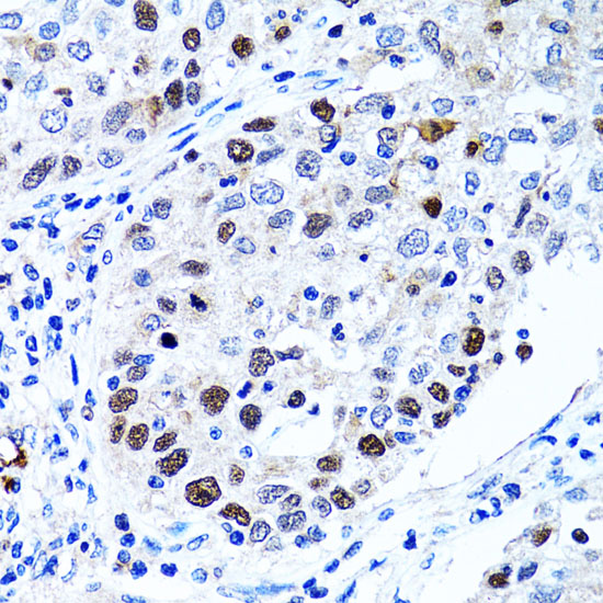
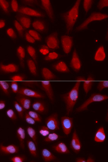
Facts about E3 ubiquitin-protein ligase UHRF1.
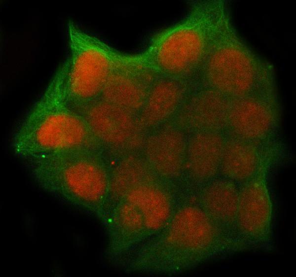
In addition to its role in maintenance of DNA methylation, also plays an integral role in chromatin modification: through its tudor-like regions and PHD- type zinc fingers, specifically recognizes and binds histone H3 trimethylated in'Lys-9' (H3K9me3) and unmethylated in'Arg-2' (H3R2me0), respectively, and recruits chromatin proteins. Enriched in pericentric heterochromatin where it recruits different chromatin modifiers required for this chromatin replication.
Mouse | |
---|---|
Gene Name: | Uhrf1 |
Uniprot: | Q8VDF2 |
Entrez: | 18140 |
Belongs to: |
---|
No superfamily |
E3 ubiquitin-protein ligase UHRF1; EC 6.3.2; EC 6.3.2.-; FLJ21925; hNP95; huNp95; ICBP90NP95; Inverted CCAAT box-binding protein of 90 kDa; Np95; Nuclear protein 95; Nuclear zinc finger protein Np95; RING finger protein 106; RNF106MGC138707; Transcription factor ICBP90; Ubiquitin-like PHD and RING finger domain-containing protein 1; ubiquitin-like with PHD and ring finger domains 1; ubiquitin-like, containing PHD and RING finger domains, 1; Ubiquitin-like-containing PHD and RING finger domains protein 1
Mass (kDA):
88.304 kDA
Mouse | |
---|---|
Location: | 17|17 D |
Sequence: | 17; |
Expressed in thymus, testis, spleen and lung. Within testis, expressed in almost all cells except elongated spermatids.
If you're searching for the best UHRF1 assay then read on. This article will help you understand how to conduct a UHRF1 test for a variety of sample types. This article will help you achieve the best results from synovial fluids to samples from patients. You'll receive the most precise and sensitive results when you follow the steps in this article.
Flow cytometric analysis may reveal new therapeutic targets for patients suffering from RA. This is possible because detailed molecular analysis can be conducted on synovial tissue to identify distinct cells. This approach will also aid in separating RA patients based on their cellular phenotypes. The synovial cytometric analysis of RA patients is hindered by the absence of protocols that can simultaneously recover and analyze synovial tissue cells. There is also no method to compare cells from patients with RA.
Flow cytometric analysis was used to determine the presence of various subsets of immune cells within synovial tissue taken from patients with RA. The synovial fluid contained immune cells called "a" cells. These cells are sensitive to side-scattered sunlight. These cells were later identified as activated macrophages and increased in number in RA fluid. Anti-CD11b and anti-CD4 antibodies were employed to stain these cells.
The high sensitivity of flow cytometric analysis allowed researchers to identify distinct cell types. The podoplanin transcript was found in fibroblasts, whereas the CD14 protein was found only in macrophages. CD3E and CD79A were distinct transcriptomes that were derived from T and B cells. This study also demonstrated that synovial tissue that has been preserved in cryopreservation is a fantastic source for high-quality transcriptomes.
The inflammatory and phagocytic cell populations in patients with RA differ. Different types of cells in the synovium are characterized by different numbers of lymphocytes infiltrated and activated macrophages. The high-macrophage group had an increased proportion of lymphocytes that exceeded 80%. The lower-macrophage-group had lower percentages, with less than 40%. The lymphocyte content of the low-macrophage groups varied between 9.6% up to 46.1%. The low-macrophage-low group had less than 40% of total macrophages. The lowest macrophage group was comprised of 10 patients.
The authors appreciate the involvement of the clinical coordinators. They also thank the patients for their time and dedication. The research was conducted by Drs. Drs. Anolik, Mina Pichavant and Bill Apruzzese were able to aid in the research. The authors thank the RA Disease Focus Group and the UR Flow Cytometry Core.
The study found that LTi-like cellular types found in RA synovial fluid show phenotypic similarities with tonsillar LTi cell. They also have higher lymphocyte infiltration which suggests that they are involved in the creation of a lymphoid tissue-like structure. They were also able to determine the presence by employing the same method.
Cryopreserved synovial tissue is an important technological advancement in synovial research. Cryopreserved tissue is similar in terms of viability to freshly prepared synovial tissues. The cells are easily functional and can be used in all studies. It is also efficient and can be used to conduct multisite consortium studies. The use of cryopreservation for tissue fragments allows for the collection of samples from various clinical sites without creating significant tissue dissociation.
The analysis of flow cytometry shows significant distinctions in synovial tissues of patients suffering from OA. The study also revealed significant differences in the number of synovial macrophages. This information will aid in the development and testing of new strategies. It will also help in the development and identification of new anti-inflammatory medicines.
A modified protocol was used to digest cells taken from synovial fluids of patients suffering from OA for flow cytometric analysis. Then, the cells were counted for their specific subsets using different gating strategies. By using the gating strategy, a cluster of DCs and monocytes was identified, as well as synovial macrophages.
These findings highlight the importance of synovial macrophages as a source of OA pathogenesis. Since macrophages constitute the primary immune cells of the synovium they are likely to be at the forefront of detecting joint injuries. OA synovial tissue macrophages generate proteins, including metalloproteinases various cytokines and other compounds that contribute to cartilage degrading. These findings show that synovial macrophages of IOA and cOA exhibit different gene expression patterns.
Flow cytometric analysis was used to study the immune response to antiinflammatory medications. The SF sample was placed in an eppendorf tube, and then mixed with fluo4 for a period of 60 min. The samples were then transferred into plastic tubes with round bottoms for analysis using flow cytometry. Synovial fluid has a variety of cell types including macrophages and monocytes as well as neutrophils.
Although immunohistochemistry is by far the most commonly used method of analysis but there are some drawbacks. The analysis is two-dimensional, requiring extensive reconstruction of the whole biopsy. Each tissue slice only provides information about the number of cells. The process of interfacing digital data and visible signals can be labor-intensive and not completely automated. In addition, the disaggregation of enzymatic enzymes of synovium may also be associated with alterations in the cell surface markers.
Despite significant improvements in the analysis of flow cytometry accuracy However, the results are not conclusive. More research is needed to establish the biomarkers that belong to specific subgroups of patients with OA. This study will help in the development of treatments to focus on specific inflammatory processes within the synovium. For example in clinical trials the presence of specific biomarkers will allow clinicians to match an appropriate anti-inflammatory medication to patients suffering from OA.
The study revealed a uniform synovium composition for patients with OA. The cells showed a uniform cell structure, with a majority of fibroblasts. These cells were analyzed by fluorescence automated cell sorting (FACS), using markers specific to bone marrow MPCs. The remaining cells from synovial tissue biopsy were then differentiated and evaluated for osteogenic potential. These cells were also significantly associated with the overall score of histological inflammation.
Recent research has revealed that the synovial fluid levels in patients suffering from rheumatoid arthritis and peripheral blood levels are strongly associated. These findings can be used to aid doctors develop better treatments for RA. Phase one of the RA Network project was completed with synovial tissues that were matched and peripheral blood samples. The results showed that synovial fluid from patients suffering from RA has higher levels of lymphocytes than the normal fluid and has more inflammation.
For the study, SF was collected from patients suffering from RA, OA, and gout. Fluo4 was then added to the synovial fluid of the patient and the samples were taken within 60 minutes. The sample preparation was straightforward and did not require washing to get rid of the dye. SF samples were placed in plastic tubes with round bottoms that facilitate the flow cytometric analysis. The SF samples are typically dilute with sheath fluid. The data are then displayed as dot plots or histograms.
Flow cytometric analysis of synovials fluid from patients suffering from RA revealed that patients suffering from the disease had a greater number of cells than patients with OA. The patients' SF was collected after informed consent was obtained. The results of the study did not show statistically significant differences in the patients' aged, SF, sex, or duration of disease. The analysis of flow cytometry revealed that synovial fluid of patients suffering from RA or OA was higher in levels of CRP, RF and ACPA.
In RA, neutrophils are activated in synovial fluid at a very high level. Inactivated neutrophils release a large amount of toxic substances and can sabotage synovial fluid's antiprotease and antioxidant shields. It is therefore important to monitor neutrophil activity in RA sufferers. The development of symptoms associated with the disease is dependent on the presence of soluble immune complexes in synovial liquid.
Flow cytometry was used to sort SF from patients suffering from RA. B cell subsets were separated using an RNeasy mini kit (Qiagen). RT-PCR was conducted in accordance with previously described methods. Then, oligo(dT)-primed cDNA was synthesized using the Fermentas RevertAid FirstStrand kit.
Adenosine is a chemical that blocks the production of TNF-a, was found in synovial fluids of patients suffering from RA. In contrast, TNF-a and adenosine did not exhibit a significant correlation in synovial fluids. The results also showed no connection between neutrophil apoptosis or adenosine.
The results indicated that RA SF is populated with B cells with specific characteristics that are vital to the development of the disease. In this study, B cells carrying CD19+CD24hiCD27+ characteristics were found in the RA the SF. This phenotype is known to enhance osteoclast development. Furthermore, this subset could play a role in inflammation.
PMID: 9880673 by Fujimori A., et al. Cloning and mapping of Np95 gene which encodes a novel nuclear protein associated with cell proliferation.
PMID: 12084726 by Muto M., et al. Targeted disruption of Np95 gene renders murine embryonic stem cells hypersensitive to DNA damaging agents and DNA replication blocks.
*More publications can be found for each product on its corresponding product page