This website uses cookies to ensure you get the best experience on our website.
- Table of Contents
and ELISA kits, proteins related to Myasthenia Gravis.
Myasthenia Gravis (MG) is a chronic autoimmune neuromuscular disorder characterized by varying degrees of skeletal muscle weakness. This condition arises when the immune system mistakenly targets and attacks the acetylcholine receptors at the neuromuscular junction, disrupting effective communication between nerves and muscles. As a result, individuals with MG often experience challenges with voluntary muscle movements, including eye movements, facial expressions, swallowing, and limb mobility. Research into MG is heavily focused on understanding the specific antibodies involved in this autoimmune response, aiming to develop targeted therapies that can better manage symptoms and improve patient outcomes. Advances in antibody-related research hold promise for more effective treatments, offering hope for those affected by this debilitating condition. Our landing page is dedicated to providing the latest insights and developments in Myasthenia Gravis research, fostering a community of support and innovation.
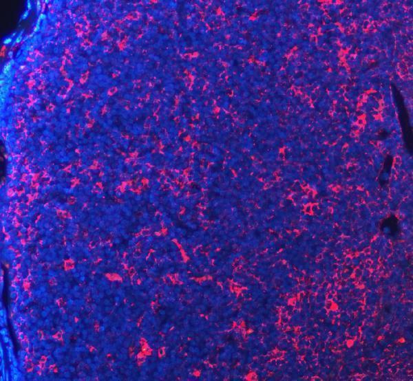
Anti-PD-1/PDCD1 Antibody Picoband®, Figure 5. IF analysis of PDCD1 using anti-PDCD1 antibody (A00178).
PDCD1 was detected in a paraffin-embedded section of mouse lymph node tissue. ...
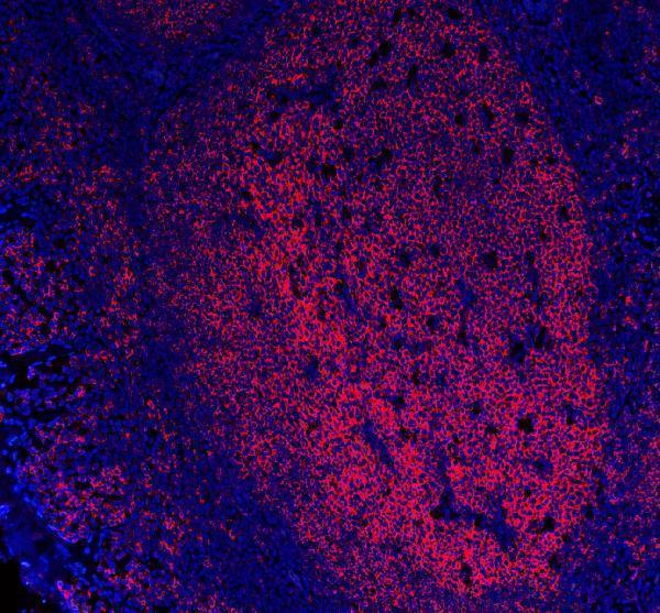
Anti-CD20/MS4A1 Antibody Picoband® (monoclonal, 4I11), Figure 1. IF analysis of CD20/MS4A1 using anti-CD20/MS4A1 antibody (M03780-4).
CD20/MS4A1 was detected in a p...

Figure 1. IHC analysis of TGF Beta 1/TGFB1 using Anti-TGF beta 1/TGFB1 Antibody (A00019-2).
TGF Beta 1/TGFB1 was detected in a paraffin-embedded section of rat li...
| Protein Name | Gene Name | Function |
|---|---|---|
| Acetylcholine Receptor (AChR) | CHRNE | Autoantibody targets AChR at neuromuscular junction, impairing signal transmission |
| Muscle-Specific Kinase (MuSK) | MUSK | Autoantibody disrupts MuSK, affecting AChR clustering |
| Low-Density Lipoprotein Receptor-Related Protein 4 (LRP4) | LRP4 | Autoantibody against LRP4 disrupts neuromuscular junction formation |
| Titin | TTN | Autoantibody associated with thymoma and late-onset Myasthenia Gravis |
| Ryanodine Receptor (RyR) | RYR1 | Autoantibody linked to thymoma in Myasthenia Gravis patients |
| Complement C3 | C3 | Involved in complement cascade activation in AChR-positive Myasthenia Gravis |
| CD20 | MS4A1 | B cell marker targeted by therapies like rituximab |
| Cytotoxic T-Lymphocyte Antigen 4 (CTLA-4) | CTLA4 | Immune checkpoint involved in regulating T cell responses |
| Interleukin-6 (IL-6) | IL6 | Pro-inflammatory cytokine elevated in Myasthenia Gravis |
| Transforming Growth Factor Beta (TGF-beta) | TGFB1 | Immunomodulatory cytokine affecting immune response |
| HLA-DR3 | HLA-DRB1 | Genetic susceptibility marker associated with Myasthenia Gravis |
| Forkhead Box P3 (FoxP3) | FOXP3 | Marker for regulatory T cells involved in immune tolerance |
| CD19 | CD19 | B cell marker important for B cell development and function |
| Programmed Death-1 (PD-1) | PDCD1 | Immune checkpoint protein regulating T cell activity |
| B-Cell Activating Factor (BAFF) | TNFSF13B | Promotes B cell survival and maturation |
| CD40 | CD40 | B cell costimulatory molecule essential for B cell activation |
| C-X-C Motif Chemokine Ligand 10 (CXCL10) | CXCL10 | Chemokine involved in recruiting immune cells to sites of inflammation |
Myasthenia Gravis (MG) is primarily characterized by its autoimmune nature, where the body's immune system mistakenly targets components critical for neuromuscular transmission. The most common antibodies implicated are those against the acetylcholine receptors (AChR) at the neuromuscular junction, leading to impaired signal transmission and muscle weakness. Additionally, a subset of patients exhibits antibodies against Muscle-Specific Kinase (MuSK), which plays a pivotal role in clustering ACh receptors and maintaining synaptic structure. Understanding these antibody profiles is crucial as they not only aid in accurate diagnosis but also influence treatment strategies. Research in this area delves into the mechanisms of autoantibody production, the role of B and T cells in sustaining the autoimmune response, and the identification of other potential autoantigens. Advances in this subfield have paved the way for targeted therapies, such as monoclonal antibodies that specifically inhibit pathogenic immune cells, thereby offering hope for more effective and personalized treatment options for individuals battling MG.
The thymus gland plays a central role in the pathogenesis of Myasthenia Gravis, making thymus pathology a critical sub-research area. In many MG patients, the thymus is abnormal—ranging from thymic hyperplasia, where the gland is enlarged with an excess of lymphocytes and plasma cells, to thymoma, a tumor of the thymic epithelial cells. The thymus is integral in T-cell development and the establishment of immune tolerance; abnormalities here can lead to the escape of autoreactive T cells that drive the autoimmune attack on neuromuscular junction components. Research focuses on elucidating how thymic abnormalities contribute to the loss of self-tolerance, the generation of autoantibodies, and the perpetuation of the disease. Additionally, studies investigate the therapeutic implications of thymectomy (surgical removal of the thymus) in MG management, assessing its efficacy in different patient subsets. Insights from thymus-related research not only enhance our understanding of MG's underlying mechanisms but also inform the development of interventions that can modulate thymic function to restore immune balance.