Click image to see more details
Product Info Summary
| SKU: | A02008 |
|---|---|
| Size: | 100ul |
| Reactive Species: | Human, Mouse, Rat |
| Host: | Rabbit |
| Application: | IHC, WB |
Customers Who Bought This Also Bought

Product info
Product Name
Anti-ACAT1 (K266) Antibody
SKU/Catalog Number
A02008
Size
100ul
Form
Liquid
Description
Boster Bio Anti-ACAT1 (K266) Antibody catalog # A02008. Tested in WB,IHC applications. This antibody reacts with Human,Mouse,Rat.
Storage & Handling
Store at -20°C for one year. For short term storage and frequent use, store at 4°C for up to one month. Avoid repeated freeze-thaw cycles.
Cite This Product
Anti-ACAT1 (K266) Antibody (Boster Biological Technology, Pleasanton CA, USA, Catalog # A02008)
Host
Rabbit
Contents
Rabbit IgG, 1mg/ml in PBS with 0.02% sodium azide, 50% glycerol, pH7.2
Clonality
Polyclonal
Isotype
IgG
Immunogen
Synthetic peptide, corresponding to amino acids 235-286 of Human ACAT1.
Reactive Species
A02008 is reactive to ACAT1 in Human, Mouse, Rat
Calculated molecular weight
45.2 kDa
Antibody Validation
Boster validates all antibodies on WB, IHC, ICC, Immunofluorescence, and ELISA with known positive control and negative samples to ensure specificity and high affinity, including thorough antibody incubations.
Application & Images
Applications
A02008 is guaranteed for IHC, WB Boster Guarantee
Recommend Dilution
WB: 1:500-1:1000
IHC: 1:50-1:200
Validation Images & Assay Conditions
Click image to see more details
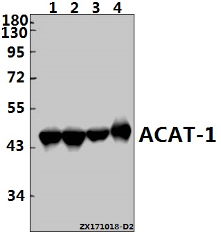
Western blot (WB) analysis of ACAT-1 (K266) pAb at 1:1000 dilution Lane1:HEK293T whole cell lysate(20ug) Lane2:HepG2 whole cell lysate(20ug) Lane3:The Lung tissue lysate of Rat(20ug) Lane4:The Lung tissue lysate of Mouse(10ug)

Click image to see more details
Immunohistochemistry (IHC) analyzes of ACAT1 (K266) pA bin paraffin-embedded human liver carcinoma tissue at 1:50,showing cytoplasmic staining.Negative control (the right)Using PBS instead of primary antibody, secondary antibody is Goat Anti-Rabbit IgG-biotin followed by avidin-peroxidase.

Click image to see more details
Immunohistochemistry (IHC) analyzes of ACAT1 (K266) pA bin paraffin-embedded human kidney carcinoma tissue at 1:50,showing cytoplasmic staining.Negative control (the right)Using PBS instead of primary antibody, secondary antibody is Goat Anti-Rabbit IgG-biotin followed by avidin-peroxidase.
Specific Publications For Anti-ACAT1 (K266) Antibody (A02008)
Loading publications
Recommended Resources
Here are featured tools and databases that you might find useful.
- Boster's Pathways Library
- Protein Databases
- Bioscience Research Protocol Resources
- Data Processing & Analysis Software
- Photo Editing Software
- Scientific Literature Resources
- Research Paper Management Tools
- Molecular Biology Software
- Primer Design Tools
- Bioinformatics Tools
- Phylogenetic Tree Analysis
Customer Reviews
Have you used Anti-ACAT1 (K266) Antibody?
Share your experimental results or join a short interview to earn up to $1,000 in product credits or other rewards.
0 Reviews For Anti-ACAT1 (K266) Antibody
Customer Q&As
Have a question?
Find answers in Q&As, reviews.
Can't find your answer?
Submit your question
