Click image to see more details
-

-
-
-
-
+4
Product Info Summary
| SKU: | PA1006 |
|---|---|
| Size: | 100 μg/vial |
| Reactive Species: | Human, Monkey |
| Host: | Rabbit |
| Application: | Flow Cytometry, IF, IHC, ICC, WB |
Customers Who Bought This Also Bought

Product info
Product Name
Anti-Annexin A1/ANXA1 Antibody Picoband®
SKU/Catalog Number
PA1006
BA3701 is an alternative SKU for this antibody, used in previous lots.
Size
100 μg/vial
Form
Lyophilized
Description
ANXA1 is a glucocorticoid-regulated mediator of inflammation resolution, acting in the ANXA1–FPR2/ALX axis to modulate neutrophil trafficking/apoptosis and pro-resolving responses (context dependent). Assay context: antibody tested for flow cytometry, IF/ICC, IHC, and WBANXA2 (surface complexes and vascular/inflammatory interfaces) and bridging opsonins like MFGE8 when distinguishing apoptotic clearance programs from sustained inflammatory recruitment (putative).
Storage & Handling
Store at -20˚C for one year from date of receipt. After reconstitution, at 4˚C for one month. It can also be aliquotted and stored frozen at -20˚C for six months. Avoid repeated freeze-thaw cycles.
Cite This Product
Anti-Annexin A1/ANXA1 Antibody Picoband® (Boster Biological Technology, Pleasanton CA, USA, Catalog # PA1006)
Host
Rabbit
Contents
Each vial contains antibody formulated with stabilizing components, 0.9mg NaCl, 0.2mg Na2HPO4, 0.05mg Thimerosal, 0.05mg NaN3.
*This antibody is supplied in a stabilized formulation.
Compatibility with conjugation reactions depends on the chemistry of the conjugation method used.
For conjugation methods that are not compatible with the stabilizing components present in this formulation, a carrier-free antibody format is required.
Clonality
Polyclonal
Isotype
Rabbit IgG
Immunogen
A synthetic peptide corresponding to a sequence at the N-terminus of human Annexin A1.
Cross-reactivity
No cross-reactivity with other proteins
Reactive Species
PA1006 is reactive to ANXA1 in Human, Monkey
Observed Molecular Weight
39 kDa
Calculated molecular weight
38.7 kDa
Background of ANXA1
Annexin I, also known as lipocortin I (Lipo1), belongs to the family of annexins. These proteins are though to control the biosynthesis of the potent mediators of inflammation, prostaglandins and leukotrienes. In two lipocortins (I and II) a short amino-terminal sequence distinct from the core structure has potential regulatory functions which are dependent on its phosphorylation state. The gene in the mouse encodes a protein of 346 amino acid residues. Mouse Lipo1 gene spans about 17 kb and is divided into 13 exons. Annexin I gene, mapped to 9q11-q22, is located on mouse chromosome 19. Annexin I acts through the formyl peptide receptor on human neutrophils. Peptides derived from the unique N-terminal domain of annexin I serve as FPR ligands and trigger different signaling pathways in a dose-dependent manner.
Antibody Validation
Boster validates all antibodies on WB, IHC, ICC, Immunofluorescence, and ELISA with known positive control and negative samples to ensure specificity and high affinity, including thorough antibody incubations.
Application & Images
Applications
PA1006 is guaranteed for Flow Cytometry, IF, IHC, ICC, WB Boster Guarantee
Recommend Dilution
| Application | Dilution | Species |
|---|---|---|
| Western blot | 0.1-0.5μg/ml | Human, Monkey |
| Immunohistochemistry (Paraffin-embedded Section) | 0.5-1μg/ml | Human |
| Immunocytochemistry/Immunofluorescence | 5μg/ml | Human |
| Flow Cytometry (Fixed) | 1-3μg/1x106 cells | Human |
Tested application
Suggested blocking solution with 5% non-fat milk or BSA; (*)Recommended protein loading: 20-40 µg per lane
Use TE buffer pH 9.0 for antigen retrieval; (*) citrate buffer pH 6.0 is an alternative.
Validation Images & Assay Conditions
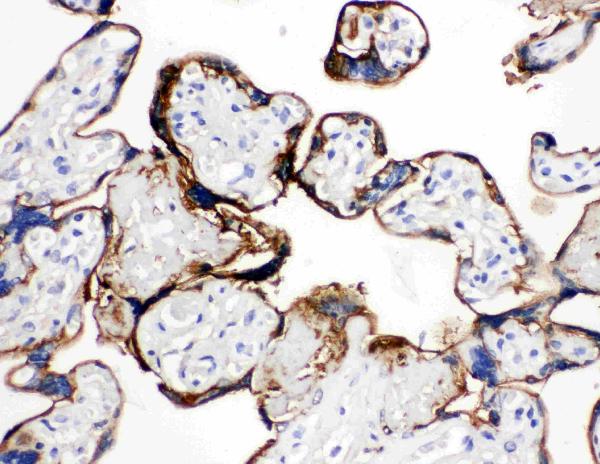
Click image to see more details
Anti-Annexin A1 antibody, PA1006, IHC(P)
IHC(P): Human Placenta Tissue

Click image to see more details
Western blot analysis of Annexin A1/ANXA1 using anti-Annexin A1/ANXA1 antibody (PA1006).
Electrophoresis was performed on a 5-20% SDS-PAGE gel at 70V (Stacking gel) / 90V (Resolving gel) for 2-3 hours. The sample well of each lane was loaded with 30ug of sample under reducing conditions.
Lane 1: human Hela whole cell lysates,
Lane 2: human K562 whole cell lysates,
Lane 3: monkey COS-7 whole cell lysates.
After Electrophoresis, proteins were transferred to a Nitrocellulose membrane at 150mA for 50-90 minutes. Blocked the membrane with 5% Non-fat Milk/ TBS for 1.5 hour at RT. The membrane was incubated with rabbit anti-Annexin A1/ANXA1 antigen affinity purified polyclonal antibody (Catalog # PA1006) at 0.5 μg/mL overnight at 4°C, then washed with TBS-0.1%Tween 3 times with 5 minutes each and probed with a goat anti-rabbit IgG-HRP secondary antibody at a dilution of 1:5000 for 1.5 hour at RT. The signal is developed using an Enhanced Chemiluminescent detection (ECL) kit (Catalog # EK1002) with Tanon 5200 system. A specific band was detected for Annexin A1/ANXA1 at approximately 39KD. The expected band size for Annexin A1/ANXA1 is at 39KD.

Click image to see more details
Germacrone induced apoptosis in gastric cancer cells. (A–D) Annexin IV-fluorescein isothiocyanate (FITC)/propidium iodide (PI) staining was used to assess the effect of germacrone on apoptosis by flow cytometry (FCM). (E) Hoechst 33258 staining was used to detect apoptosis. (F, G) Changes in the expression levels of BAX, Bcl-2, caspase-3, and cleaved caspase-3 were detected by western blot. (H, I) Changes in the BAX/Bcl-2 and cleaved caspase-3/caspase-3 ratios were analyzed after germacrone treatment. Data are the means ± SD of three experiments. * P < 0.05; ns, not significant.
Index in PubMed under a CC BY license. PMID: 33244453

Click image to see more details
Label-free proteomic and bioinformatic analysis of proteins associated with the cell cycle, apoptosis, and autophagy. (A–C) The DAVID database was used for analysis of molecular function, cellular component, and biological process. (D) A heat map was constructed based on the abundance of 111 proteins. D1, D2, and D3 represent the DMSO 1, DMSO 2, and DMSO 3 groups; G1, G2, and G3 represent the germacrone 1, germacrone 2, and germacrone 3 groups. (E) A protein interaction network was constructed in Cytoscape based on the information provided by the STRING database. Red indicates increased expression of the protein in the germacrone group; green indicates reduced expression of the protein in the germacrone group. (F, G) . The expression of HBXIP, HSP70, and ANXA1 was detected to verify the accuracy of the proteomics results.
Index in PubMed under a CC BY license. PMID: 33244453

Click image to see more details
Overexpression of HBXIP regulated autophagy and reversed the germacrone-induced cell cycle arrest and apoptosis. (A–D) Propidium iodide (PI) staining was used for flow cytometric (FCM) analysis of the effect of germacrone and overexpression of HBXIP on the cell cycle. (E–H) Annexin IV-fluorescein isothiocyanate (FITC)/PI staining was used to detect the effect of germacrone and overexpression of HBXIP on apoptosis by FCM. (I, J) The expression of HBXIP, p-62, LC3I, and LC3II was detected by western blot. (K) A schematic of the effect of germacrone on gastric cancer cells. Germacrone inhibits gastric cancer cell proliferation involving HBXIP-mediated regulation of the cell cycle, apoptosis, and autophagy. Data are the means ± SD of three experiments. * P < 0.05; ns, not significant.
Index in PubMed under a CC BY license. PMID: 33244453

Click image to see more details
Anti-Annexin A1 antibody, PA1006, IHC(P)
IHC(P): Human Tonsil Tissue

Click image to see more details
IF analysis of Annexin A1/ANXA1 using anti-Annexin A1/ANXA1 antibody (PA1006).
Annexin A1/ANXA1 was detected in immunocytochemical section of A431 cells. Enzyme antigen retrieval was performed using IHC enzyme antigen retrieval reagent (AR0022) for 15 mins. The cells were blocked with 10% goat serum. And then incubated with 5μg/mL rabbit anti-Annexin A1/ANXA1 Antibody (PA1006) overnight at 4°C. DyLight®488 Conjugated Goat Anti-Rabbit IgG (BA1127) was used as secondary antibody at 1:100 dilution and incubated for 30 minutes at 37°C. The section was counterstained with DAPI. Visualize using a fluorescence microscope and filter sets appropriate for the label used.

Click image to see more details
Flow Cytometry analysis of A431 cells using anti-Annexin A1/ANXA1 antibody (PA1006).
Overlay histogram showing A431 cells stained with PA1006 (Blue line). To facilitate intracellular staining, cells were fixed with 4% paraformaldehyde and permeabilized with permeabilization buffer. The cells were blocked with 10% normal goat serum. And then incubated with rabbit anti-Annexin A1/ANXA1 Antibody (PA1006, 1μg/1x106 cells) for 30 min at 20°C. DyLight®488 conjugated goat anti-rabbit IgG (BA1127, 5-10μg/1x106 cells) was used as secondary antibody for 30 minutes at 20°C. Isotype control antibody (Green line) was rabbit IgG (1μg/1x106) used under the same conditions. Unlabelled sample without incubation with primary antibody and secondary antibody (Red line) was used as a blank control.
Specific Publications For Anti-Annexin A1/ANXA1 Antibody Picoband® (PA1006)
Loading publications
Recommended Resources
Here are featured tools and databases that you might find useful.
- Boster's Pathways Library
- Protein Databases
- Bioscience Research Protocol Resources
- Data Processing & Analysis Software
- Photo Editing Software
- Scientific Literature Resources
- Research Paper Management Tools
- Molecular Biology Software
- Primer Design Tools
- Bioinformatics Tools
- Phylogenetic Tree Analysis
Customer Reviews
Have you used Anti-Annexin A1/ANXA1 Antibody Picoband®?
Share your experimental results or join a short interview to earn up to $1,000 in product credits or other rewards.
0 Reviews For Anti-Annexin A1/ANXA1 Antibody Picoband®
Customer Q&As
Have a question?
Find answers in Q&As, reviews.
Can't find your answer?
Submit your question
19 Customer Q&As for Anti-Annexin A1/ANXA1 Antibody Picoband®
Question
Does PA1006 require dry ice during shipping?
Verified customer
Asked: 2022-06-03
Answer
The Anti-Annexin A1/ANXA1 Antibody (PA1006) doesn't require dry ice during shipping.
Boster Scientific Support
Answered: 2022-06-03
Question
Is a blocking peptide available for product anti-Annexin A1/ANXA1 antibody (PA1006)?
Verified Customer
Verified customer
Asked: 2020-04-23
Answer
We do provide the blocking peptide for product anti-Annexin A1/ANXA1 antibody (PA1006). If you would like to place an order for it please contact support@bosterbio.com and make a special request.
Boster Scientific Support
Answered: 2020-04-23
Question
We have tried in the past anti-Annexin A1/ANXA1 antibody for WB on cervix carcinoma erythroleukemia last year. I am using human, and I plan to use the antibody for IHC next. We need examining cervix carcinoma erythroleukemia as well as liver in our next experiment. Do you have any suggestion on which antibody would work the best for IHC?
Verified Customer
Verified customer
Asked: 2020-02-18
Answer
I have checked the website and datasheets of our anti-Annexin A1/ANXA1 antibody and it appears that PA1006 has been tested on human in both WB and IHC. Thus PA1006 should work for your application. Our Boster satisfaction guarantee will cover this product for IHC in human even if the specific tissue type has not been validated. We do have a comprehensive range of products for IHC detection and you can check out our website bosterbio.com to find out more information about them.
Boster Scientific Support
Answered: 2020-02-18
Question
I see that the anti-Annexin A1/ANXA1 antibody PA1006 works with WB, what is the protocol used to produce the result images on the product page?
Verified Customer
Verified customer
Asked: 2019-10-30
Answer
You can find protocols for WB on the "support/technical resources" section of our navigation menu. If you have any further questions, please send an email to support@bosterbio.com
Boster Scientific Support
Answered: 2019-10-30
Question
Please see the WB image, lot number and protocol we used for cervix carcinoma using anti-Annexin A1/ANXA1 antibody PA1006. Please let me know if you require anything else.
Verified Customer
Verified customer
Asked: 2019-10-18
Answer
Thank you very much for the data. Our lab team are working to resolve this as quickly as possible, and we appreciate your patience and understanding! You have provided everything we needed. Please let me know if there is anything you need in the meantime.
Boster Scientific Support
Answered: 2019-10-18
Question
I would like to test anti-Annexin A1/ANXA1 antibody PA1006 on human cervix carcinoma for research purposes, then I may be interested in using anti-Annexin A1/ANXA1 antibody PA1006 for diagnostic purposes as well. Is the antibody suitable for diagnostic purposes?
Verified Customer
Verified customer
Asked: 2019-10-17
Answer
The products we sell, including anti-Annexin A1/ANXA1 antibody PA1006, are only intended for research use. They would not be suitable for use in diagnostic work. If you have the means to develop a product into diagnostic use, and are interested in collaborating with us and develop our product into an IVD product, please contact us for more discussions.
Boster Scientific Support
Answered: 2019-10-17
Question
We have seen staining in human cervix lung. Do you have any suggestions? Is anti-Annexin A1/ANXA1 antibody supposed to stain cervix lung positively?
Verified Customer
Verified customer
Asked: 2019-09-05
Answer
From literature cervix lung does express ANXA1. From Uniprot.org, ANXA1 is expressed in mouth mucosa, cervix lung, cervix carcinoma, cervix carcinoma erythroleukemia, liver, among other tissues. Regarding which tissues have ANXA1 expression, here are a few articles citing expression in various tissues:
Cervix carcinoma, Pubmed ID: 18669648, 20068231
Cervix carcinoma, and Erythroleukemia, Pubmed ID: 23186163
Cervix, and Lung, Pubmed ID: 15489334
Liver, Pubmed ID: 24275569
Boster Scientific Support
Answered: 2019-09-05
Question
Can you help my question with product PA1006, anti-Annexin A1/ANXA1 antibody. I was wondering if it would be possible to conjugate this antibody with biotin. I would need it to be without BSA or sodium azide. I am planning on using a buffer exchange of sodium azide with PBS only. Would there be problems for me to conjugate the antibody and store it in -20 degrees in small aliquots?
R. Krishna
Verified customer
Asked: 2019-07-11
Answer
It is not recommended storing this antibody with PBS buffer only in -20 degrees. If you want to store it in -20 degrees it is best to add some cryoprotectant like glycerol. If you want carrier free PA1006 anti-Annexin A1/ANXA1 antibody, we can provide it to you in a special formula with trehalose and/or glycerol. These molecules will not interfere with conjugation chemistry and provide a good level of protection for the antibody from degradation. Please be sure to specify this in your purchase order.
Boster Scientific Support
Answered: 2019-07-11
Question
Have you mapped the epitope for PA1006?
Verified customer
Asked: 2019-05-20
Answer
Unfortunately, we do not map the epitope of our antibodies.
Boster Scientific Support
Answered: 2019-05-20
Question
Does anti-Annexin A1/ANXA1 antibody PA1006 work for WB with cervix carcinoma?
Verified Customer
Verified customer
Asked: 2019-05-10
Answer
According to the expression profile of cervix carcinoma, ANXA1 is highly expressed in cervix carcinoma. So, it is likely that anti-Annexin A1/ANXA1 antibody PA1006 will work for WB with cervix carcinoma.
Boster Scientific Support
Answered: 2019-05-10
Question
Will PA1006 anti-Annexin A1/ANXA1 antibody work on parafin embedded sections? If so, which fixation method do you recommend we use (PFA, paraformaldehyde, other)?
S. Jha
Verified customer
Asked: 2018-06-26
Answer
You can see on the product datasheet, PA1006 anti-Annexin A1/ANXA1 antibody as been validated on WB. It is best to use PFA for fixation because it has better tissue penetration ability. PFA needs to be prepared fresh before use. Long term stored PFA turns into formalin, as the PFA molecules congregate and become formalin.
Boster Scientific Support
Answered: 2018-06-26
Question
My team were well pleased with the WB result of your anti-Annexin A1/ANXA1 antibody. However we have observed positive staining in cervix lung nucleus using this antibody. Is that expected? Could you tell me where is ANXA1 supposed to be expressed?
Verified Customer
Verified customer
Asked: 2018-04-25
Answer
From what I have seen in literature, cervix lung does express ANXA1. Generally ANXA1 expresses in nucleus. Regarding which tissues have ANXA1 expression, here are a few articles citing expression in various tissues:
Cervix carcinoma, Pubmed ID: 18669648, 20068231
Cervix carcinoma, and Erythroleukemia, Pubmed ID: 23186163
Cervix, and Lung, Pubmed ID: 15489334
Liver, Pubmed ID: 24275569
Boster Scientific Support
Answered: 2018-04-25
Question
Will anti-Annexin A1/ANXA1 antibody PA1006 work on zebrafish WB with cervix carcinoma erythroleukemia?
Verified Customer
Verified customer
Asked: 2018-02-06
Answer
Our lab technicians have not validated anti-Annexin A1/ANXA1 antibody PA1006 on zebrafish. You can run a BLAST between zebrafish and the immunogen sequence of anti-Annexin A1/ANXA1 antibody PA1006 to see if they may cross-react. If the sequence homology is close, then you can perform a pilot test. Keep in mind that since we have not validated zebrafish samples, this use of the antibody is not covered by our guarantee. However we have an innovator award program that if you test this antibody and show it works in zebrafish cervix carcinoma erythroleukemia in WB, you can get your next antibody for free.
Boster Scientific Support
Answered: 2018-02-06
Question
I was wanting to use using your anti-Annexin A1/ANXA1 antibody for arachidonic acid secretion studies. Has this antibody been tested with western blotting on colo320 cell lysate? We would like to see some validation images before ordering.
Verified Customer
Verified customer
Asked: 2017-07-04
Answer
Thanks for your inquiry. This PA1006 anti-Annexin A1/ANXA1 antibody is validated on u87 cell lysate, hela cell lysate, panc cell lysate, colo320 cell lysate, smmc cell lysate, human tonsil tissue, placenta tissue. It is guaranteed to work for IHC, WB in human. Our Boster guarantee will cover your intended experiment even if the sample type has not been be directly tested.
Boster Scientific Support
Answered: 2017-07-04
Question
I was wanting to use your anti-Annexin A1/ANXA1 antibody for WB for human cervix carcinoma on frozen tissues, but I want to know if it has been tested for this particular application. Has this antibody been tested and is this antibody a good choice for human cervix carcinoma identification?
J. Jones
Verified customer
Asked: 2017-05-04
Answer
It shows on the product datasheet, PA1006 anti-Annexin A1/ANXA1 antibody has been validated for IHC, WB on human tissues. We have an innovator award program that if you test this antibody and show it works in human cervix carcinoma in IHC-frozen, you can get your next antibody for free.
Boster Scientific Support
Answered: 2017-05-04
Question
We are currently using anti-Annexin A1/ANXA1 antibody PA1006 for human tissue, and we are satisfied with the IHC results. The species of reactivity given in the datasheet says human. Is it likely that the antibody can work on monkey tissues as well?
R. Walker
Verified customer
Asked: 2015-12-28
Answer
The anti-Annexin A1/ANXA1 antibody (PA1006) has not been tested for cross reactivity specifically with monkey tissues, though there is a good chance of cross reactivity. We have an innovator award program that if you test this antibody and show it works in monkey you can get your next antibody for free. Please contact me if I can help you with anything.
Boster Scientific Support
Answered: 2015-12-28
Question
Is this PA1006 anti-Annexin A1/ANXA1 antibody reactive to the isotypes of ANXA1?
N. Li
Verified customer
Asked: 2014-09-12
Answer
The immunogen of PA1006 anti-Annexin A1/ANXA1 antibody is A synthetic peptide corresponding to a sequence at the N-terminus of human Annexin A1(3-24aa MVSEFLKQAWFIENEEQEYVQT). Could you tell me which isotype you are interested in so I can help see if the immunogen is part of this isotype?
Boster Scientific Support
Answered: 2014-09-12
Question
Is there a BSA free version of anti-Annexin A1/ANXA1 antibody PA1006 available?
V. Krishna
Verified customer
Asked: 2013-09-02
Answer
Thanks for your recent telephone inquiry. I can confirm that some lots of this anti-Annexin A1/ANXA1 antibody PA1006 are BSA free. For now, these lots are available and we can make a BSA free formula for you free of charge. It will take 3 extra days to prepare. If you require this antibody BSA free again in future, please do not hesitate to contact me and I will be pleased to check which lots we have in stock that are BSA free.
Boster Scientific Support
Answered: 2013-09-02
Question
Thanks for helping with my inquiry over the phone. Here are the WB image, lot number and protocol we used for cervix carcinoma using anti-Annexin A1/ANXA1 antibody PA1006. Let me know if you need anything else.
C. Baker
Verified customer
Asked: 2013-08-08
Answer
Thank you for the data. You have provided everything we needed. Our lab team are working to resolve your inquiry as quickly as possible, and we appreciate your patience and understanding! Please let me know if there is anything you need in the meantime.
Boster Scientific Support
Answered: 2013-08-08
