Click image to see more details
-

-
-
-
-
+1
Product Info Summary
| SKU: | A03085 |
|---|---|
| Size: | 100 μg/vial |
| Reactive Species: | Human, Mouse, Rat |
| Host: | Rabbit |
| Application: | IHC, WB |
Customers Who Bought This Also Bought

Product info
Product Name
Anti-Aquaporin 5/AQP5 Antibody Picoband®
SKU/Catalog Number
A03085
Size
100 μg/vial
Form
Lyophilized
Description
Boster Bio Anti-Aquaporin 5/AQP5 Antibody Picoband® catalog # A03085. Tested in IHC, WB applications. This antibody reacts with Human, Mouse, Rat. The brand Picoband indicates this is a premium antibody that guarantees superior quality, high affinity, and strong signals with minimal background in Western blot applications. Only our best-performing antibodies are designated as Picoband, ensuring unmatched performance.
Storage & Handling
Store at -20˚C for one year from date of receipt. After reconstitution, at 4˚C for one month. It can also be aliquotted and stored frozen at -20˚C for six months. Avoid repeated freeze-thaw cycles.
Cite This Product
Anti-Aquaporin 5/AQP5 Antibody Picoband® (Boster Biological Technology, Pleasanton CA, USA, Catalog # A03085)
Host
Rabbit
Contents
Each vial contains 4mg Trehalose, 0.9mg NaCl, 0.2mg Na2HPO4, 0.05mg NaN3.
Clonality
Polyclonal
Isotype
Rabbit IgG
Immunogen
A synthetic peptide corresponding to a sequence at the C-terminus of human Aquaporin 5, which shares 71.7% and 73% amino acid (aa) sequence identity with mouse and rat Aquaporin 5, respectively.
Cross-reactivity
No cross-reactivity with other proteins.
Reactive Species
A03085 is reactive to AQP5 in Human, Mouse, Rat
Observed Molecular Weight
28 kDa
Calculated molecular weight
28.3 kDa
Background of AQP5
Aquaporin 5, also known as AQP5, is a water channel protein. The aquaporins (AQPs) are a family of more than 10 homologous water transporting proteins expressed in many mammalian epithelia and endothelia. At least five AQPs are expressed in the eye: AQP0 (MIP) in lens fiber, AQP1 in cornea endothelium, ciliary and lens epithelia and trabecular meshwork, AQP3 in conjunctiva, AQP4 in ciliary epithelium and retinal Müller cells, and AQP5 in corneal and lacrimal gland epithelia. Among the seven human aquaporins cloned to date (AQPs 0-6), genes encoding the four most closely related aquaporins (AQP0, AQP2, AQP5, and AQP6) have been mapped to chromosome band 12q13, suggesting an aquaporin family gene cluster at this locus. Aquaporin 5 plays a role in the generation of saliva, tears and pulmonary secretions.
Antibody Validation
Boster validates all antibodies on WB, IHC, ICC, Immunofluorescence, and ELISA with known positive control and negative samples to ensure specificity and high affinity, including thorough antibody incubations.
Application & Images
Applications
A03085 is guaranteed for IHC, WB Boster Guarantee
Recommend Dilution
| Application | Dilution | Species |
|---|---|---|
| Western blot | 0.1-0.5μg/ml | |
| Immunohistochemistry (Paraffin-embedded Section) | 0.5-1μg/ml |
Tested application
Suggested blocking solution with 5% non-fat milk or BSA; (*)Recommended protein loading: 20-40 µg per lane
Use TE buffer pH 9.0 for antigen retrieval; (*) citrate buffer pH 6.0 is an alternative.
Validation Images & Assay Conditions
Click image to see more details
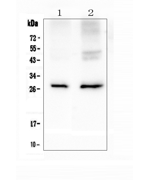
Western blot analysis of Aquaporin 5 using anti-Aquaporin 5 antibody (A03085).
Electrophoresis was performed on a 5-20% SDS-PAGE gel at 70V (Stacking gel) / 90V (Resolving gel) for 2-3 hours. The sample well of each lane was loaded with 50ug of sample under reducing conditions.
Lane 1: rat lung tissue lysates,
Lane 2: mouse lung tissue lysates.
After Electrophoresis, proteins were transferred to a Nitrocellulose membrane at 150mA for 50-90 minutes. Blocked the membrane with 5% Non-fat Milk/ TBS for 1.5 hour at RT. The membrane was incubated with rabbit anti-Aquaporin 5 antigen affinity purified polyclonal antibody (Catalog # A03085) at 0.5 μg/mL overnight at 4°C, then washed with TBS-0.1%Tween 3 times with 5 minutes each and probed with a goat anti-rabbit IgG-HRP secondary antibody at a dilution of 1:10000 for 1.5 hour at RT. The signal is developed using an Enhanced Chemiluminescent detection (ECL) kit (Catalog # EK1002) with Tanon 5200 system. A specific band was detected for Aquaporin 5 at approximately 28KD. The expected band size for Aquaporin 5 is at 28KD.

Click image to see more details
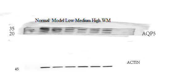
Western blot analysis of Aquaporin 5 using anti-Aquaporin 5 antibody (A03085).
Electrophoresis was performed on a 5-20% SDS-PAGE gel at 70V (Stacking gel) / 90V (Resolving gel) for 2-3 hours. The sample well of each lane was loaded with 50ug of sample under reducing conditions.
Lane 1: Normal group-rat colon tissue lysates,
Lane 2: Model group-rat colon tissue lysates,
Lane 3: Traditional Chinese medicine treatment (low concetration)-rat colon tissue lysates,
Lane 4: Traditional Chinese medicine treatment (medium concentration)-rat colon tissue lysates,
Lane 5: Traditional Chinese medicine treatment (High concentration)-rat colon tissue lysates,
Lane 6: Western medicine treatment-rat colon tissue lysates.
After Electrophoresis, proteins were transferred to a Nitrocellulose membrane at 150mA for 50-90 minutes. Blocked the membrane with 5% Non-fat Milk/ TBS for 1.5 hour at RT. The membrane was incubated with rabbit anti-Aquaporin 5 antigen affinity purified polyclonal antibody (Catalog # A03085) at 1:1000 overnight at 4°C, then washed with TBS-0.1%Tween 3 times with 5 minutes each and probed with a goat anti-rabbit IgG-HRP secondary antibody at RT. The signal is developed using an Enhanced Chemiluminescent detection (ECL) kit (Catalog # EK1002) with ChemiDoc MP system. The expected band size for Aquaporin 5 is at 28KD.

Click image to see more details
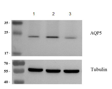
Western blot analysis of Aquaporin 5 using anti-Aquaporin 5 antibody (A03085).
Electrophoresis was performed on a 5-20% SDS-PAGE gel at 70V (Stacking gel) / 90V (Resolving gel) for 2-3 hours. The sample well of each lane was loaded with 50ug of sample under reducing conditions.
Lane 1: MADB106 cells under normal culture conditions,
Lane 2: MADB106 cells treated with agonist,
Lane 3: MADB106 cells treated with inhibitor,
After Electrophoresis, proteins were transferred to a Nitrocellulose membrane at 150mA for 50-90 minutes. Blocked the membrane with 5% Non-fat Milk/ TBS for 1.5 hour at RT. The membrane was incubated with rabbit anti-Aquaporin 5 antigen affinity purified polyclonal antibody (Catalog # A03085) at 1:2000 overnight at 4°C, then washed with TBS-0.1%Tween 3 times with 5 minutes each and probed with a goat anti-rabbit IgG-HRP secondary antibody at a dilution of 1:10000 for 1 hour at RT. The signal is developed using an Enhanced Chemiluminescent detection (ECL) kit (Catalog # EK1002) with ChemiDoc MP system. A specific band was detected for Aquaporin 5 at approximately 23KD. The expected band size for Aquaporin 5 is at 28KD.

Click image to see more details
IHC analysis of Aquaporin 5 using anti-Aquaporin 5 antibody (A03085).
Aquaporin 5 was detected in paraffin-embedded section of mouse lung tissue. Heat mediated antigen retrieval was performed in citrate buffer (pH6, epitope retrieval solution) for 20 mins. The tissue section was blocked with 10% goat serum. The tissue section was then incubated with 2μg/ml rabbit anti-Aquaporin 5 Antibody (A03085) overnight at 4°C. Biotinylated goat anti-rabbit IgG was used as secondary antibody and incubated for 30 minutes at 37°C. The tissue section was developed using Strepavidin-Biotin-Complex (SABC)(Catalog # SA1022) with DAB as the chromogen.

Click image to see more details
IHC analysis of Aquaporin 5 using anti-Aquaporin 5 antibody (A03085).
Aquaporin 5 was detected in paraffin-embedded section of rat lung tissue. Heat mediated antigen retrieval was performed in citrate buffer (pH6, epitope retrieval solution) for 20 mins. The tissue section was blocked with 10% goat serum. The tissue section was then incubated with 2μg/ml rabbit anti-Aquaporin 5 Antibody (A03085) overnight at 4°C. Biotinylated goat anti-rabbit IgG was used as secondary antibody and incubated for 30 minutes at 37°C. The tissue section was developed using Strepavidin-Biotin-Complex (SABC)(Catalog # SA1022) with DAB as the chromogen.
Specific Publications For Anti-Aquaporin 5/AQP5 Antibody Picoband® (A03085)
Loading publications
Recommended Resources
Here are featured tools and databases that you might find useful.
- Boster's Pathways Library
- Protein Databases
- Bioscience Research Protocol Resources
- Data Processing & Analysis Software
- Photo Editing Software
- Scientific Literature Resources
- Research Paper Management Tools
- Molecular Biology Software
- Primer Design Tools
- Bioinformatics Tools
- Phylogenetic Tree Analysis
Customer Reviews
Have you used Anti-Aquaporin 5/AQP5 Antibody Picoband®?
Share your experimental results or join a short interview to earn up to $1,000 in product credits or other rewards.
2 Reviews For Anti-Aquaporin 5/AQP5 Antibody Picoband®
In this Western blot (WB) experiment using Anti-AQP5 antibody (Cat# A03085) on MADB106 rat mammary carcinoma cells, AQP5 protein levels were significantly increased with agonist treatment and markedly reduced with inhibitor treatment.
Excellent
| SKU | A03085 |
|---|---|
| Application | Western blot |
| Sample | MADB106 rat mammary carcinoma cells |
| Sample Processing Description | MADB106 cells under normal culture conditions , MADB106 cells treated with agonist , MADB106 cells treated with inhibitor |
| Other Reagents | RIPA Lysis Buffer, Protease Inhibitor, Resolving Gel Solution ,Transfer Buffer ,Blocking Buffer |
| Primary Antibody | Aquaporin 5/AQP5 Antibody Picoband® |
| Primary Incubation | 1:2000, overnight at 4 ℃ |
| Secondary Antibody | 1:10,000, HRP-conjugated Goat Anti-Rabbit IgG |
| Secondary Incubation | 1h at 37℃ |
| Detection | Substrate: ECL substrate, Imaging system:ChemiDoc MP |
| Results Summary | AQP5 (Aquaporin 5) is a transmembrane water channel protein that primarily regulates cellular water transport and plays a critical and complex role in tumor progression. In breast cancer, high AQP5 expression is often associated with increased tumor invasiveness, lymph node metastasis, and poor patient prognosis. In this experiment, AQP5 protein levels were markedly increased in cells treated with the agonist, whereas they were significantly reduced in cells treated with the inhibitor. |
Xinshuo Wang, Xi’an Jiaotong University
Verified customer
Submitted 2026-02-25
This antibody is highly efficient and specific, suitable for detecting AQP5 protein in rat colon by Western blot, with only minor nonspecific bands.
Excellent
| SKU | A03085 |
|---|---|
| Application | Western Blot |
| Sample | rat colon tissue |
| Sample Processing Description | RIPA lysis buffer with protease inhibitor PMSF (100:1) was used to lyse the sample for 10 minutes, followed by centrifugation at 12,000 rpm for 15 minutes. The supernatant was mixed with 5× loading buffer, denatured at 100°C for 10 minutes, and then loaded onto SDS-PAGE. |
| Other Reagents | Blocking buffer |
| Primary Antibody | Aquaporin 5/AQP5 Antibody Picoband® |
| Primary Incubation | 1:1000, overnight at 4 ℃ |
| Secondary Antibody | HRP Conjugated AffiniPure Goat Anti-Rabbit IgG (H+L) |
| Secondary Incubation | 1 hour in room temperature |
| Detection | Substrate: ECL, Imaging system:ChemiDoc MP |
| Results Summary | This antibody is highly specific and efficient, suitable for detecting AQP5 protein in rat colon by Western blot, with only minimal nonspecific bands. |
Shiyu Zhang, LUTCM
Verified customer
Submitted 2025-12-30
Customer Q&As
Have a question?
Find answers in Q&As, reviews.
Can't find your answer?
Submit your question
