Click image to see more details
-

-
-
-
-
+1
Product Info Summary
| SKU: | A01767-4 |
|---|---|
| Size: | 100 μg/vial |
| Reactive Species: | Mouse, Rat |
| Host: | Rabbit |
| Application: | ELISA, IHC, WB |
Customers Who Bought This Also Bought

Product info
Product Name
Anti-CD133/Prom1 Antibody Picoband®
SKU/Catalog Number
A01767-4
Size
100 μg/vial
Form
Lyophilized
Description
Boster Bio Anti-CD133/Prom1 Antibody Picoband® catalog # A01767-4. Tested in ELISA, IHC, WB applications. This antibody reacts with Mouse, Rat. The brand Picoband indicates this is a premium antibody that guarantees superior quality, high affinity, and strong signals with minimal background in Western blot applications. Only our best-performing antibodies are designated as Picoband, ensuring unmatched performance.
Storage & Handling
Store at -20˚C for one year from date of receipt. After reconstitution, at 4˚C for one month. It can also be aliquotted and stored frozen at -20˚C for six months. Avoid repeated freeze-thaw cycles.
Cite This Product
Anti-CD133/Prom1 Antibody Picoband® (Boster Biological Technology, Pleasanton CA, USA, Catalog # A01767-4)
Host
Rabbit
Contents
Each vial contains 4mg Trehalose, 0.9mg NaCl, 0.2mg Na2HPO4.
Clonality
Polyclonal
Isotype
Rabbit IgG
Immunogen
E.coli-derived mouse Prom1 recombinant protein (Position: E20-Y867).
Cross-reactivity
No cross-reactivity with other proteins.
Reactive Species
A01767-4 is reactive to Prom1 in Mouse, Rat
Observed Molecular Weight
130 kDa
Calculated molecular weight
97.1 kDa
Background of Prom1
Prominin-1, also known as CD133, is a glycoprotein that in humans is encoded by the PROM1 gene. It is mapped to 4p15.32. Prominin-1 is a member of pentaspan transmembrane glycoproteins (5-transmembrane, 5-TM), which specifically localize to cellular protrusions. This gene encodes a pentaspan transmembrane glycoprotein. The protein localizes to membrane protrusions and is often expressed on adult stem cells, where it is thought to function in maintaining stem cell properties by suppressing differentiation. It has been proposed to act as an organizer of cell membrane topology. Prominin-1 was expressed not only on metastatic colon cancer cells, but also on differentiated colonic epithelium in both adult mice and humans.
Antibody Validation
Boster validates all antibodies on WB, IHC, ICC, Immunofluorescence, and ELISA with known positive control and negative samples to ensure specificity and high affinity, including thorough antibody incubations.
Application & Images
Applications
A01767-4 is guaranteed for ELISA, IHC, WB Boster Guarantee
Recommend Dilution
| Application | Dilution | Species |
|---|---|---|
| Western blot | 0.1-0.25μg/ml | Mouse, Rat |
| Immunohistochemistry (Paraffin-embedded Section) | 2-5μg/ml | Mouse, Rat |
| ELISA | 0.1-0.5μg/ml | - |
Tested application
Suggested blocking solution with 5% non-fat milk or BSA; (*)Recommended protein loading: 20-40 µg per lane
Use TE buffer pH 9.0 for antigen retrieval; (*) citrate buffer pH 6.0 is an alternative.
Validation Images & Assay Conditions
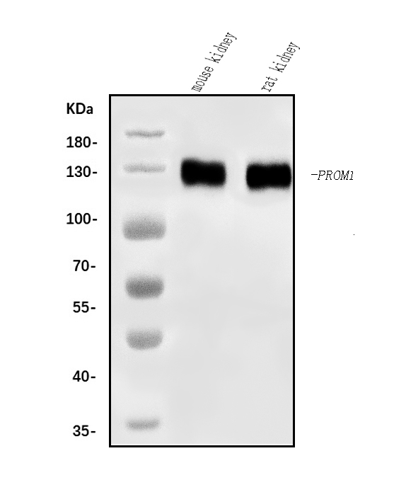
Click image to see more details
Western blot analysis of CD133/Prom1 using anti-CD133/Prom1 antibody (A01767-4).
Electrophoresis was performed on a 5-20% SDS-PAGE gel at 70V (Stacking gel) / 90V (Resolving gel) for 2-3 hours. The sample well of each lane was loaded with 30 ug of sample under reducing conditions.
Lane 1: mouse kidney tissue lysates,
Lane 2: rat kidney tissue lysates.
After electrophoresis, proteins were transferred to a nitrocellulose membrane at 150 mA for 50-90 minutes. Blocked the membrane with 5% non-fat milk/TBS for 1.5 hour at RT. The membrane was incubated with rabbit anti-CD133/Prom1 antigen affinity purified polyclonal antibody (Catalog # A01767-4) at 0.25 μg/mL overnight at 4°C, then washed with TBS-0.1%Tween 3 times with 5 minutes each and probed with a goat anti-rabbit IgG-HRP secondary antibody at a dilution of 1:5000 for 1.5 hour at RT. The signal is developed using an Enhanced Chemiluminescent detection (ECL) kit (Catalog # EK1002) with Tanon 5200 system. A specific band was detected for CD133/Prom1 at approximately 130 kDa. The expected band size for CD133/Prom1 is at 130 kDa.

Click image to see more details
Analysis of tumor infiltrating lymphocytes by Flowcytometer showed that a population of Th1 cells (CD3 + CD4 + CD25 − ) was significantly increased inside tumors of 1-MT, 1-MT+TL and 1-MT+DC-TL treated groups in comparison to PBS ( A , B ). Meanwhile, Treg + cells (CD25 + FOXP3 + ) were significantly reduced in comparison to PBS. Cancer stem cells (CSC) CD133 + viability in tumor microenvironments of all tested groups showed that 1-MT, 1-MT+TL and 1-MT+DC-TL extremely reduced CD133 viability in comparison to PBS group ( C , D ), as well as the analysis of relative mRNA showed that CD133 gene expression was significantly declined in 1-MT+TL and 1-MT+DC-TL treated groups in comparison to PBS group ( E ). These results indicated that 1-MT significantly enhanced TL to elicit immunosurveillance that recognizes and effectively reduces CSC prognosis, which inhibits tumor growth and tumorgensis. ***( P < 0.0001), and **( P < 0.001).
Index in PubMed under a CC BY license. PMID: 29959375

Click image to see more details
Analysis of tumor microenvironments’ mediators, markers and signaling pathways under the effect of 1-MT-TL/DCs-TL regimens by western blot presented that TGF-β, PDL-1, and NF-κβ2 were significantly inhibited in comparison to PBS ( A ). Meanwhile, IDO, β-catenin, and CD133 showed significant reduction under the effect of 1-MT, 1-MT+TL, and 1-MT+DCs-TL in comparison to PBS ( B ). These results clearly evidenced that 1-MT extremely prohibited main tumor growth, progression, and immune escaping pathways, which activates immune surveillance restore and recruits cellular and humoral immune responses triggering tumor tissues.
Index in PubMed under a CC BY license. PMID: 29959375

Click image to see more details
IHC analysis of CD133/Prom1 using anti-CD133/Prom1 antibody (A01767-4).
CD133/Prom1 was detected in a paraffin-embedded section of mouse kidney tissue. Heat mediated antigen retrieval was performed in EDTA buffer (pH 8.0, epitope retrieval solution). The tissue section was blocked with 10% goat serum. The tissue section was then incubated with 2 μg/ml rabbit anti-CD133/Prom1 Antibody (A01767-4) overnight at 4°C. Biotinylated goat anti-rabbit IgG was used as secondary antibody and incubated for 30 minutes at 37°C. The tissue section was developed using Strepavidin-Biotin-Complex (SABC) (Catalog # SA1022) with DAB as the chromogen.

Click image to see more details
IHC analysis of CD133/Prom1 using anti-CD133/Prom1 antibody (A01767-4).
CD133/Prom1 was detected in a paraffin-embedded section of rat kidney tissue. Heat mediated antigen retrieval was performed in EDTA buffer (pH 8.0, epitope retrieval solution). The tissue section was blocked with 10% goat serum. The tissue section was then incubated with 2 μg/ml rabbit anti-CD133/Prom1 Antibody (A01767-4) overnight at 4°C. Biotinylated goat anti-rabbit IgG was used as secondary antibody and incubated for 30 minutes at 37°C. The tissue section was developed using Strepavidin-Biotin-Complex (SABC) (Catalog # SA1022) with DAB as the chromogen.
Specific Publications For Anti-CD133/Prom1 Antibody Picoband® (A01767-4)
Loading publications
Recommended Resources
Here are featured tools and databases that you might find useful.
- Boster's Pathways Library
- Protein Databases
- Bioscience Research Protocol Resources
- Data Processing & Analysis Software
- Photo Editing Software
- Scientific Literature Resources
- Research Paper Management Tools
- Molecular Biology Software
- Primer Design Tools
- Bioinformatics Tools
- Phylogenetic Tree Analysis
Customer Reviews
Have you used Anti-CD133/Prom1 Antibody Picoband®?
Share your experimental results or join a short interview to earn up to $1,000 in product credits or other rewards.
0 Reviews For Anti-CD133/Prom1 Antibody Picoband®
Customer Q&As
Have a question?
Find answers in Q&As, reviews.
Can't find your answer?
Submit your question
