Click image to see more details
-

-
-
-
-
+3
Product Info Summary
| SKU: | PB9156 |
|---|---|
| Size: | 100 μg/vial |
| Reactive Species: | Human |
| Host: | Rabbit |
| Application: | Flow Cytometry, IHC, WB |
Customers Who Bought This Also Bought

Product info
Product Name
Anti-PROM1 Antibody Picoband®
SKU/Catalog Number
PB9156
Size
100 μg/vial
Form
Lyophilized
Description
Boster Bio Anti-PROM1 Antibody Picoband® catalog # PB9156. Tested in Flow Cytometry, IHC, WB applications. This antibody reacts with Human. The brand Picoband indicates this is a premium antibody that guarantees superior quality, high affinity, and strong signals with minimal background in Western blot applications. Only our best-performing antibodies are designated as Picoband, ensuring unmatched performance.
Storage & Handling
Store at -20˚C for one year from date of receipt. After reconstitution, at 4˚C for one month. It can also be aliquotted and stored frozen at -20˚C for six months. Avoid repeated freeze-thaw cycles.
Cite This Product
Anti-PROM1 Antibody Picoband® (Boster Biological Technology, Pleasanton CA, USA, Catalog # PB9156)
Host
Rabbit
Contents
Each vial contains antibody formulated with stabilizing components, 0.9 mg NaCl, 0.2 mg Na2HPO4, and 0.05 mg NaN3.
*This antibody is supplied in a stabilized formulation.
Compatibility with conjugation reactions depends on the chemistry of the conjugation method used.
For conjugation methods that are not compatible with the stabilizing components present in this formulation, a carrier-free antibody format is required.
Clonality
Polyclonal
Isotype
Rabbit IgG
Immunogen
E.coli-derived human PROM1 recombinant protein (Position: P531-H865). Human PROM1 shares 61% amino acid (aa) sequence identity with mouse PROM1.
Cross-reactivity
No cross-reactivity with other proteins
Reactive Species
PB9156 is reactive to PROM1 in Human
Observed Molecular Weight
120 kDa
Calculated molecular weight
97.2 kDa
Background of PROM1
Prominin-1, also known as CD133, is a glycoprotein that in humans is encoded by the PROM1 gene. It is mapped to 4p15.32. Prominin-1 is a member of pentaspan transmembrane glycoproteins (5-transmembrane, 5-TM), which specifically localize to cellular protrusions. This gene encodes a pentaspan transmembrane glycoprotein. The protein localizes to membrane protrusions and is often expressed on adult stem cells, where it is thought to function in maintaining stem cell properties by suppressing differentiation. It has been proposed to act as an organizer of cell membrane topology. Prominin-1 was expressed not only on metastatic colon cancer cells, but also on differentiated colonic epithelium in both adult mice and humans.
Antibody Validation
Boster validates all antibodies on WB, IHC, ICC, Immunofluorescence, and ELISA with known positive control and negative samples to ensure specificity and high affinity, including thorough antibody incubations.
Application & Images
Applications
PB9156 is guaranteed for Flow Cytometry, IHC, WB Boster Guarantee
Recommend Dilution
| Application | Dilution | Species |
|---|---|---|
| Western blot | 0.1-0.5μg/ml | Human |
| Immunohistochemistry (Paraffin-embedded Section) | 0.5-1μg/ml | Human |
| Flow Cytometry (Fixed) | 1-3μg/1x106 cells | Human |
Tested application
Suggested blocking solution with 5% non-fat milk or BSA; (*)Recommended protein loading: 20-40 µg per lane
Use TE buffer pH 9.0 for antigen retrieval; (*) citrate buffer pH 6.0 is an alternative.
Validation Images & Assay Conditions
Click image to see more details
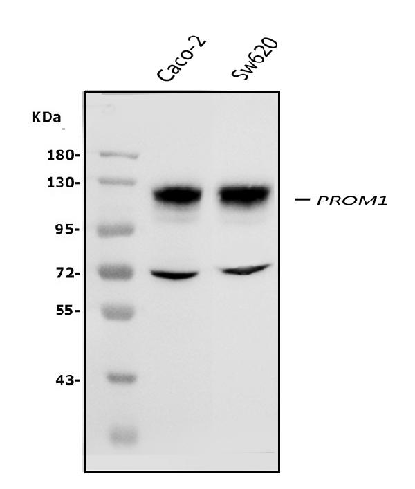
Western blot analysis of PROM1 using anti-PROM1 antibody (PB9156).
Electrophoresis was performed on a 5-20% SDS-PAGE gel at 70V (Stacking gel) / 90V (Resolving gel) for 2-3 hours. The sample well of each lane was loaded with 50ug of sample under reducing conditions.
Lane 1: human CACO-2 whole cell lysates,
Lane 2: human SW620 whole cell lysates.
After Electrophoresis, proteins were transferred to a Nitrocellulose membrane at 150mA for 50-90 minutes. Blocked the membrane with 5% Non-fat Milk/ TBS for 1.5 hour at RT. The membrane was incubated with rabbit anti-PROM1 antigen affinity purified polyclonal antibody (Catalog # PB9156) at 0.5 μg/mL overnight at 4°C, then washed with TBS-0.1%Tween 3 times with 5 minutes each and probed with a goat anti-rabbit IgG-HRP secondary antibody at a dilution of 1:10000 for 1.5 hour at RT. The signal is developed using an Enhanced Chemiluminescent detection (ECL) kit (Catalog # EK1002) with Tanon 5200 system. A specific band was detected for PROM1 at approximately 120KD. The expected band size for PROM1 is at 120KD.

Click image to see more details
MCF-7 ALDH1A1 affects in vitro stemness. a Representative images of tumorspheres (4x magnification) showing morphology of spheroids grown on ultra-low attachment plate. Scale bar, 100 μm. b Representative images of tumorspheres (4x magnification) of MCF-7 Scr, MCF-7 ALDH1A1KD and MCF-7 ALDH1A1 + , showing morphology of spheroids grown on ultra-low attachment plate. Scale bar, 100 μm. b1, b2, b3. Representative images of tumorspheres (10x magnification) of MCF-1 Scr, MCF-7 ALDH1A1KD and MCF-7 ALDH1A1 + , showing morphology of spheroids grown on ultra-low attachment plate. Scale bar, 100 μm. c Quantification of MCF-7 tumorspheres. Tumorspheres area were calculated using ImageJ Software. Ten pictures for each well were quantified. Tumorspheres> 10.000 pixel square were considered. ** p < 0.01 vs MCF-7 Scr. ### p < 0.001 vs MCF-7 ALDH1A1KD. ( n = 3). d Western blot analysis of stemness markers CD133 and KLF4 in MCF-7 Scr, MCF-7 ALDH1A1KD, and MCF-7 ALDH1A1 + tumorspheres. e Western blot analysis of ALDH1A1, HIF-1α and VEGF in MCF-7 Scr, MCF-7 ALDH1A1KD and MCF-7 ALDH1A1 + tumorspheres
Index in PubMed under a CC BY license. PMID: 30541574

Click image to see more details
MCF-7 ALDH1A1 regulates angiogenic factor output via retinoic acid signalling. a Angiogenic factor release evaluated by ELISA plate array in supernatants of MCF-7 treated with CM037 (1 μM) for 48 h. The experiment was performed 2 times in duplicate. b MCF-7 cells were exposed to CM037 at different concentrations (1 and 10 μM) for 18 h and western blot was carried out. β-Actin was used to normalize loading. c Cells were treated with CM037 (1 μM, 18 h) and VEGF levels were measured by ELISA assay in MCF-7 conditioned media. After 18 h supernatants were harvested and cells fixed, stained and counted. The number of counted cells was not significantly different. Data are reported as pg/ml. ** p < 0.01 vs untreated cells. d RT-PCR analysis of VEGF in MCF-7 Scr, MCF-7 ALDH1A1KD and MCF-7 ALDH1A1 + cultured in medium with 1% FBS for 48 h. Data are reported as ΔCt (Ct gene of interest-Ct Housekeeping gene). *** p < 0.001 vs MCF-7 Scr. ### p < 0.001 vs MCF-7 ALDH1A1KD. e Western blot analysis of VEGF and HIF-1α in MCF-7 exposed or not to CoCl 2 (100 μM, 72 h, 1% FBS). β-Actin was used as loading control. Gel shown is representative of three experiments with similar results. f Quantification of blots reported in e . * p < 0.05 vs MCF-7 Scr. ** p < 0.01 vs MCF-7 Scr. ### p < 0.001 vs MCF-7 ALDH1A1KD. g Soluble VEGF was detected by ELISA in media conditioned by MCF-7 cells. Cells were seeded in 24-well plates at density 3 × 10 4 cells/well. After 48 h the supernatants were harvested and cells fixed, stained and counted. The number of counted cells was not significantly different. Data are reported as pg/ml. ** p < 0.01 vs MCF-7 Scr. ## p < 0.01 vs MCF-7 ALDH1A1KD. h HIF-1α and VEGF expression evaluated by western blot in MCF-7 ALDH1A1KD cells exposed for 48 h (1 μM) to exogenous retinoic acid. i HIF-1α and VEGF expression in MCF-7 ALDH1A1 + treated with RAR antagonist (AGN193109) and RXR antagonist (UVI 3003) for 48 h (each at 1 μM). β-Actin was used as loading control. Gel shown is representative of three experiments with similar results. j VEGF and CD133 expression in MCF-7 transiently silenced for HIF-1α. β-Actin was used as loading control. Gel shown is representative of three experiments with similar results
Index in PubMed under a CC BY license. PMID: 30541574

Click image to see more details
ALDH1A1 influences tumor angiogenesis and VEGF production in vivo. a Evaluation of VEGF, HIF-1α and ALDH1A1 RNA in tumor samples. Frozen tumors were homogenized and RNA was extracted to perform RT-PCR analysis of VEGF, HIF-1α and ALDH1A1 mRNA. Data are reported as ΔCt (Ct gene of interest-Ct Housekeeping gene). Each bar is the mean of 6 different tumors. The experiment was repeated two times. * p < 0.05 vs Scr group. ** p < 0.01 vs Scr group. # p < 0.05 vs ALDH1A1KD group. ### p < 0.001 vs ALDH1A1KD group. b Evaluation of VEGF and ALDH1A1 proteins in tumor samples. Tissues were harvested, homogenized and sonicated. Subsequently, proteins were extracted and western blot was performed. β-Actin was used as loading control. The experiment was repeated two times. c Evaluation of mRNA for CAIX (HIF-1α target gene) and stemness markers (SOX2, NANOG, OCT-4 and TWIST) in tumor samples. Each bar is the mean of 6 different tumors. The experiment was repeated two times. # p < 0.05 vs ALDH1A1KD group. ## p < 0.01 vs ALDH1A1KD group. ### p < 0.001 vs ALDH1A1KD group. d Evaluation of HIF-1α and stemness markers (CD133, KLF4 and SOX2) proteins in tumor samples. The experiment was repeated two times. e Quantification of blots reported in d . * p < 0.05 vs Scr group. # p < 0.05 vs ALDH1A1KD group. ## p < 0.01 vs ALDH1A1KD group. f Quantification of microvessel density by human CD31 staining (magnification 20x) was done counting 5 random fields for section, each slide having five sections. ** p < 0.01 vs Scr group. ## p < 0.01 vs ALDH1A1 + group. g Representative images of immunostaining for CD31 (red) and DAPI (blue) in tumor sections from Scr (left), ALDH1A1KD (center) or ALDH1A1 + (right) mice. Pictures report different vessel densities in tumors. Magnification 20x. Scale bar, 50 μm. h Representative images of double-immunostaining for CD31 (red) and NG2 (green) in tumor sections from Scr (left), ALDH1A1KD (center) or ALDH1A1 + (right) mice. DAPI staining is blue. Magnification 40x. Scale bars, 50 μm
Index in PubMed under a CC BY license. PMID: 30541574

Click image to see more details
IHC analysis of PROM1 using anti-PROM1 antibody (PB9156).
PROM1 was detected in paraffin-embedded section of human liver cancer tissue. Heat mediated antigen retrieval was performed in EDTA buffer (pH8.0, epitope retrieval solution). The tissue section was blocked with 10% goat serum. The tissue section was then incubated with 1μg/ml rabbit anti-PROM1 Antibody (PB9156) overnight at 4°C. Biotinylated goat anti-rabbit IgG was used as secondary antibody and incubated for 30 minutes at 37°C. The tissue section was developed using Strepavidin-Biotin-Complex (SABC) (Catalog # SA1022) with DAB as the chromogen.

Click image to see more details
IHC analysis of PROM1 using anti-PROM1 antibody (PB9156).
PROM1 was detected in paraffin-embedded section of human mammary cancer tissue. Heat mediated antigen retrieval was performed in EDTA buffer (pH8.0, epitope retrieval solution). The tissue section was blocked with 10% goat serum. The tissue section was then incubated with 1μg/ml rabbit anti-PROM1 Antibody (PB9156) overnight at 4°C. Biotinylated goat anti-rabbit IgG was used as secondary antibody and incubated for 30 minutes at 37°C. The tissue section was developed using Strepavidin-Biotin-Complex (SABC) (Catalog # SA1022) with DAB as the chromogen.

Click image to see more details
Flow Cytometry analysis of CACO-2 cells using anti-PROM1 antibody (PB9156).
Overlay histogram showing CACO-2 cells stained with PB9156 (Blue line). The cells were fixed with 4% paraformaldehyde and blocked with 10% normal goat serum. And then incubated with rabbit anti-PROM1 Antibody (PB9156,1μg/1x106 cells) for 30 min at 20°C. DyLight®488 conjugated goat anti-rabbit IgG (BA1127, 5-10μg/1x106 cells) was used as secondary antibody for 30 minutes at 20°C. Isotype control antibody (Green line) was rabbit IgG (1μg/1x106) used under the same conditions. Unlabelled sample without incubation with primary antibody and secondary antibody (Red line) was used as a blank control.
Specific Publications For Anti-PROM1 Antibody Picoband® (PB9156)
Loading publications
Recommended Resources
Here are featured tools and databases that you might find useful.
- Boster's Pathways Library
- Protein Databases
- Bioscience Research Protocol Resources
- Data Processing & Analysis Software
- Photo Editing Software
- Scientific Literature Resources
- Research Paper Management Tools
- Molecular Biology Software
- Primer Design Tools
- Bioinformatics Tools
- Phylogenetic Tree Analysis
Customer Reviews
Have you used Anti-PROM1 Antibody Picoband®?
Share your experimental results or join a short interview to earn up to $1,000 in product credits or other rewards.
0 Reviews For Anti-PROM1 Antibody Picoband®
Customer Q&As
Have a question?
Find answers in Q&As, reviews.
Can't find your answer?
Submit your question
1 Customer Q&As for Anti-PROM1 Antibody Picoband®
Question
We are currently using anti-PROM1 antibody PB9156 for human tissue, and we are satisfied with the WB results. The species of reactivity given in the datasheet says human. Is it likely that the antibody can work on bovine tissues as well?
Verified Customer
Verified customer
Asked: 2020-03-27
Answer
The anti-PROM1 antibody (PB9156) has not been tested for cross reactivity specifically with bovine tissues, but there is a good chance of cross reactivity. We have an innovator award program that if you test this antibody and show it works in bovine you can get your next antibody for free. Please contact me if I can help you with anything.
Boster Scientific Support
Answered: 2020-03-27
