Click image to see more details
-

-
-
-
-
+7
Product Info Summary
| SKU: | A01513-2 |
|---|---|
| Size: | 100 μg/vial |
| Reactive Species: | Mouse, Rat |
| Host: | Rabbit |
| Application: | ELISA, IHC, WB |
Customers Who Bought This Also Bought

Product info
Product Name
Anti-CD31/Pecam1 Antibody Picoband®
SKU/Catalog Number
A01513-2
Size
100 μg/vial
Form
Lyophilized
Description
Boster Bio Anti-CD31/Pecam1 Antibody Picoband® catalog # A01513-2. Tested in ELISA, IHC, WB applications. This antibody reacts with Mouse, Rat. The brand Picoband indicates this is a premium antibody that guarantees superior quality, high affinity, and strong signals with minimal background in Western blot applications. Only our best-performing antibodies are designated as Picoband, ensuring unmatched performance.
Storage & Handling
Store at -20˚C for one year from date of receipt. After reconstitution, at 4˚C for one month. It can also be aliquotted and stored frozen at -20˚C for six months. Avoid repeated freeze-thaw cycles.
Cite This Product
Anti-CD31/Pecam1 Antibody Picoband® (Boster Biological Technology, Pleasanton CA, USA, Catalog # A01513-2)
Host
Rabbit
Contents
Each vial contains 4 mg Trehalose, 0.9 mg NaCl and 0.2 mg Na2HPO4.
Clonality
Polyclonal
Isotype
Rabbit IgG
Immunogen
E.coli-derived mouse CD31/Pecam1 recombinant protein (Position: Q41-E258).
Cross-reactivity
No cross-reactivity with other proteins.
Reactive Species
A01513-2 is reactive to Pecam1 in Mouse, Rat
Observed Molecular Weight
100-120 kDa
Calculated molecular weight
81.3 kDa
Background of Pecam1
CD31 also known as Platelet endothelial cell adhesion molecule (PECAM-1), is a protein that in human is encoded by the PECAM1 gene. Encoded protein is a member of the immunoglobulin superfamily and this gene is mapped to 17q23.3. CD31 is found on the surface of platelets, monocytes, neutrophils, and some types of T-cells, and makes up a large portion of endothelial cell intercellular junctions. It is demonstrated that CD31 expression on human PBSCs may positively affect both neutrophil and platelet engraftment. Meanwhile, CD31 is involved in leukocyte migration and angiogenesis, which are key components of venous thrombus resolution.
Antibody Validation
Boster validates all antibodies on WB, IHC, ICC, Immunofluorescence, and ELISA with known positive control and negative samples to ensure specificity and high affinity, including thorough antibody incubations.
Application & Images
Applications
A01513-2 is guaranteed for ELISA, IHC, WB Boster Guarantee
Recommend Dilution
| Application | Dilution | Species |
|---|---|---|
| Western blot | 0.1-0.25μg/ml | Mouse, Rat |
| Immunohistochemistry (Paraffin-embedded Section) | 2-5μg/ml | Mouse, Rat |
| ELISA | 0.1-0.5μg/ml | - |
Tested application
Suggested blocking solution with 5% non-fat milk or BSA; (*)Recommended protein loading: 20-40 µg per lane
Use TE buffer pH 9.0 for antigen retrieval; (*) citrate buffer pH 6.0 is an alternative.
Validation Images & Assay Conditions
Click image to see more details
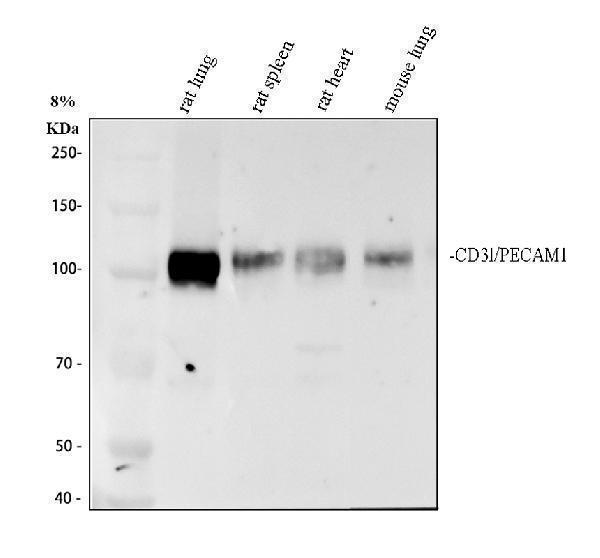
Western blot analysis of CD31/Pecam1 using anti-CD31/Pecam1 antibody (A01513-2).
Electrophoresis was performed on a 8% SDS-PAGE gel at 80V (Stacking gel) / 120V (Resolving gel) for 2 hours. The sample well of each lane was loaded with 30 ug of sample under reducing conditions.
Lane 1: rat lung tissue lysates,
Lane 2: rat spleen tissue lysates,
Lane 3: rat heart tissue lysates,
Lane 4: mouse lung tissue lysates.
After electrophoresis, proteins were transferred to a nitrocellulose membrane at 150 mA for 50-90 minutes. Blocked the membrane with 5% non-fat milk/TBS for 1.5 hour at RT. The membrane was incubated with rabbit anti-CD31/Pecam1 antigen affinity purified polyclonal antibody (A01513-2) at 0.5 μg/mL overnight at 4°C, then washed with TBS-0.1%Tween 3 times with 5 minutes each and probed with a goat anti-rabbit IgG-HRP secondary antibody (Catalog # BA1054) at a dilution of 1:5000 for 1.5 hour at RT. The signal is developed using an ECL Plus Western Blotting Substrate (Catalog # AR1196-200) with Tanon 5200 system. A specific band was detected for CD31/Pecam1 at approximately 100-120 kDa. The expected band size for CD31/Pecam1 is at 81 kDa.

Click image to see more details
IHC analysis of CD31/Pecam1 using anti-CD31/Pecam1 antibody (A01513-2).
CD31/Pecam1 was detected in a paraffin-embedded section of mouse colon tissue. Heat mediated antigen retrieval was performed in EDTA buffer (pH 8.0, epitope retrieval solution). The tissue section was blocked with 10% goat serum. The tissue section was then incubated with 2 μg/ml rabbit anti-CD31/Pecam1 Antibody (A01513-2) overnight at 4°C. Peroxidase Conjugated Goat Anti-rabbit IgG was used as secondary antibody and incubated for 30 minutes at 37°C. The tissue section was developed using HRP Conjugated Rabbit IgG Super Vision Assay Kit (Catalog # SV0002) with DAB as the chromogen.

Click image to see more details
IHC analysis of CD31/Pecam1 using anti-CD31/Pecam1 antibody (A01513-2).
CD31/Pecam1 was detected in a paraffin-embedded section of mouse kidney tissue. Heat mediated antigen retrieval was performed in EDTA buffer (pH 8.0, epitope retrieval solution). The tissue section was blocked with 10% goat serum. The tissue section was then incubated with 2 μg/ml rabbit anti-CD31/Pecam1 Antibody (A01513-2) overnight at 4°C. Peroxidase Conjugated Goat Anti-rabbit IgG was used as secondary antibody and incubated for 30 minutes at 37°C. The tissue section was developed using HRP Conjugated Rabbit IgG Super Vision Assay Kit (Catalog # SV0002) with DAB as the chromogen.

Click image to see more details
IHC analysis of CD31/Pecam1 using anti-CD31/Pecam1 antibody (A01513-2).
CD31/Pecam1 was detected in a paraffin-embedded section of rat colon tissue. Heat mediated antigen retrieval was performed in EDTA buffer (pH 8.0, epitope retrieval solution). The tissue section was blocked with 10% goat serum. The tissue section was then incubated with 2 μg/ml rabbit anti-CD31/Pecam1 Antibody (A01513-2) overnight at 4°C. Peroxidase Conjugated Goat Anti-rabbit IgG was used as secondary antibody and incubated for 30 minutes at 37°C. The tissue section was developed using HRP Conjugated Rabbit IgG Super Vision Assay Kit (Catalog # SV0002) with DAB as the chromogen.

Click image to see more details
IHC analysis of CD31/Pecam1 using anti-CD31/Pecam1 antibody (A01513-2).
CD31/Pecam1 was detected in a paraffin-embedded section of rat heart tissue. Heat mediated antigen retrieval was performed in EDTA buffer (pH 8.0, epitope retrieval solution). The tissue section was blocked with 10% goat serum. The tissue section was then incubated with 2 μg/ml rabbit anti-CD31/Pecam1 Antibody (A01513-2) overnight at 4°C. Peroxidase Conjugated Goat Anti-rabbit IgG was used as secondary antibody and incubated for 30 minutes at 37°C. The tissue section was developed using HRP Conjugated Rabbit IgG Super Vision Assay Kit (Catalog # SV0002) with DAB as the chromogen.

Click image to see more details
IHC analysis of CD31/Pecam1 using anti-CD31/Pecam1 antibody (A01513-2).
CD31/Pecam1 was detected in a paraffin-embedded section of rat kidney tissue. Heat mediated antigen retrieval was performed in EDTA buffer (pH 8.0, epitope retrieval solution). The tissue section was blocked with 10% goat serum. The tissue section was then incubated with 2 μg/ml rabbit anti-CD31/Pecam1 Antibody (A01513-2) overnight at 4°C. Peroxidase Conjugated Goat Anti-rabbit IgG was used as secondary antibody and incubated for 30 minutes at 37°C. The tissue section was developed using HRP Conjugated Rabbit IgG Super Vision Assay Kit (Catalog # SV0002) with DAB as the chromogen.

Click image to see more details
IHC analysis of CD31/Pecam1 using anti-CD31/Pecam1 antibody (A01513-2).
CD31/Pecam1 was detected in a paraffin-embedded section of rat liver tissue. Heat mediated antigen retrieval was performed in EDTA buffer (pH 8.0, epitope retrieval solution). The tissue section was blocked with 10% goat serum. The tissue section was then incubated with 2 μg/ml rabbit anti-CD31/Pecam1 Antibody (A01513-2) overnight at 4°C. Peroxidase Conjugated Goat Anti-rabbit IgG was used as secondary antibody and incubated for 30 minutes at 37°C. The tissue section was developed using HRP Conjugated Rabbit IgG Super Vision Assay Kit (Catalog # SV0002) with DAB as the chromogen.

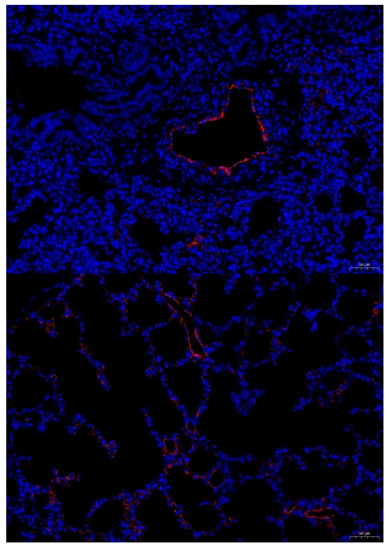
Click image to see more details
IF analysis of CD31 using anti-CD31 antibody (A01513-2).
Paraffin-embedded mouse skin sections were stained for CD31 in three experimental groups: (1) normal control, (2) congestion group, in which the skin was treated with ethanol, and (3) inflammation group, stimulated with LPS. Heat mediated antigen retrieval was performed in EDTA buffer (pH 8.0, epitope retrieval solution). The tissue section was blocked with 10% goat serum. The tissue section was then incubated with 1:200 rabbit anti-CD31 Antibody (A01513-2) overnight at 4°C. DyLight®488 Conjugated Goat Anti-Rabbit IgG (BA1127) was used as secondary antibody incubated with 1:500 for 45 minutes at 37°C. The section was counterstained with DAPI. Visualize using a fluorescence microscope and filter sets appropriate for the label used.

Click image to see more details
IHC analysis of CD31/Pecam1 using anti-CD31/Pecam1 antibody (A01513-2).
CD31/Pecam1 was detected in a paraffin-embedded section of rat lung tissue. Heat mediated antigen retrieval was performed in EDTA buffer (pH 8.0, epitope retrieval solution). The tissue section was blocked with 10% goat serum. The tissue section was then incubated with 2 μg/ml rabbit anti-CD31/Pecam1 Antibody (A01513-2) overnight at 4°C. Peroxidase Conjugated Goat Anti-rabbit IgG was used as secondary antibody and incubated for 30 minutes at 37°C. The tissue section was developed using HRP Conjugated Rabbit IgG Super Vision Assay Kit (Catalog # SV0002) with DAB as the chromogen.

Click image to see more details
IF analysis of CD31 using anti-CD31 antibody (A01513-2).
CD31/Pecam1 was detected in a paraffin-embedded section of mouse lung tissue. Heat mediated antigen retrieval was performed in EDTA buffer (pH 8.0, epitope retrieval solution). The tissue section was blocked with 10% goat serum. The tissue section was then incubated with 1:200 rabbit anti-CD31 Antibody (A01513-2) overnight at 4°C. DyLight®594 Conjugated Goat Anti-Rabbit IgG (BA1142) was used as secondary antibody incubated with 1:500 for 45 minutes at 37°C. The section was counterstained with DAPI. Visualize using a fluorescence microscope and filter sets appropriate for the label used.
Specific Publications For Anti-CD31/Pecam1 Antibody Picoband® (A01513-2)
Loading publications
Recommended Resources
Here are featured tools and databases that you might find useful.
- Boster's Pathways Library
- Protein Databases
- Bioscience Research Protocol Resources
- Data Processing & Analysis Software
- Photo Editing Software
- Scientific Literature Resources
- Research Paper Management Tools
- Molecular Biology Software
- Primer Design Tools
- Bioinformatics Tools
- Phylogenetic Tree Analysis
Customer Reviews
Have you used Anti-CD31/Pecam1 Antibody Picoband®?
Share your experimental results or join a short interview to earn up to $1,000 in product credits or other rewards.
2 Reviews For Anti-CD31/Pecam1 Antibody Picoband®
The Anti-CD31 antibody (Cat# A01513-2) shows high specificity in mouse lung IF, clearly labeling endothelial cells with accurate localization.
Excellent
| SKU | A01513-2 |
|---|---|
| Application | Immunofluorescence |
| Sample | mouse lung tissue |
| Sample Processing Description | Left lung from normal mouse was washed 3 times with PBS to remove blood cells, fixed in formaldehyde for 24 h, and paraffin-embedded for sectioning. |
| Other Reagents | Goat serum, DAPI (Cat# AR1176, Boster Bio) |
| Primary Antibody | CD31/Pecam1 Antibody Picoband® |
| Primary Incubation | 1:200, overnight at 4 ℃ |
| Secondary Antibody | Goat Anti-Rabbit IgG (H+L) Secondary Antibody, Fluoro594 Conjugated (Cat# BA1142, Boster Bio) |
| Secondary Incubation | 1:500, 45min in 37℃ |
| Detection | Image system:Leica DM2500 |
| Results Summary | CD31, also known as PECAM-1, is a key marker of vascular endothelial cells and plays a central role in biomedical research, especially in angiogenesis and tumor microenvironment studies. In this experiment, CD31 IF was performed to label endothelial cells, facilitating subsequent evaluation of vascular density distribution. The results show clear endothelial staining with precise localization, yielding excellent outcomes. |
Fei Liu, The First Affiliated Hospital of Nanchang University
Verified customer
Submitted 2026-03-24
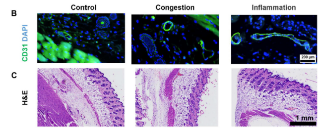
IF using Anti-CD31 antibody (A01513-2) showed specific, strong, and stable endothelial staining, revealing expanded and inflamed microvessels in treated mouse skin compared to controls.
Excellent
| SKU | A01513-2 |
|---|---|
| Application | Immunofluorescence |
| Sample | Paraffin-embedded mouse skin sections |
| Sample Processing Description | Mouse skin samples were divided into three groups: (i) normal control, (ii) congestion group, treated with ethanol, and (iii) inflammation group, stimulated with LPS. |
| Other Reagents | Goat serum, DAPI, Anti-fade mounting medium |
| Primary Antibody | CD31/Pecam1 Antibody Picoband® |
| Primary Incubation | 1:200, overnight at 4 ℃ |
| Secondary Antibody | DyLight 488–conjugated Goat Anti-Rabbit IgG (H+L)) (Boster, BA1127) |
| Secondary Incubation | 1:500, 45 min at 37℃ |
| Detection | Imaging system:Leica DM2500 |
| Results Summary | IF staining with Boster CD31 antibody produced high-quality vascular images. In the control group, vessels were small and well-defined. In the congestion group (ethanol-treated), CD31 staining revealed markedly dilated vascular lumens. In the inflammation group (LPS-treated), vessel dilation was accompanied by changes in tissue spacing. Combined with Prussian blue staining and magnetic signal measurements, we observed that despite extreme vessel dilation in the congestion group, magnetic signals were not significantly elevated; only the inflammation group, with extensive macrophage infiltration and uptake, showed high magnetic signals. |
Wenbo Wang, Shandong University
Verified customer
Submitted 2026-01-30
Customer Q&As
Have a question?
Find answers in Q&As, reviews.
Can't find your answer?
Submit your question
