Click image to see more details
-

-
-
-
-
+12
Product Info Summary
| SKU: | A01357-1 |
|---|---|
| Size: | 100 μg/vial |
| Reactive Species: | Human, Mouse, Rat |
| Host: | Rabbit |
| Application: | Flow Cytometry, IF, IHC, WB |
Customers Who Bought This Also Bought

Product info
Product Name
Anti-Cytokeratin 18/KRT18 Antibody Picoband®
SKU/Catalog Number
A01357-1
Size
100 μg/vial
Form
Lyophilized
Description
Boster Bio Anti-Cytokeratin 18/KRT18 Antibody Picoband® catalog # A01357-1. Tested in Flow Cytometry, IF, IHC, WB applications. This antibody reacts with Human, Mouse, Rat. The brand Picoband indicates this is a premium antibody that guarantees superior quality, high affinity, and strong signals with minimal background in Western blot applications. Only our best-performing antibodies are designated as Picoband, ensuring unmatched performance.
Storage & Handling
Store at -20˚C for one year from date of receipt. After reconstitution, at 4˚C for one month. It can also be aliquotted and stored frozen at -20˚C for six months. Avoid repeated freeze-thaw cycles.
Cite This Product
Anti-Cytokeratin 18/KRT18 Antibody Picoband® (Boster Biological Technology, Pleasanton CA, USA, Catalog # A01357-1)
Host
Rabbit
Contents
Each vial contains 4mg Trehalose, 0.9mg NaCl, 0.2mg Na2HPO4, 0.05mg NaN3.
Clonality
Polyclonal
Isotype
Rabbit IgG
Immunogen
E.coli-derived human Cytokeratin 18 recombinant protein (Position: E204-H430). Human Cytokeratin 18 shares 87.7% and 85.9% amino acid (aa) sequence identity with mouse and rat Cytokeratin 18, respectively.
Cross-reactivity
No cross-reactivity with other proteins
Reactive Species
A01357-1 is reactive to KRT18 in Human, Mouse, Rat
Observed Molecular Weight
48 kDa
Calculated molecular weight
48.1 kDa
Background of KRT18
Keratin 18, mapped to 12q13.13, is a type I cytokeratin. It is, together with its filament partner keratin 8, perhaps the most commonly found products of the intermediate filament gene family. They are expressed in single layer epithelial tissues of the body. Mutations in this gene have been linked to cryptogenic cirrhosis. Two transcript variants encoding the same protein have been found for this gene.
Antibody Validation
Boster validates all antibodies on WB, IHC, ICC, Immunofluorescence, and ELISA with known positive control and negative samples to ensure specificity and high affinity, including thorough antibody incubations.
Application & Images
Applications
A01357-1 is guaranteed for Flow Cytometry, IF, IHC, WB Boster Guarantee
Recommend Dilution
| Application | Dilution | Species |
|---|---|---|
| Western blot | 0.1-0.5μg/ml | Human, Mouse, Rat |
| Immunohistochemistry (Paraffin-embedded Section) | 2-5μg/ml | Human, Mouse, Rat |
| Immunofluorescence | 5 μg/ml | Human |
| Flow Cytometry (Fixed) | 1-3 μg/1x106 cells | Human |
Tested application
Suggested blocking solution with 5% non-fat milk or BSA; (*)Recommended protein loading: 20-40 µg per lane
Use TE buffer pH 9.0 for antigen retrieval; (*) citrate buffer pH 6.0 is an alternative.
Validation Images & Assay Conditions
Click image to see more details
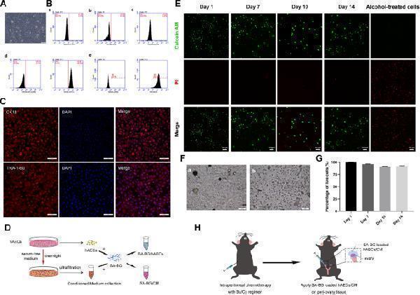
Characterization of hAECs, detection of the survival hAECs encapsulated in SA-BG, and schematic illustration of the surgical procedure. a The morphology of cultured hAECs was observed under a microscope. Scale bar 100 μm. b Flow cytometry analysis of cell surface markers on hAECs. The isotypes (ISO) of SSEA4 and CD324 were used as negative controls. c Immunostaining images showed the high expression of epithelial marker (CK18) and stem cell marker (TRA-1-60). Scale bar 100 μm. d The fabrication method of SA-BG-loaded hAECs and CM. e Representative live/dead images of hAECs encapsulated in SA-BG at days 1, 7, 10, and 14, respectively. hAECs encapsulated in SA-BG treated with 70% alcohol were positive for PI. Live cells were shown green color and dead cells were red color. Scale bar 100 μm. f Bright field image of hAECs capsulated in SA-BG at day 1 ( a ) and 14 ( b ). Scale bar 100 μm. g The percentage of live cells to total cells. h Schematic of the experimental procedure for the transplantation of SA-BG-loaded hAECs/CM into mice with chemotherapy-induced POF
Index in PubMed under a CC BY license. PMID: 33794993

Click image to see more details
The effect of SA-BG extracts on the biological characterization and paracrine capacity of hAECs. a The viability of hAECs cultured with SA-BG extracts was detected by CCK-8 assay. b – d Expression of stemness (Oct-4 and Nanog) and epithelial (CK18) genes of hAECs cultured with SA-BG extracts at different time points. e , f The results of cytokine array of CM from hAECs and SA-BG extract-treated hAECs, respectively. g Column displayed the higher expression of cytokines in the SA-BG extract-treated hAECs than in the control hAECs. h , i The results of quantification of angiogenic factors released from SA-BG extract-treated hAECs and control hAECs by ELISA
Index in PubMed under a CC BY license. PMID: 33794993

Click image to see more details
hAECs express specific surface markers and have stem cell characteristics with low immunogenicity. a hAECs presented an epithelial morphology under bright-field microscopy. Scale bar = 100 μm. b hAECs were labeled with CFSE to track implanted cells. The expression rate of green fluorescence staining was nearly 100%. Scale bar = 100 μm. c-f By flow cytometry, hAECs were positive for stem cell marker SSEA-4 ( c ) and epithelial marker CD324 ( d ) and were negative for mesenchymal markers CD146 ( e ) and HLA-DR ( f ). g Immunofluorescence staining for CK18 (an epithelial marker) expression and vimentin (a mesenchymal marker) in hAECs. Scale bar = 200 μm
Index in PubMed under a CC BY license. PMID: 33762002

Click image to see more details
Western blot analysis of KRT18 using anti-KRT18 antibody (A01357-1).
Electrophoresis was performed on a 10% SDS-PAGE gel at 80V (Stacking gel) / 120V (Resolving gel) for 2 hours. The sample well of each lane was loaded with 30 ug of sample under reducing conditions.
Lane 1: human placenta tissue lysates,
Lane 2: human CACO2 whole cell lysates,
Lane 3: human A549 whole cell lysates,
Lane 4: human A431 whole cell lysates,
Lane 5: rat liver tissue lysates,
Lane 6: rat thymus tissue lysates,
Lane 7: mouse liver tissue lysates,
Lane 8: mouse thymus tissue lysates.
After electrophoresis, proteins were transferred to a nitrocellulose membrane at 150 mA for 50-90 minutes. Blocked the membrane with 5% non-fat milk/TBS for 1.5 hour at RT. The membrane was incubated with rabbit anti-KRT18 antigen affinity purified polyclonal antibody (A01357-1) at 0.5 μg/mL overnight at 4°C, then washed with TBS-0.1%Tween 3 times with 5 minutes each and probed with a goat anti-rabbit IgG-HRP secondary antibody (Catalog # BA1054) at a dilution of 1:5000 for 1.5 hour at RT. The signal is developed using an ECL Plus Western Blotting Substrate (Catalog # AR1196-200) with Tanon 5200 system. A specific band was detected for KRT18 at approximately 48 kDa. The expected band size for KRT18 is at 48 kDa.

Click image to see more details
IHC analysis of KRT18 using anti-KRT18 antibody (A01357-1).
KRT18 was detected in a paraffin-embedded section of human intestinal cancer tissue. Heat mediated antigen retrieval was performed in EDTA buffer (pH 8.0, epitope retrieval solution). The tissue section was blocked with 10% goat serum. The tissue section was then incubated with 2 μg/ml rabbit anti-KRT18 Antibody (A01357-1) overnight at 4°C. Peroxidase Conjugated Goat Anti-rabbit IgG was used as secondary antibody and incubated for 30 minutes at 37°C. The tissue section was developed using HRP Conjugated Rabbit IgG Super Vision Assay Kit (Catalog # SV0002) with DAB as the chromogen.

Click image to see more details
IHC analysis of KRT18 using anti-KRT18 antibody (A01357-1).
KRT18 was detected in a paraffin-embedded section of human breast cancer tissue. Heat mediated antigen retrieval was performed in EDTA buffer (pH 8.0, epitope retrieval solution). The tissue section was blocked with 10% goat serum. The tissue section was then incubated with 2 μg/ml rabbit anti-KRT18 Antibody (A01357-1) overnight at 4°C. Peroxidase Conjugated Goat Anti-rabbit IgG was used as secondary antibody and incubated for 30 minutes at 37°C. The tissue section was developed using HRP Conjugated Rabbit IgG Super Vision Assay Kit (Catalog # SV0002) with DAB as the chromogen.

Click image to see more details
IHC analysis of KRT18 using anti-KRT18 antibody (A01357-1).
KRT18 was detected in a paraffin-embedded section of human placenta tissue. Heat mediated antigen retrieval was performed in EDTA buffer (pH 8.0, epitope retrieval solution). The tissue section was blocked with 10% goat serum. The tissue section was then incubated with 2 μg/ml rabbit anti-KRT18 Antibody (A01357-1) overnight at 4°C. Peroxidase Conjugated Goat Anti-rabbit IgG was used as secondary antibody and incubated for 30 minutes at 37°C. The tissue section was developed using HRP Conjugated Rabbit IgG Super Vision Assay Kit (Catalog # SV0002) with DAB as the chromogen.

Click image to see more details
IHC analysis of KRT18 using anti-KRT18 antibody (A01357-1).
KRT18 was detected in a paraffin-embedded section of mouse intestine tissue. Heat mediated antigen retrieval was performed in EDTA buffer (pH 8.0, epitope retrieval solution). The tissue section was blocked with 10% goat serum. The tissue section was then incubated with 2 μg/ml rabbit anti-KRT18 Antibody (A01357-1) overnight at 4°C. Peroxidase Conjugated Goat Anti-rabbit IgG was used as secondary antibody and incubated for 30 minutes at 37°C. The tissue section was developed using HRP Conjugated Rabbit IgG Super Vision Assay Kit (Catalog # SV0002) with DAB as the chromogen.

Click image to see more details
IHC analysis of KRT18 using anti-KRT18 antibody (A01357-1).
KRT18 was detected in a paraffin-embedded section of rat colon tissue. Heat mediated antigen retrieval was performed in EDTA buffer (pH 8.0, epitope retrieval solution). The tissue section was blocked with 10% goat serum. The tissue section was then incubated with 2 μg/ml rabbit anti-KRT18 Antibody (A01357-1) overnight at 4°C. Peroxidase Conjugated Goat Anti-rabbit IgG was used as secondary antibody and incubated for 30 minutes at 37°C. The tissue section was developed using HRP Conjugated Rabbit IgG Super Vision Assay Kit (Catalog # SV0002) with DAB as the chromogen.

Click image to see more details
IF analysis of KRT18 using anti-KRT18 antibody (A01357-1)
KRT18 was detected in paraffin-embedded section of human intestinal cancer tissues. Heat mediated antigen retrieval was performed in EDTA buffer (pH 8.0, epitope retrieval solution). The tissue section was blocked with 10% goat serum. The tissue section was then incubated with 5μg/mL rabbit anti-KRT18 Antibody (A01357-1) overnight at 4°C. Biotin conjugated goat anti-rabbit IgG (BA1003) was used as secondary antibody and incubated for 30 minutes at 37°C. The tissue section was developed using Cy3 Conjugated Avidin (BA1037). The section was counterstained with DAPI. Visualize using a fluorescence microscope and filter sets appropriate for the label used.

Click image to see more details
IF analysis of KRT18 using anti-KRT18 antibody (A01357-1)
KRT18 was detected in paraffin-embedded section of human gastric cancer tissues. Heat mediated antigen retrieval was performed in EDTA buffer (pH 8.0, epitope retrieval solution). The tissue section was blocked with 10% goat serum. The tissue section was then incubated with 5μg/mL rabbit anti-KRT18 Antibody (A01357-1) overnight at 4°C. Biotin conjugated goat anti-rabbit IgG (BA1003) was used as secondary antibody and incubated for 30 minutes at 37°C. The tissue section was developed using Cy3 Conjugated Avidin (BA1037). The section was counterstained with DAPI. Visualize using a fluorescence microscope and filter sets appropriate for the label used.

Click image to see more details
IF analysis of KRT18 using anti-KRT18 antibody (A01357-1)
KRT18 was detected in paraffin-embedded section of human lung cancer tissues. Heat mediated antigen retrieval was performed in EDTA buffer (pH 8.0, epitope retrieval solution). The tissue section was blocked with 10% goat serum. The tissue section was then incubated with 5μg/mL rabbit anti-KRT18 Antibody (A01357-1) overnight at 4°C. Biotin conjugated goat anti-rabbit IgG (BA1003) was used as secondary antibody and incubated for 30 minutes at 37°C. The tissue section was developed using Cy3 Conjugated Avidin (BA1037). The section was counterstained with DAPI. Visualize using a fluorescence microscope and filter sets appropriate for the label used.

Click image to see more details
IF analysis of KRT18 using anti-KRT18 antibody (A01357-1)
KRT18 was detected in paraffin-embedded section of human prostatic cancer tissues. Heat mediated antigen retrieval was performed in EDTA buffer (pH 8.0, epitope retrieval solution). The tissue section was blocked with 10% goat serum. The tissue section was then incubated with 5μg/mL rabbit anti-KRT18 Antibody (A01357-1) overnight at 4°C. Biotin conjugated goat anti-rabbit IgG (BA1003) was used as secondary antibody and incubated for 30 minutes at 37°C. The tissue section was developed using Cy3 Conjugated Avidin (BA1037). The section was counterstained with DAPI. Visualize using a fluorescence microscope and filter sets appropriate for the label used.

Click image to see more details
IF analysis of KRT18 using anti-KRT18 antibody (A01357-1)
KRT18 was detected in paraffin-embedded section of human ovarian cancer tissues. Heat mediated antigen retrieval was performed in EDTA buffer (pH 8.0, epitope retrieval solution). The tissue section was blocked with 10% goat serum. The tissue section was then incubated with 5μg/mL rabbit anti-KRT18 Antibody (A01357-1) overnight at 4°C. Biotin conjugated goat anti-rabbit IgG (BA1003) was used as secondary antibody and incubated for 30 minutes at 37°C. The tissue section was developed using Cy3 Conjugated Avidin (BA1037). The section was counterstained with DAPI. Visualize using a fluorescence microscope and filter sets appropriate for the label used.

Click image to see more details
Flow Cytometry analysis of A431 cells using anti-KRT18 antibody (A01357-1).
Overlay histogram showing A431 cells stained with A01357-1 (Blue line). To facilitate intracellular staining, cells were fixed with 4% paraformaldehyde and permeabilized with permeabilization buffer. The cells were blocked with 10% normal goat serum. And then incubated with rabbit anti-KRT18 Antibody (A01357-1, 1 μg/1x106 cells) for 30 min at 20°C. Fluoro488 conjugated goat anti-rabbit IgG (BA1127, 5-10 μg/1x106 cells) was used as secondary antibody for 30 minutes at 20°C. Isotype control antibody (Green line) was rabbit IgG (1 μg/1x106) used under the same conditions. Unlabelled sample without incubation with primary antibody and secondary antibody (Red line) was used as a blank control.
Specific Publications For Anti-Cytokeratin 18/KRT18 Antibody Picoband® (A01357-1)
Loading publications
Recommended Resources
Here are featured tools and databases that you might find useful.
- Boster's Pathways Library
- Protein Databases
- Bioscience Research Protocol Resources
- Data Processing & Analysis Software
- Photo Editing Software
- Scientific Literature Resources
- Research Paper Management Tools
- Molecular Biology Software
- Primer Design Tools
- Bioinformatics Tools
- Phylogenetic Tree Analysis
Customer Reviews
Have you used Anti-Cytokeratin 18/KRT18 Antibody Picoband®?
Share your experimental results or join a short interview to earn up to $1,000 in product credits or other rewards.
0 Reviews For Anti-Cytokeratin 18/KRT18 Antibody Picoband®
Customer Q&As
Have a question?
Find answers in Q&As, reviews.
Can't find your answer?
Submit your question
16 Customer Q&As for Anti-Cytokeratin 18/KRT18 Antibody Picoband®
Question
I was wanting to use your anti-Cytokeratin 18/KRT18 antibody for IHC-P for rat liver on frozen tissues, but I want to know if it has been validated for this particular application. Has this antibody been validated and is this antibody a good choice for rat liver identification?
Verified Customer
Verified customer
Asked: 2020-04-28
Answer
You can see on the product datasheet, A01357-1 anti-Cytokeratin 18/KRT18 antibody has been tested for IF, IHC-P, WB on human, mouse, rat tissues. We have an innovator award program that if you test this antibody and show it works in rat liver in IHC-frozen, you can get your next antibody for free.
Boster Scientific Support
Answered: 2020-04-28
Question
We need to test anti-Cytokeratin 18/KRT18 antibody A01357-1 on rat liver for research purposes, then I may be interested in using anti-Cytokeratin 18/KRT18 antibody A01357-1 for diagnostic purposes as well. Is the antibody suitable for diagnostic purposes?
Verified Customer
Verified customer
Asked: 2020-04-28
Answer
The products we sell, including anti-Cytokeratin 18/KRT18 antibody A01357-1, are only intended for research use. They would not be suitable for use in diagnostic work. If you have the means to develop a product into diagnostic use, and are interested in collaborating with us and develop our product into an IVD product, please contact us for more discussions.
Boster Scientific Support
Answered: 2020-04-28
Question
We have tried in the past anti-Cytokeratin 18/KRT18 antibody for IHC-P on pancreas last year. I am using mouse, and We intend to use the antibody for IF next. We are interested in examining pancreas as well as vulva in our next experiment. Could you please give me some suggestion on which antibody would work the best for IF?
Verified Customer
Verified customer
Asked: 2020-04-20
Answer
I viewed the website and datasheets of our anti-Cytokeratin 18/KRT18 antibody and it appears that A01357-1 has been tested on mouse in both IHC-P and IF. Thus A01357-1 should work for your application. Our Boster satisfaction guarantee will cover this product for IF in mouse even if the specific tissue type has not been validated. We do have a comprehensive range of products for IF detection and you can check out our website bosterbio.com to find out more information about them.
Boster Scientific Support
Answered: 2020-04-20
Question
We appreciate helping with my inquiry over the phone. Here are the WB image, lot number and protocol we used for liver using anti-Cytokeratin 18/KRT18 antibody A01357-1. Let me know if you need anything else.
Verified Customer
Verified customer
Asked: 2020-03-30
Answer
I appreciate the data. You have provided everything we needed. Our lab team are working to resolve your inquiry as quickly as possible, and we appreciate your patience and understanding! Please let me know if there is anything you need in the meantime.
Boster Scientific Support
Answered: 2020-03-30
Question
We have observed staining in mouse placenta. Are there any suggestions? Is anti-Cytokeratin 18/KRT18 antibody supposed to stain placenta positively?
Verified Customer
Verified customer
Asked: 2020-03-11
Answer
According to literature placenta does express KRT18. According to Uniprot.org, KRT18 is expressed in adenohypophysis, placenta, cervix, colon, pancreas, placenta uterus, vulva, liver, colon carcinoma, cervix carcinoma, cervix carcinoma erythroleukemia, among other tissues. Regarding which tissues have KRT18 expression, here are a few articles citing expression in various tissues:
Cervix carcinoma, Pubmed ID: 17081983, 17924679, 18669648, 18691976, 20068231
Cervix carcinoma, and Erythroleukemia, Pubmed ID: 23186163
Cervix, Colon, Pancreas, Placenta, and Uterus, Pubmed ID: 15489334
Colon carcinoma, Pubmed ID: 9150948, 24129315
Liver, Pubmed ID: 2422083, 24275569
Placenta, Pubmed ID: 2434380
Vulva, Pubmed ID: 2434381
Boster Scientific Support
Answered: 2020-03-11
Question
Can you help my question with product A01357-1, anti-Cytokeratin 18/KRT18 antibody. I was wondering if it would be possible to conjugate this antibody with biotin. I would need it to be without BSA or sodium azide. I am planning on using a buffer exchange of sodium azide with PBS only. Would there be problems for me to conjugate the antibody and store it in -20 degrees in small aliquots?
Verified Customer
Verified customer
Asked: 2020-02-27
Answer
It is not recommended storing this antibody with PBS buffer only in -20 degrees. If you want to store it in -20 degrees it is best to add some cryoprotectant like glycerol. If you want carrier free A01357-1 anti-Cytokeratin 18/KRT18 antibody, we can provide it to you in a special formula with trehalose and/or glycerol. These molecules will not interfere with conjugation chemistry and provide a good level of protection for the antibody from degradation. Please be sure to specify this in your purchase order.
Boster Scientific Support
Answered: 2020-02-27
Question
We are currently using anti-Cytokeratin 18/KRT18 antibody A01357-1 for rat tissue, and we are content with the IHC-P results. The species of reactivity given in the datasheet says human, mouse, rat. Is it true that the antibody can work on goat tissues as well?
Verified Customer
Verified customer
Asked: 2019-12-25
Answer
The anti-Cytokeratin 18/KRT18 antibody (A01357-1) has not been tested for cross reactivity specifically with goat tissues, though there is a good chance of cross reactivity. We have an innovator award program that if you test this antibody and show it works in goat you can get your next antibody for free. Please contact me if I can help you with anything.
Boster Scientific Support
Answered: 2019-12-25
Question
Will anti-Cytokeratin 18/KRT18 antibody A01357-1 work for IHC-P with liver?
Verified Customer
Verified customer
Asked: 2019-12-04
Answer
According to the expression profile of liver, KRT18 is highly expressed in liver. So, it is likely that anti-Cytokeratin 18/KRT18 antibody A01357-1 will work for IHC-P with liver.
Boster Scientific Support
Answered: 2019-12-04
Question
I see that the anti-Cytokeratin 18/KRT18 antibody A01357-1 works with IHC-P, what is the protocol used to produce the result images on the product page?
Verified Customer
Verified customer
Asked: 2019-11-11
Answer
You can find protocols for IHC-P on the "support/technical resources" section of our navigation menu. If you have any further questions, please send an email to support@bosterbio.com
Boster Scientific Support
Answered: 2019-11-11
Question
Please see the WB image, lot number and protocol we used for liver using anti-Cytokeratin 18/KRT18 antibody A01357-1. Please let me know if you require anything else.
Verified Customer
Verified customer
Asked: 2019-09-20
Answer
Thank you very much for the data. Our lab team are working to resolve this as quickly as possible, and we appreciate your patience and understanding! You have provided everything we needed. Please let me know if there is anything you need in the meantime.
Boster Scientific Support
Answered: 2019-09-20
Question
Does A01357-1 anti-Cytokeratin 18/KRT18 antibody work on parafin embedded sections? If so, which fixation method do you recommend we use (PFA, paraformaldehyde, other)?
Verified Customer
Verified customer
Asked: 2019-06-14
Answer
It shows on the product datasheet, A01357-1 anti-Cytokeratin 18/KRT18 antibody as been tested on IHC-P. It is best to use PFA for fixation because it has better tissue penetration ability. PFA needs to be prepared fresh before use. Long term stored PFA turns into formalin, as the PFA molecules congregate and become formalin.
Boster Scientific Support
Answered: 2019-06-14
Question
Our team were satisfied with the WB result of your anti-Cytokeratin 18/KRT18 antibody. However we have seen positive staining in vulva cytoplasm using this antibody. Is that expected? Could you tell me where is KRT18 supposed to be expressed?
Verified Customer
Verified customer
Asked: 2018-11-16
Answer
Based on literature, vulva does express KRT18. Generally KRT18 expresses in cytoplasm, perinuclear region. nucleus,. Regarding which tissues have KRT18 expression, here are a few articles citing expression in various tissues:
Cervix carcinoma, Pubmed ID: 17081983, 17924679, 18669648, 18691976, 20068231
Cervix carcinoma, and Erythroleukemia, Pubmed ID: 23186163
Cervix, Colon, Pancreas, Placenta, and Uterus, Pubmed ID: 15489334
Colon carcinoma, Pubmed ID: 9150948, 24129315
Liver, Pubmed ID: 2422083, 24275569
Placenta, Pubmed ID: 2434380
Vulva, Pubmed ID: 2434381
Boster Scientific Support
Answered: 2018-11-16
Question
Is this A01357-1 anti-Cytokeratin 18/KRT18 antibody reactive to the isotypes of KRT18?
Verified Customer
Verified customer
Asked: 2018-03-30
Answer
The immunogen of A01357-1 anti-Cytokeratin 18/KRT18 antibody is E.coli-derived human Cytokeratin 18 recombinant protein (Position: E204-H430). Human Cytokeratin 18 shares 87.7% and 85.9% amino acid (aa) sequence identity with mouse and rat Cytokeratin 18, respectively. Could you tell me which isotype you are interested in so I can help see if the immunogen is part of this isotype?
Boster Scientific Support
Answered: 2018-03-30
Question
Is a blocking peptide available for product anti-Cytokeratin 18/KRT18 antibody (A01357-1)?
Verified Customer
Verified customer
Asked: 2017-08-04
Answer
We do provide the blocking peptide for product anti-Cytokeratin 18/KRT18 antibody (A01357-1). If you would like to place an order for it please contact support@bosterbio.com and make a special request.
Boster Scientific Support
Answered: 2017-08-04
Question
Is there a BSA free version of anti-Cytokeratin 18/KRT18 antibody A01357-1 available?
R. Anderson
Verified customer
Asked: 2013-04-05
Answer
Thanks for your recent telephone inquiry. I can confirm that some lots of this anti-Cytokeratin 18/KRT18 antibody A01357-1 are BSA free. For now, these lots are available and we can make a BSA free formula for you free of charge. It will take 3 extra days to prepare. If you require this antibody BSA free again in future, please do not hesitate to contact me and I will be pleased to check which lots we have in stock that are BSA free.
Boster Scientific Support
Answered: 2013-04-05
Question
I am interested in using your anti-Cytokeratin 18/KRT18 antibody for cell cycle studies. Has this antibody been tested with western blotting on hepg2 whole cell lysates? We would like to see some validation images before ordering.
G. Gonzalez
Verified customer
Asked: 2013-01-22
Answer
Thanks for your inquiry. This A01357-1 anti-Cytokeratin 18/KRT18 antibody is validated on rat kidney, mouse liver, hela whole cell lysates, hepg2 whole cell lysates, a431 cells, a549 cells. It is guaranteed to work for IF, IHC-P, WB in human, mouse, rat. Our Boster guarantee will cover your intended experiment even if the sample type has not been be directly tested.
Boster Scientific Support
Answered: 2013-01-22
