Click image to see more details
-

-
-
-
-
+3
Product Info Summary
| SKU: | PB9585 |
|---|---|
| Size: | 100 μg/vial |
| Reactive Species: | Human, Mouse, Rat |
| Host: | Rabbit |
| Application: | Flow Cytometry, IP, IF, IHC, ICC, WB |
Customers Who Bought This Also Bought

Product info
Product Name
Anti-EWSR1 Antibody Picoband®
SKU/Catalog Number
PB9585
PB0616 is an alternative SKU for this antibody, used in previous lots.
Size
100 μg/vial
Form
Lyophilized
Description
Boster Bio Anti-EWSR1 Antibody Picoband® catalog # PB9585. Tested in Flow Cytometry, IP, IF, IHC, ICC, WB applications. This antibody reacts with Human, Mouse, Rat. The brand Picoband indicates this is a premium antibody that guarantees superior quality, high affinity, and strong signals with minimal background in Western blot applications. Only our best-performing antibodies are designated as Picoband, ensuring unmatched performance.
Storage & Handling
Store at -20˚C for one year from date of receipt. After reconstitution, at 4˚C for one month. It can also be aliquotted and stored frozen at -20˚C for six months. Avoid repeated freeze-thaw cycles.
Cite This Product
Anti-EWSR1 Antibody Picoband® (Boster Biological Technology, Pleasanton CA, USA, Catalog # PB9585)
Host
Rabbit
Contents
Each vial contains 4 mg Trehalose, 0.9 mg NaCl and 0.2 mg Na2HPO4.
Clonality
Polyclonal
Isotype
Rabbit IgG
Immunogen
A synthetic peptide corresponding to a sequence in the middle region of human EWSR1, different from the related mouse sequence by one amino acid.
Cross-reactivity
No cross-reactivity with other proteins
Reactive Species
PB9585 is reactive to EWSR1 in Human, Mouse, Rat
Observed Molecular Weight
90-95 kDa
Calculated molecular weight
68.5 kDa
Background of EWSR1
This gene encodes a multifunctional protein that is involved in various cellular processes, including gene expression, cell signaling, and RNA processing and transport. The protein includes an N-terminal transcriptional activation domain and a C-terminal RNA-binding domain. Chromosomal translocations between this gene and various genes encoding transcription factors result in the production of chimeric proteins that are involved in tumorigenesis. These chimeric proteins usually consist of the N-terminal transcriptional activation domain of this protein fused to the C-terminal DNA-binding domain of the transcription factor protein. Mutations in this gene, specifically a t (11;22) (q24;q12) translocation, are known to cause Ewing sarcoma as well as neuroectodermal and various other tumors. Alternative splicing of this gene results in multiple transcript variants. Related pseudogenes have been identified on chromosomes 1 and 14.
Antibody Validation
Boster validates all antibodies on WB, IHC, ICC, Immunofluorescence, and ELISA with known positive control and negative samples to ensure specificity and high affinity, including thorough antibody incubations.
Application & Images
Applications
PB9585 is guaranteed for Flow Cytometry, IP, IF, IHC, ICC, WB Boster Guarantee
Assay Dilutions Recommendation
The recommendations below provide a starting point for assay optimization. The actual working concentration varies and should be decided by the user.
Western blot, 0.1-0.5μg/ml, Human, Mouse, Rat
Immunohistochemistry (Paraffin-embedded Section), 2-5μg/ml, Human, Rat
Immunocytochemistry/Immunofluorescence, 5μg/ml, Human
Immunoprecipitation, 0.5-2 μg/ml, Human
Flow Cytometry (Fixed), 1-3μg/1x106 cells, Human
Positive Control
WB: human Jurkat whole cell, human K562 whole cell, human Hela whole cell, human RT4 whole cell, rat testis tissue, rat C6 whole cell, mouse testis tissue
IHC: human testis cancer tissue, human thyroid cancer tissue, rat spleen tissue
ICC/IF: Hela cell
IP: Hela cell
FCM: RT4 cell
Validation Images & Assay Conditions
Click image to see more details
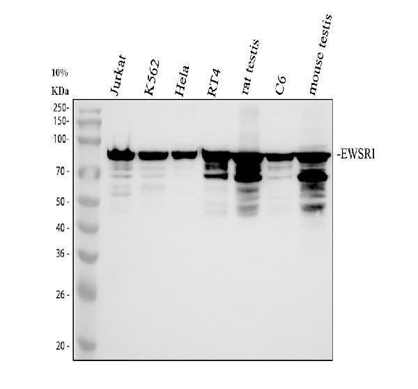
Western blot analysis of EWSR1 using anti-EWSR1 antibody (PB9585).
Electrophoresis was performed on a 10% SDS-PAGE gel at 80V (Stacking gel) / 120V (Resolving gel) for 2 hours. The sample well of each lane was loaded with 30 ug of sample under reducing conditions.
Lane 1: human Jurkat whole cell lysates,
Lane 2: human K562 whole cell lysates,
Lane 3: human Hela whole cell lysates,
Lane 4: human RT4 whole cell lysates,
Lane 5: rat testis tissue lysates,
Lane 6: rat C6 whole cell lysates,
Lane 7: mouse testis tissue lysates.
After electrophoresis, proteins were transferred to a nitrocellulose membrane at 150 mA for 50-90 minutes. Blocked the membrane with 5% non-fat milk/TBS for 1.5 hour at RT. The membrane was incubated with rabbit anti-EWSR1 antigen affinity purified polyclonal antibody (PB9585) at 0.5 μg/mL overnight at 4°C, then washed with TBS-0.1%Tween 3 times with 5 minutes each and probed with a goat anti-rabbit IgG-HRP secondary antibody (Catalog # BA1054) at a dilution of 1:5000 for 1.5 hour at RT. The signal is developed using an ECL Plus Western Blotting Substrate (Catalog # AR1196-200) with Tanon 5200 system. A specific band was detected for EWSR1 at approximately 90-95 kDa. The expected band size for EWSR1 is at 69 kDa.

Click image to see more details
IHC analysis of EWSR1 using anti-EWSR1 antibody (PB9585).
EWSR1 was detected in a paraffin-embedded section of human testis cancer tissue. Heat mediated antigen retrieval was performed in EDTA buffer (pH 8.0, epitope retrieval solution). The tissue section was blocked with 10% goat serum. The tissue section was then incubated with 2 μg/ml rabbit anti-EWSR1 Antibody (PB9585) overnight at 4°C. Peroxidase Conjugated Goat Anti-rabbit IgG was used as secondary antibody and incubated for 30 minutes at 37°C. The tissue section was developed using HRP Conjugated Rabbit IgG Super Vision Assay Kit (Catalog # SV0002) with DAB as the chromogen.

Click image to see more details
IHC analysis of EWSR1 using anti-EWSR1 antibody (PB9585).
EWSR1 was detected in a paraffin-embedded section of human thyroid cancer tissue. Heat mediated antigen retrieval was performed in EDTA buffer (pH 8.0, epitope retrieval solution). The tissue section was blocked with 10% goat serum. The tissue section was then incubated with 2 μg/ml rabbit anti-EWSR1 Antibody (PB9585) overnight at 4°C. Peroxidase Conjugated Goat Anti-rabbit IgG was used as secondary antibody and incubated for 30 minutes at 37°C. The tissue section was developed using HRP Conjugated Rabbit IgG Super Vision Assay Kit (Catalog # SV0002) with DAB as the chromogen.

Click image to see more details
IHC analysis of EWSR1 using anti-EWSR1 antibody (PB9585).
EWSR1 was detected in a paraffin-embedded section of rat spleen tissue. Heat mediated antigen retrieval was performed in EDTA buffer (pH 8.0, epitope retrieval solution). The tissue section was blocked with 10% goat serum. The tissue section was then incubated with 2 μg/ml rabbit anti-EWSR1 Antibody (PB9585) overnight at 4°C. Peroxidase Conjugated Goat Anti-rabbit IgG was used as secondary antibody and incubated for 30 minutes at 37°C. The tissue section was developed using HRP Conjugated Rabbit IgG Super Vision Assay Kit (Catalog # SV0002) with DAB as the chromogen.

Click image to see more details
IF analysis of EWSR1 using anti-EWSR1 antibody (PB9585) and anti-Tubulin Alpha antibody (M03989-3).
EWSR1 was detected in immunocytochemical section of Hela cell. Enzyme antigen retrieval was performed using IHC enzyme antigen retrieval reagent (AR0022) for 15 mins. The cells were blocked with 10% goat serum. And then incubated with 5 μg/mL rabbit anti-EWSR1 Antibody (PB9585) and mouse anti-Tubulin Alpha antibody (M03989-3) overnight at 4°C. Fluoro488 Conjugated Goat Anti-Rabbit IgG (BA11245) and Cy3 Conjugated Goat Anti-Mouse IgG (BA1031) were used as secondary antibody at 1:500 dilution and incubated for 30 minutes at 37°C. Visualize using a fluorescence microscope and filter sets appropriate for the label used.

Click image to see more details
Immunoprecipitating EWSR1 in Hela whole cell lysate.
Western blot analysis of EWSR1 using anti-EWSR1 antibody (PB9585);
Lane 1: Hela whole cell lysates (30ug);
Lane 2: Rabbit control IgG instead of anti-EWSR1 antibody in Hela whole cell lysate;
Lane 3: anti-EWSR1 antibody (2μg) + Hela whole cell lysate (500μg).
After electrophoresis, proteins were transferred to a membrane. Then the membrane was incubated with rabbit anti-EWSR1 antigen affinity purified polyclonal antibody (PB9585) at a dilution of 0.5 μg/mL and probed with a goat anti-rabbit IgG-HRP secondary antibody (Catalog # BA1054). The signal is developed using ECL Plus Western Blotting Substrate (Catalog # AR1196-200). A specific band was detected for EWSR1 at approximately 90-95 kDa. The expected band size for EWSR1 is at 69 kDa.

Click image to see more details
Flow Cytometry analysis of RT4 cells using anti-EWSR1 antibody (PB9585).
Overlay histogram showing RT4 cells stained with PB9585 (Blue line). The cells were fixed with 4% paraformaldehyde and blocked with 10% normal goat serum. And then incubated with rabbit anti-EWSR1 Antibody (PB9585, 1 μg/1x106 cells) for 30 min at 20°C. Fluoro488 conjugated goat anti-rabbit IgG (BA1127, 5-10 μg/1x106 cells) was used as secondary antibody for 30 minutes at 20°C. Isotype control antibody (Green line) was rabbit IgG (1 μg/1x106) used under the same conditions. Unlabelled sample without incubation with primary antibody and secondary antibody (Red line) was used as a blank control.
Specific Publications For Anti-EWSR1 Antibody Picoband® (PB9585)
Loading publications
Recommended Resources
Here are featured tools and databases that you might find useful.
- Boster's Pathways Library
- Protein Databases
- Bioscience Research Protocol Resources
- Data Processing & Analysis Software
- Photo Editing Software
- Scientific Literature Resources
- Research Paper Management Tools
- Molecular Biology Software
- Primer Design Tools
- Bioinformatics Tools
- Phylogenetic Tree Analysis
Customer Reviews
Have you used Anti-EWSR1 Antibody Picoband®?
Share your experimental results or join a short interview to earn up to $1,000 in product credits or other rewards.
0 Reviews For Anti-EWSR1 Antibody Picoband®
Customer Q&As
Have a question?
Find answers in Q&As, reviews.
Can't find your answer?
Submit your question
5 Customer Q&As for Anti-EWSR1 Antibody Picoband®
Question
Has PB9585 antibody been tested on mouse samples with Western Blot, Immunocytochemistry and Immunofluorescence applications?
Verified customer
Asked: 2019-09-19
Answer
The Anti-EWSR1 Antibody Picoband PB9585 was tested on mouse samples with WB only. Our lab hasn't worked on mouse samples with Immunocytochemistry and Immunofluorescence applications due to sample limitation. It is suggested to run pilot tests for this.
Boster Scientific Support
Answered: 2019-09-20
Question
I was wanting to use your anti-EWSR1 antibody for Flow Cytometry for rat testis on frozen tissues, but I want to know if it has been validated for this particular application. Has this antibody been validated and is this antibody a good choice for rat testis identification?
Verified Customer
Verified customer
Asked: 2019-09-18
Answer
You can see on the product datasheet, PB9585 anti-EWSR1 antibody has been tested for Flow Cytometry, IF, IHC-P, IHC-F, ICC, WB on human, mouse, rat tissues. We have an innovator award program that if you test this antibody and show it works in rat testis in IHC-frozen, you can get your next antibody for free.
Boster Scientific Support
Answered: 2019-09-18
Question
We are currently using anti-EWSR1 antibody PB9585 for rat tissue, and we are satisfied with the IHC-F results. The species of reactivity given in the datasheet says human, mouse, rat. Is it true that the antibody can work on zebrafish tissues as well?
B. Jones
Verified customer
Asked: 2018-10-09
Answer
The anti-EWSR1 antibody (PB9585) has not been tested for cross reactivity specifically with zebrafish tissues, though there is a good chance of cross reactivity. We have an innovator award program that if you test this antibody and show it works in zebrafish you can get your next antibody for free. Please contact me if I can help you with anything.
Boster Scientific Support
Answered: 2018-10-09
Question
See below the WB image, lot number and protocol we used for testis using anti-EWSR1 antibody PB9585. Please let me know if you require anything else.
Verified Customer
Verified customer
Asked: 2017-11-27
Answer
Thank you very much for the data. Our lab team are working to resolve this as quickly as possible, and we appreciate your patience and understanding! You have provided everything we needed. Please let me know if there is anything you need in the meantime.
Boster Scientific Support
Answered: 2017-11-27
Question
Do you have a BSA free version of anti-EWSR1 antibody PB9585 available?
S. Carter
Verified customer
Asked: 2016-09-26
Answer
Thank you for your recent telephone inquiry. I can confirm that some lots of this anti-EWSR1 antibody PB9585 are BSA free. For now, these lots are available and we can make a BSA free formula for you free of charge. It will take 3 extra days to prepare. If you require this antibody BSA free again in future, please do not hesitate to contact me and I will be pleased to check which lots we have in stock that are BSA free.
Boster Scientific Support
Answered: 2016-09-26
