Click image to see more details
-
 Index in PubMed under a CC BY license. PMID: 31920652" alt="pb9085 fphar 10 01459 g007">
-
-
-
-
+3
Product Info Summary
| SKU: | PB9085 |
|---|---|
| Size: | 100 μg/vial |
| Reactive Species: | Mouse, Rat |
| Host: | Rabbit |
| Application: | IHC, WB |
Customers Who Bought This Also Bought

Product info
Product Name
Anti-Heme Oxygenase 1/HMOX1 Antibody Picoband®
SKU/Catalog Number
PB9085
PB0050 is an alternative SKU for this antibody, used in previous lots.
Size
100 μg/vial
Form
Lyophilized
Description
Boster Bio Anti-Heme Oxygenase 1/HMOX1 Antibody Picoband® catalog # PB9085. Tested in IHC, WB applications. This antibody reacts with Mouse, Rat. The brand Picoband indicates this is a premium antibody that guarantees superior quality, high affinity, and strong signals with minimal background in Western blot applications. Only our best-performing antibodies are designated as Picoband, ensuring unmatched performance.
Storage & Handling
At -20°C for one year from date of receipt. After reconstitution, at 4°C for one month. It can also be aliquotted and stored frozen at -20°C for six months. Avoid repeated freezing and thawing.
Cite This Product
Anti-Heme Oxygenase 1/HMOX1 Antibody Picoband® (Boster Biological Technology, Pleasanton CA, USA, Catalog # PB9085)
Host
Rabbit
Contents
Each vial contains 4 mg Trehalose, 0.9 mg NaCl and 0.2 mg Na2HPO4.
Clonality
Polyclonal
Isotype
Rabbit IgG
Immunogen
E.coli-derived mouse HMOX1 recombinant protein (Position: E2-T261). Mouse HMOX1 shares 82% and 93% amino acid (aa) sequences identity with human and rat HMOX1, respectively.
Cross-reactivity
No cross-reactivity with other proteins
Reactive Species
PB9085 is reactive to Hmox1 in Mouse, Rat
Observed Molecular Weight
33 kDa
Calculated molecular weight
32.9 kDa
Background of Hmox1
HMOX1 (heme oxygenase (decycling) 1) is a human gene that encodes for the enzyme heme oxygenase 1. It is an essential enzyme in heme catabolism, it cleaves heme to form biliverdin. HMOX1 belongs to the heme oxygenase family. The HMOX1 gene is located on the long (q) arm of chromosome 22 at position 12.3, from base pair 34,101,636 to base pair 34,114,748. HMOX1, an essential enzyme in heme catabolism, cleaves heme to form biliverdin, which is subsequently converted to bilirubin by biliverdin reductase, and carbon monoxide, a putative neurotransmitter. HMOX1 activity is induced by its substrate heme and by various nonheme substances.
Antibody Validation
Boster validates all antibodies on WB, IHC, ICC, Immunofluorescence, and ELISA with known positive control and negative samples to ensure specificity and high affinity, including thorough antibody incubations.
Application & Images
Applications
PB9085 is guaranteed for IHC, WB Boster Guarantee
Assay Dilutions Recommendation
The recommendations below provide a starting point for assay optimization. The actual working concentration varies and should be decided by the user.
Western blot, 0.25-0.5μg/ml, Mouse, Rat
Immunohistochemistry(Paraffin-embedded Section), 2-5 μg/ml, Mouse, Rat
Positive Control
WB: rat spleen tissue, rat liver tissue, rat PC-12 whole cell, mouse spleen tissue, mouse NIH/3T3 whole cell, mouse RAW264.7 whole cell
IHC: mouse liver tissue, rat alcoholic hepatitis tissue
Validation Images & Assay Conditions
Click image to see more details
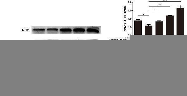
Effects of magnolol on mice alcohol-induced liver damage in the Nrf2/HO-1 signaling pathway. After the completion of modeling and samples were collected, the liver of mice was lysed to detect the proteins by western blotting analysis. The levels of Nrf2 and HO-1 were compared with GAPDH. The data were demonstrated as means ± SD. (*P < 0.05, ***P < 0.001 and "ns" means not significant).
Index in PubMed under a CC BY license. PMID: 31920652

Click image to see more details
Trimetazidine restrained EE-induced activation of NF-κB and inactivation of HO-1/Nrf2 signaling pathways. (A–C) Western blot analysis of the protein levels of nuclear factor of kappa light polypeptide gene enhancer in B-cells inhibitor alpha (IκBα), P-IκBα, and nuclear factor kappa-light-chain-enhancer of activated B-cells (NF-κB) in myocardial tissues. GAPDH and Histone H3 were used as loading controls. (D,E) The DNA binding activity of NF-κB was evaluated by EMSA assay. (F–G) Western blot analysis of the protein levels of heme oxygenase 1 (HO-1) and nuclear factor (erythroid-derived 2)-like 2 (Nrf2) in myocardial tissues. Each value is shown as mean ± SD ( n = 6). ∗ P < 0.05, ∗∗ P < 0.01, ∗∗∗ P < 0.001, versus the indicated group.
Index in PubMed under a CC BY license. PMID: 30890937

Click image to see more details
Involvement of Nrf2/HO-1 signaling pathway in therapeutic effects of NMN. ( A ) Representative images and quantitative analysis of HO-1 protein in peri-hemorrhagic brain tissues at 24 hours after cICH. ( B ) Representative images and quantitative analysis of Nrf2 protein in peri-hemorrhagic brain tissues at 24 hours after cICH. ( C ) Representative images and quantitative analysis of Nrf2 protein distribution in cytosolic and nuclear extracts of brain peri-hemorrhagic tissues at 24 hours after cICH. * P < 0.01, n = 5 per group. NS, no significance.
Index in PubMed under a CC BY license. PMID: 28386082

Click image to see more details
Western blot analysis of HO-1/HMOX1 using anti-HO-1/HMOX1 antibody (PB9085).
Electrophoresis was performed on a 10% SDS-PAGE gel at 80V (Stacking gel) / 120V (Resolving gel) for 2 hours. The sample well of each lane was loaded with 30 ug of sample under reducing conditions.
Lane 1: mouse RAW264.7- WT whole cell lysates,
Lane 2: mouse RAW264.7-HMOX1 KO whole cell lysates.
After electrophoresis, proteins were transferred to a nitrocellulose membrane at 150 mA for 50-90 minutes. Blocked the membrane with 5% non-fat milk/TBS for 1.5 hour at RT. The membrane was incubated with rabbit anti-HO-1/HMOX1 antigen affinity purified polyclonal antibody (PB9085) at 0.5 μg/mL overnight at 4°C, then washed with TBS-0.1%Tween 3 times with 5 minutes each and probed with a goat anti-rabbit IgG-HRP secondary antibody at a dilution of 1:5000 for 1.5 hour at RT. The signal is developed using an ECL Plus Western Blotting Substrate (Catalog # AR1196-200) with Tanon 5200 system. A specific band was detected for HO-1/HMOX1 at approximately 33 kDa. The expected band size for HO-1/HMOX1 is at 33 kDa.

Click image to see more details
Western blot analysis of HMOX1 using anti-HMOX1 antibody (PB9085).
Electrophoresis was performed on a 10% SDS-PAGE gel at 80V (Stacking gel) / 120V (Resolving gel) for 2 hours. The sample well of each lane was loaded with 30 ug of sample under reducing conditions.
Lane 1: rat spleen tissue lysates,
Lane 2: rat liver tissue lysates,
Lane 3: rat PC-12 whole cell lysates,
Lane 4: mouse spleen tissue lysates,
Lane 5: mouse NIH/3T3 whole cell lysates,
Lane 6: mouse RAW264.7 whole cell lysates.
After electrophoresis, proteins were transferred to a nitrocellulose membrane at 150 mA for 50-90 minutes. Blocked the membrane with 5% non-fat milk/TBS for 1.5 hour at RT. The membrane was incubated with rabbit anti-HMOX1 antigen affinity purified polyclonal antibody (PB9085) at 0.5 μg/mL overnight at 4°C, then washed with TBS-0.1%Tween 3 times with 5 minutes each and probed with a goat anti-rabbit IgG-HRP secondary antibody (Catalog # BA1054) at a dilution of 1:5000 for 1.5 hour at RT. The signal is developed using an ECL Plus Western Blotting Substrate (Catalog # AR1196-200) with Tanon 5200 system. A specific band was detected for HMOX1 at approximately 33 kDa. The expected band size for HMOX1 is at 33 kDa.

Click image to see more details
IHC analysis of HMOX1 using anti-HMOX1 antibody (PB9085).
HMOX1 was detected in a paraffin-embedded section of mouse liver tissue. Heat mediated antigen retrieval was performed in EDTA buffer (pH 8.0, epitope retrieval solution). The tissue section was blocked with 10% goat serum. The tissue section was then incubated with 2 μg/ml rabbit anti-HMOX1 Antibody (PB9085) overnight at 4°C. Peroxidase Conjugated Goat Anti-rabbit IgG was used as secondary antibody and incubated for 30 minutes at 37°C. The tissue section was developed using HRP Conjugated Rabbit IgG Super Vision Assay Kit (Catalog # SV0002) with DAB as the chromogen.

Click image to see more details
IHC analysis of HMOX1 using anti-HMOX1 antibody (PB9085).
HMOX1 was detected in a paraffin-embedded section of rat alcoholic hepatitis tissue. Heat mediated antigen retrieval was performed in EDTA buffer (pH 8.0, epitope retrieval solution). The tissue section was blocked with 10% goat serum. The tissue section was then incubated with 2 μg/ml rabbit anti-HMOX1 Antibody (PB9085) overnight at 4°C. Peroxidase Conjugated Goat Anti-rabbit IgG was used as secondary antibody and incubated for 30 minutes at 37°C. The tissue section was developed using HRP Conjugated Rabbit IgG Super Vision Assay Kit (Catalog # SV0002) with DAB as the chromogen.
Specific Publications For Anti-Heme Oxygenase 1/HMOX1 Antibody Picoband® (PB9085)
Loading publications
Recommended Resources
Here are featured tools and databases that you might find useful.
- Boster's Pathways Library
- Protein Databases
- Bioscience Research Protocol Resources
- Data Processing & Analysis Software
- Photo Editing Software
- Scientific Literature Resources
- Research Paper Management Tools
- Molecular Biology Software
- Primer Design Tools
- Bioinformatics Tools
- Phylogenetic Tree Analysis
Customer Reviews
Have you used Anti-Heme Oxygenase 1/HMOX1 Antibody Picoband®?
Share your experimental results or join a short interview to earn up to $1,000 in product credits or other rewards.
0 Reviews For Anti-Heme Oxygenase 1/HMOX1 Antibody Picoband®
Customer Q&As
Have a question?
Find answers in Q&As, reviews.
Can't find your answer?
Submit your question
