Click image to see more details
-

-
-
-
-
+8
Product Info Summary
| SKU: | PB9025 |
|---|---|
| Size: | 100 μg/vial |
| Reactive Species: | Mouse, Rat |
| Host: | Rabbit |
| Application: | ELISA, IHC, WB |
Customers Who Bought This Also Bought

Product info
Product Name
Anti-IL1 beta/IL1B Antibody Picoband®
SKU/Catalog Number
PB9025
PB0055 is an alternative SKU for this antibody, used in previous lots.
Size
100 μg/vial
Form
Lyophilized
Description
Boster Bio Anti-IL1 beta/IL1B Antibody Picoband® catalog # PB9025. Tested in ELISA, IHC, WB applications. This antibody reacts with Mouse, Rat. The brand Picoband indicates this is a premium antibody that guarantees superior quality, high affinity, and strong signals with minimal background in Western blot applications. Only our best-performing antibodies are designated as Picoband, ensuring unmatched performance.
Storage & Handling
Store at -20˚C for one year from date of receipt. After reconstitution, at 4˚C for one month. It can also be aliquotted and stored frozen at -20˚C for six months. Avoid repeated freeze-thaw cycles.
Cite This Product
Anti-IL1 beta/IL1B Antibody Picoband® (Boster Biological Technology, Pleasanton CA, USA, Catalog # PB9025)
Host
Rabbit
Contents
Each vial contains 4 mg Trehalose, 0.9 mg NaCl and 0.2 mg Na2HPO4.
Clonality
Polyclonal
Isotype
Rabbit IgG
Immunogen
E.coli-derived rat IL-1 beta recombinant protein (Position: V117-S268). Rat IL-1 beta shares 78% and 90% amino acid (aa) sequences identity with human and mouse IL-1 beta, respectively.
Cross-reactivity
No cross-reactivity with other proteins
Reactive Species
PB9025 is reactive to Il1b in Mouse, Rat
Observed Molecular Weight
17, 35 kDa
Calculated molecular weight
30.6 kDa
Background of Il1b
Interleukin-1beta (IL-1beta) is a potent stimulator of bone resorption whose gene is mapped to 2q14, and has been implicated in the pathogenesis of high bone turnover and osteoporosis. IL-1beta, a prominent microglia-derived cytokine, caused oligodendrocyte death in coculture with astrocytes and microglia, but not in pure culture of oligodendrocytes alone. It also can cause nuclear export of a specific NCOR corepressor complex, resulting in derepression of a specific subset of nuclear factor-kappa-B (NFKB)-regulated genes. Furthermore, Microenvironmental IL-1beta and, to a lesser extent, IL-1alpha are required for in vivo angiogenesis and invasiveness of different tumor cells. Additional, the cooperation of IL-1beta and PDGFB induces contractile-to-synthetic phenotype modulation of human aortic smooth muscle cells in culture. Moreover, the association with disease may be explained by the biologic properties of IL-1beta, which is an important proinflammatory cytokine and a powerful inhibitor of gastric acid secretion.
Antibody Validation
Boster validates all antibodies on WB, IHC, ICC, Immunofluorescence, and ELISA with known positive control and negative samples to ensure specificity and high affinity, including thorough antibody incubations.
Application & Images
Applications
PB9025 is guaranteed for ELISA, IHC, WB Boster Guarantee
Recommend Dilution
| Application | Dilution | Species |
|---|---|---|
| Western blot | 0.1-0.5μg/ml | Mouse, Rat |
| Immunohistochemistry(Paraffin-embedded Section) | 2-5 μg/ml | Mouse, Rat |
| ELISA | 0.1-0.5μg/ml |
Tested application
Suggested blocking solution with 5% non-fat milk or BSA; (*)Recommended protein loading: 20-40 µg per lane
Use TE buffer pH 9.0 for antigen retrieval; (*) citrate buffer pH 6.0 is an alternative.
Validation Images & Assay Conditions
Click image to see more details
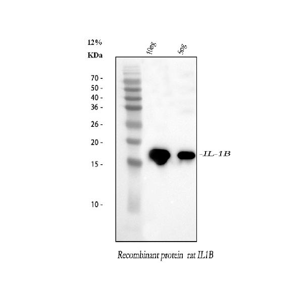
Western blot analysis of IL1 beta using anti-IL1 beta antibody (PB9025).
Electrophoresis was performed on a 5-20% SDS-PAGE gel at 70V (Stacking gel) / 90V (Resolving gel) for 2-3 hours.
Lane 1: recombinant rat IL1 beta protein 10 ng,
Lane 2: recombinant rat IL1 beta protein 5 ng.
After electrophoresis, proteins were transferred to a nitrocellulose membrane at 150 mA for 50-90 minutes. Blocked the membrane with 5% non-fat milk/TBS for 1.5 hour at RT. The membrane was incubated with rabbit anti-IL1 beta antigen affinity purified polyclonal antibody (Catalog # PB9025) at 0.5 μg/mL overnight at 4°C, then washed with TBS-0.1%Tween 3 times with 5 minutes each and probed with a goat anti-rabbit IgG-HRP secondary antibody at a dilution of 1:5000 for 1.5 hour at RT. The signal is developed using an Enhanced Chemiluminescent detection (ECL) kit (Catalog # EK1002) with Tanon 5200 system. A specific band was detected for IL1 beta at approximately 17 kDa.

Click image to see more details
IHC analysis of IL1 beta using anti-IL1 beta antibody (PB9025).
IL1 beta was detected in a paraffin-embedded section of mouse spleen tissue. Heat mediated antigen retrieval was performed in EDTA buffer (pH 8.0, epitope retrieval solution). The tissue section was blocked with 10% goat serum. The tissue section was then incubated with 2 μg/ml rabbit anti-IL1 beta Antibody (PB9025) overnight at 4°C. Peroxidase Conjugated Goat Anti-rabbit IgG was used as secondary antibody and incubated for 30 minutes at 37°C. The tissue section was developed using HRP Conjugated Rabbit IgG Super Vision Assay Kit (Catalog # SV0002) with DAB as the chromogen.

Click image to see more details
Western blot analysis of IL1 beta using anti-IL1 beta antibody (PB9025).
Electrophoresis was performed on a 5-20% SDS-PAGE gel at 70V (Stacking gel) / 90V (Resolving gel) for 2-3 hours.The sample well of each lane was loaded with 30 ug of sample under reducing conditions.
Lane 1: mouse RAW264.7 whole cell lysates,
Lane 2: mouse RAW264.7 whole cell lysates.
After electrophoresis, proteins were transferred to a nitrocellulose membrane at 150 mA for 50-90 minutes. Blocked the membrane with 5% non-fat milk/TBS for 1.5 hour at RT. The membrane was incubated with rabbit anti-IL1 beta antigen affinity purified polyclonal antibody (Catalog # PB9025) at 0.5 μg/mL overnight at 4°C, then washed with TBS-0.1%Tween 3 times with 5 minutes each and probed with a goat anti-rabbit IgG-HRP secondary antibody at a dilution of 1:5000 for 1.5 hour at RT. The signal is developed using an Enhanced Chemiluminescent detection (ECL) kit (Catalog # EK1002) with Tanon 5200 system. A specific band was detected for IL1 beta at approximately 35 kDa.

Click image to see more details
IHC analysis of IL1 beta using anti-IL1 beta antibody (PB9025).
IL1 beta was detected in a paraffin-embedded section of rat spleen tissue. Heat mediated antigen retrieval was performed in EDTA buffer (pH 8.0, epitope retrieval solution). The tissue section was blocked with 10% goat serum. The tissue section was then incubated with 2 μg/ml rabbit anti-IL1 beta Antibody (PB9025) overnight at 4°C. Peroxidase Conjugated Goat Anti-rabbit IgG was used as secondary antibody and incubated for 30 minutes at 37°C. The tissue section was developed using HRP Conjugated Rabbit IgG Super Vision Assay Kit (Catalog # SV0002) with DAB as the chromogen.

Click image to see more details
TP corrected inflammation levels in the aging with DKD model rats. (A) Representative WB images and quantification of the expression of TNF‐α, IL‐1β, and IL‐18 ( n = 6). (B–D) Levels of TNF‐α, IL‐18, and IL‐1β in serum ( n = 6). (E) Representative WB images and quantification of the expression of IL‐4 and IL‐10 ( n = 6). (F, G) Levels of IL‐4 and IL‐10 in serum ( n = 6). Compared with the CON group, a p < 0.05; compared with the MOD group, b p < 0.05; compared with the TP‐L group, c p < 0.05; compared with the TP‐M group, d p < 0.05.
Index in PubMed under a CC BY license. PMID: 40801050

Click image to see more details
SIRT1 agonist SRT1720 increased M2‐like macrophage and decreased lipid deposition by EGCG in the model cells. (A) Determination of the optimal concentration of SRT1720 ( n = 3). (B) Representative WB images and quantitative analysis of the level of iNOS, Arg‐1, SIRT1, IL‐4, P‐STAT6, and P‐AKT1 in the RAW264.7 cells ( n = 3). (C) Representative WB images and quantitative analysis of the level of SREBP‐1 and SREBP‐2 in the MPC5 cells ( n = 3). (D) Levels of TNF‐α, IL‐18, and IL‐1β in supernatant ( n = 6). (E, F) Levels of IL‐4 and IL‐10 in supernatant ( n = 6). Compared with the CON group, a p < 0.05; compared with the MOD group, b p < 0.05; compared with the EGCG group, c p < 0.05.
Index in PubMed under a CC BY license. PMID: 40801050

Click image to see more details
DJ-1 regulated the disassociation of NLRX1 from TRAF6 after cerebral I/R injury via SHP-1. a , g After virus and TPI-1 were used to overexpress DJ-1 and inhibit SHP-1, respectively, Western blotting was used to detect NLRX1, TRAF6, and SHP-1 in rats. b , h After virus and TPI-1 were used to overexpress DJ-1 and inhibit SHP-1, respectively, Western blotting was used to detect NLRX1, TRAF6, and SHP-1 in astrocytes. c , i After treatment with an SHP-1 inhibitor, Western blotting was used to detect the cytokines IL-1β, IL-6, and TNF-α in rats. d , j After treatment with an SHP-1 inhibitor, Western blotting was used to detect the cytokines IL-1β, IL-6, and TNF-α in astrocytes. e Immunoprecipitation and immunoblot analyses of NLRX1-TRAF6 in rats. f Immunoprecipitation and immunoblot analyses of SHP-1-TRAF6 in rats. The data are expressed as the mean ± SEM. * p < 0.05 vs. the sham group; # p < 0.05 vs. the MCAO group; & p < 0.05 vs. the overexpression group; ★ p < 0.05 vs. the DMSO group; △ p < 0.05 vs. the DMSO group. n = 6 per group. The data are expressed as the mean ± SEM. * p < 0.05 vs. the control group; # p < 0.05 vs. the OGD/R group; & p < 0.05 vs. the overexpression group; ★ p < 0.05 vs. the DMSO group; △ p < 0.05 vs. the DMSO group. n = 6 per group
Index in PubMed under a CC BY license. PMID: 32151250

Click image to see more details
SIRT1 inhibitor EX‐527 increased M1‐like macrophage and lipid accumulation by EGCG in the model cells. (A) Determination of the optimal concentration of EX‐527. n = 3. (B) Representative WB images and quantitative analysis of the level of iNOS, Arg‐1, SIRT1, IL‐4, P‐STAT6, and P‐AKT1 in the RAW264.7 cells. n = 3 (C) Representative WB images and quantitative analysis of the level of SREBP‐1 and SREBP‐2 in the MPC5 cells. n = 3. (D) Levels of TNF‐α, IL‐18, and IL‐1β in supernatant. n = 6. (E, F) Levels of IL‐4 and IL‐10 in supernatant. n = 6. Compared with the CON group, a p < 0.05; compared with the MOD group, b p < 0.05; compared with the EGCG group, c p < 0.05.
Index in PubMed under a CC BY license. PMID: 40801050

Click image to see more details
DJ-1 interference increased the expression of TNF-α, IL-1β, and IL-6 after cerebral I/R injury. a and c Western blot detecting DJ-1 and the cytokines TNF-α, IL-1β, and IL-6 in rats. b , d Western blot detecting DJ-1 and the cytokines TNF-α, IL-1β, and IL-6 in astrocytes. e–g Quantification of TNF-α, IL-1β, and IL-6 in rats by ELISA. h‑j Quantification of TNF-α, IL-1β, and IL-6 in astrocytes by ELISA. The data are expressed as the mean ± SEM. * p < 0.05, ** p < 0.01, *** p < 0.001, **** p < 0.0001. n = 6 per group
Index in PubMed under a CC BY license. PMID: 32151250

Click image to see more details
DJ-1 inhibited the expression of TNF-α, IL-1β, and IL-6 after cerebral I/R injury via SHP-1. a , c After virus and TPI-1 were used to overexpress DJ-1 and inhibit SHP-1, respectively, Western blotting was used to detect the cytokines IL-1β, IL-6, and TNF-α in rats. b , d After virus and TPI-1 were used to overexpress DJ-1 and inhibit SHP-1, respectively, Western blotting was used to detect the cytokines IL-1β, IL-6, and TNF-α in astrocytes. e , g After treatment with an SHP-1 inhibitor, Western blotting was used to detect the cytokines IL-1β, IL-6, and TNF-α in rats. f , h After treatment with an SHP-1 inhibitor, Western blotting was used to detect the cytokines IL-1β, IL-6, and TNF-α in astrocytes. i , k , m Quantification of TNF-α, IL-1β, and IL-6 in rats by ELISA. j , l , n Quantification of TNF-α, IL-1β, and IL-6 in astrocytes by ELISA. The data are expressed as the mean ± SEM. * p < 0.05 vs. the sham group; # p < 0.05 vs. the MCAO group; & p < 0.05 vs. the overexpression group; ★ p < 0.05 vs. the DMSO group; △ p < 0.05 vs. the DMSO group; ◆ p < 0.05 vs. the TPI-1 group. n = 6 per group. The data are expressed as the mean ± SEM. * p < 0.05 vs. the control group; # p < 0.05 vs. the OGD/R group; & p < 0.05 vs. the overexpression group; ★ p < 0.05 vs. the DMSO group; △ p < 0.05 vs. the DMSO group; ◆ p < 0.05 vs. the TPI-1 group. n = 6 per group. The groups in a and b and their corresponding groups in the statistical graphs are as follows: sham (−−−), MCAO or OGD/R (+−−), scramble (+−−), overexpression (++−), overexpression + DMSO (++−), and overexpression + TPI-1 (+++). The groups in e and f and the corresponding groups in the statistical graphs are as follows: DMSO (+−−), TPI-1 (+−+), overexpression + DMSO (++−), overexpression + TPI-1 (+++)
Index in PubMed under a CC BY license. PMID: 32151250

Click image to see more details
The pretreatment of RD-6 inhibited the IL-17 signaling pathway in indomethacin-induced GU rats. The expression of IL17RA, FOS, IL1B, and PTGS2 determined in gastric tissue by qRT-PCR (A) and western bloting (B) . Data are expressed as mean ± S.E.M ( n = 3). One-way ANOVA with the uncorrected Fisher’s LSD test was used to evaluate multiple comparisons. # p < 0.05, ## p < 0.01 vs. NC group; * p < 0.05, ** p < 0.01 vs. IND group. NC, normal control; IND, indomethacin; RD-6-L, M, and H represent Ruda-6 at low, medium and high doses, respectively.
Index in PubMed under a CC BY license. PMID: 37637418

Click image to see more details
The pretreatment of RD-6 exerts anti-inflammatory effects against indomethacin-induced GU rats. The levels of IL-1β (A) , IL-6 (B) , IL-17 (C) , and PGE2 (D) in gastric tissue. Data are expressed as mean ± S.E.M ( n = 6). One-way ANOVA with the uncorrected Fisher’s LSD test was used to evaluate multiple comparisons. # p < 0.05, and ## p < 0.01 vs. the NC group; * p < 0.05, and ** p < 0.01 vs. the IND group. NC, normal control; IND, indomethacin; RAN, ranitidine; RD-6-L, M, and H represent Ruda-6 at low, medium and high doses, respectively.
Index in PubMed under a CC BY license. PMID: 37637418
Specific Publications For Anti-IL1 beta/IL1B Antibody Picoband® (PB9025)
Loading publications
Recommended Resources
Here are featured tools and databases that you might find useful.
- Boster's Pathways Library
- Protein Databases
- Bioscience Research Protocol Resources
- Data Processing & Analysis Software
- Photo Editing Software
- Scientific Literature Resources
- Research Paper Management Tools
- Molecular Biology Software
- Primer Design Tools
- Bioinformatics Tools
- Phylogenetic Tree Analysis
Customer Reviews
Have you used Anti-IL1 beta/IL1B Antibody Picoband®?
Share your experimental results or join a short interview to earn up to $1,000 in product credits or other rewards.
0 Reviews For Anti-IL1 beta/IL1B Antibody Picoband®
Customer Q&As
Have a question?
Find answers in Q&As, reviews.
Can't find your answer?
Submit your question
16 Customer Q&As for Anti-IL1 beta/IL1B Antibody Picoband®
Question
Does anti-IL1 beta/IL1B antibody PB9025 work for WB with lung?
Verified Customer
Verified customer
Asked: 2020-02-21
Answer
According to the expression profile of lung, IL1B is highly expressed in lung. So, it is likely that anti-IL1 beta/IL1B antibody PB9025 will work for WB with lung.
Boster Scientific Support
Answered: 2020-02-21
Question
Is there a BSA free version of anti-IL1 beta/IL1B antibody PB9025 available?
Verified Customer
Verified customer
Asked: 2020-01-24
Answer
We appreciate your recent telephone inquiry. I can confirm that some lots of this anti-IL1 beta/IL1B antibody PB9025 are BSA free. For now, these lots are available and we can make a BSA free formula for you free of charge. It will take 3 extra days to prepare. If you require this antibody BSA free again in future, please do not hesitate to contact me and I will be pleased to check which lots we have in stock that are BSA free.
Boster Scientific Support
Answered: 2020-01-24
Question
I was wanting to use your anti-IL1 beta/IL1B antibody for WB for mouse lung on frozen tissues, but I want to know if it has been validated for this particular application. Has this antibody been validated and is this antibody a good choice for mouse lung identification?
Verified Customer
Verified customer
Asked: 2020-01-07
Answer
As indicated on the product datasheet, PB9025 anti-IL1 beta/IL1B antibody has been validated for ELISA, WB on mouse, rat tissues. We have an innovator award program that if you test this antibody and show it works in mouse lung in IHC-frozen, you can get your next antibody for free.
Boster Scientific Support
Answered: 2020-01-07
Question
Would PB9025 anti-IL1 beta/IL1B antibody work on parafin embedded sections? If so, which fixation method do you recommend we use (PFA, paraformaldehyde, other)?
Verified Customer
Verified customer
Asked: 2019-12-31
Answer
As indicated on the product datasheet, PB9025 anti-IL1 beta/IL1B antibody as been validated on WB. It is best to use PFA for fixation because it has better tissue penetration ability. PFA needs to be prepared fresh before use. Long term stored PFA turns into formalin, as the PFA molecules congregate and become formalin.
Boster Scientific Support
Answered: 2019-12-31
Question
We have seen staining in mouse histiocytic lymphoma. Do you have any suggestions? Is anti-IL1 beta/IL1B antibody supposed to stain histiocytic lymphoma positively?
Verified Customer
Verified customer
Asked: 2019-11-07
Answer
From what I have seen in literature histiocytic lymphoma does express IL1B. From what I have seen in Uniprot.org, IL1B is expressed in smooth muscle tissue, leukocyte, histiocytic lymphoma, monocyte, lung, skin, macrophage, among other tissues. Regarding which tissues have IL1B expression, here are a few articles citing expression in various tissues:
Histiocytic lymphoma, Pubmed ID: 3493774
Leukocyte, Pubmed ID: 3490654
Lung, Pubmed ID: 15489334
Macrophage, Pubmed ID: 20148899
Monocyte, Pubmed ID: 2635664, 11991722
Skin, Pubmed ID: 1919436
Boster Scientific Support
Answered: 2019-11-07
Question
We purchased anti-IL1 beta/IL1B antibody for WB on macrophage in the past. I am using mouse, and We are going to use the antibody for ELISA next. I am looking for examining macrophage as well as skin in our next experiment. Could you please give me some suggestion on which antibody would work the best for ELISA?
Verified Customer
Verified customer
Asked: 2019-09-13
Answer
I viewed the website and datasheets of our anti-IL1 beta/IL1B antibody and it seems that PB9025 has been tested on mouse in both WB and ELISA. Thus PB9025 should work for your application. Our Boster satisfaction guarantee will cover this product for ELISA in mouse even if the specific tissue type has not been validated. We do have a comprehensive range of products for ELISA detection and you can check out our website bosterbio.com to find out more information about them.
Boster Scientific Support
Answered: 2019-09-13
Question
I see that the anti-IL1 beta/IL1B antibody PB9025 works with WB, what is the protocol used to produce the result images on the product page?
Verified Customer
Verified customer
Asked: 2019-06-14
Answer
You can find protocols for WB on the "support/technical resources" section of our navigation menu. If you have any further questions, please send an email to support@bosterbio.com
Boster Scientific Support
Answered: 2019-06-14
Question
I would like to test anti-IL1 beta/IL1B antibody PB9025 on mouse lung for research purposes, then I may be interested in using anti-IL1 beta/IL1B antibody PB9025 for diagnostic purposes as well. Is the antibody suitable for diagnostic purposes?
E. Kulkarni
Verified customer
Asked: 2019-04-01
Answer
The products we sell, including anti-IL1 beta/IL1B antibody PB9025, are only intended for research use. They would not be suitable for use in diagnostic work. If you have the means to develop a product into diagnostic use, and are interested in collaborating with us and develop our product into an IVD product, please contact us for more discussions.
Boster Scientific Support
Answered: 2019-04-01
Question
We are currently using anti-IL1 beta/IL1B antibody PB9025 for rat tissue, and we are well pleased with the WB results. The species of reactivity given in the datasheet says mouse, rat. Is it true that the antibody can work on horse tissues as well?
Verified Customer
Verified customer
Asked: 2018-08-06
Answer
The anti-IL1 beta/IL1B antibody (PB9025) has not been tested for cross reactivity specifically with horse tissues, but there is a good chance of cross reactivity. We have an innovator award program that if you test this antibody and show it works in horse you can get your next antibody for free. Please contact me if I can help you with anything.
Boster Scientific Support
Answered: 2018-08-06
Question
Is this PB9025 anti-IL1 beta/IL1B antibody reactive to the isotypes of IL1B?
Verified Customer
Verified customer
Asked: 2018-07-24
Answer
The immunogen of PB9025 anti-IL1 beta/IL1B antibody is E.coli-derived rat IL-1 beta recombinant protein (Position: V117-S268). Rat IL-1 beta shares 78% and 90% amino acid (aa) sequences identity with human and mouse IL-1 beta, respectively. Could you tell me which isotype you are interested in so I can help see if the immunogen is part of this isotype?
Boster Scientific Support
Answered: 2018-07-24
Question
My question regards using your anti-IL1 beta/IL1B antibody for r-rno-9020702; interleukin-1 signaling studies. Has this antibody been tested with western blotting on tissue lysate? We would like to see some validation images before ordering.
Verified Customer
Verified customer
Asked: 2018-06-22
Answer
Thank you for your inquiry. This PB9025 anti-IL1 beta/IL1B antibody is tested on rat testis tissue, tissue lysate. It is guaranteed to work for ELISA, WB in mouse, rat. Our Boster guarantee will cover your intended experiment even if the sample type has not been be directly tested.
Boster Scientific Support
Answered: 2018-06-22
Question
Is a blocking peptide available for product anti-IL1 beta/IL1B antibody (PB9025)?
Verified Customer
Verified customer
Asked: 2018-04-10
Answer
We do provide the blocking peptide for product anti-IL1 beta/IL1B antibody (PB9025). If you would like to place an order for it please contact support@bosterbio.com and make a special request.
Boster Scientific Support
Answered: 2018-04-10
Question
We appreciate helping with my inquiry over the phone. Here are the WB image, lot number and protocol we used for lung using anti-IL1 beta/IL1B antibody PB9025. Let me know if you need anything else.
G. Rodriguez
Verified customer
Asked: 2016-10-05
Answer
Thank you for the data. You have provided everything we needed. Our lab team are working to resolve your inquiry as quickly as possible, and we appreciate your patience and understanding! Please let me know if there is anything you need in the meantime.
Boster Scientific Support
Answered: 2016-10-05
Question
My question regarding product PB9025, anti-IL1 beta/IL1B antibody. I was wondering if it would be possible to conjugate this antibody with biotin. I would need it to be without BSA or sodium azide. I am planning on using a buffer exchange of sodium azide with PBS only. Would there be problems for me to conjugate the antibody and store it in -20 degrees in small aliquots?
T. Krishna
Verified customer
Asked: 2014-12-01
Answer
We suggest not storing this antibody with PBS buffer only in -20 degrees. If you want to store it in -20 degrees it is best to add some cryoprotectant like glycerol. If you want carrier free PB9025 anti-IL1 beta/IL1B antibody, we can provide it to you in a special formula with trehalose and/or glycerol. These molecules will not interfere with conjugation chemistry and provide a good level of protection for the antibody from degradation. Please be sure to specify this in your purchase order.
Boster Scientific Support
Answered: 2014-12-01
Question
My team were happy with the WB result of your anti-IL1 beta/IL1B antibody. However we have been able to see positive staining in macrophage cytosol using this antibody. Is that expected? Could you tell me where is IL1B supposed to be expressed?
J. Jones
Verified customer
Asked: 2014-06-04
Answer
From what I have seen in literature, macrophage does express IL1B. Generally IL1B expresses in cytoplasm, cytosol. Regarding which tissues have IL1B expression, here are a few articles citing expression in various tissues:
Histiocytic lymphoma, Pubmed ID: 3493774
Leukocyte, Pubmed ID: 3490654
Lung, Pubmed ID: 15489334
Macrophage, Pubmed ID: 20148899
Monocyte, Pubmed ID: 2635664, 11991722
Skin, Pubmed ID: 1919436
Boster Scientific Support
Answered: 2014-06-04
Question
See attached the WB image, lot number and protocol we used for lung using anti-IL1 beta/IL1B antibody PB9025. Please let me know if you require anything else.
S. Miller
Verified customer
Asked: 2014-01-15
Answer
Thank you very much for the data. Our lab team are working to resolve this as quickly as possible, and we appreciate your patience and understanding! You have provided everything we needed. Please let me know if there is anything you need in the meantime.
Boster Scientific Support
Answered: 2014-01-15
