Click image to see more details
Product Info Summary
| SKU: | A00102-1 |
|---|---|
| Size: | 100ug |
| Reactive Species: | Human |
| Host: | Rabbit |
| Application: | ELISA, IHC, WB |
Customers Who Bought This Also Bought

Product info
Product Name
Anti-IL-6 Antibody
SKU/Catalog Number
A00102-1
Size
100ug
Form
Lyophilized
Description
Boster Bio Anti-IL-6 Antibody (Catalog # A00102-1). Tested in IHC, WB applications. This antibody reacts with Human.
Storage & Handling
Store vial at 4°C prior to restoration. For extended storage aliquot contents and freeze at -20°C or below. Avoid cycles of freezing and thawing. Centrifuge product if not completely clear after standing at room temperature. This product is stable for several weeks at 4°C as an undiluted liquid. Dilute only prior to immediate use. Expiration date is one (1) year from date of opening. (Ship at ambient temperature.)
Cite This Product
Anti-IL-6 Antibody (Boster Biological Technology, Pleasanton CA, USA, Catalog # A00102-1)
Host
Rabbit
Contents
0.02 M Potassium Phosphate, 0.15 M Sodium Chloride, pH 7.2
Clonality
Polyclonal
Isotype
IgG
Immunogen
This purified antibody was prepared from whole rabbit serum produced by repeated immunizations with mature length recombinant human IL-6 produced in E.coli.
Reactive Species
A00102-1 is reactive to IL6 in Human
Observed Molecular Weight
42 kDa
Calculated molecular weight
23.7 kDa
Background of IL6
Anti-IL-6 antibody is validated by ELISA, Immunohistochemistry and Western Blot Assays. IL-6 is a secreted cytokine with a wide variety of biological functions. It is a potent inducer of the acute phase response and plays an essential role in the final differentiation of B-cells into Ig-secreting cells Involved in lymphocyte and monocyte differentiation. IL-6 induces myeloma and plasmacytoma growth and induces nerve cells differentiation and acts on B-cells, T-cells, hepatocytes, hematopoeitic progenitor cells and cells of the CNS. IL-6 also acts as a myokine. It is discharged into the bloodstream after muscle contraction and acts to increase the breakdown of fats and to improve insulin resistance.
Antibody Validation
Boster validates all antibodies on WB, IHC, ICC, Immunofluorescence, and ELISA with known positive control and negative samples to ensure specificity and high affinity, including thorough antibody incubations.
Application & Images
Applications
A00102-1 is guaranteed for ELISA, IHC, WB Boster Guarantee
Recommend Dilution
| Application | Dilution | Species |
|---|---|---|
| ELISA: 1:10 | 000 | |
| IHC: 1:200 - 1:1 | 000 | |
| WB: 1:300 - 1:1 | 000 | |
| This purified antibody has been tested for use in IHC and western blotting. Reactivity is also expected in ELISA | neutralizations | radioimmunoassay and immunohistochemistry. The endotoxin content is estimated to be |
Validation Images & Assay Conditions
Click image to see more details
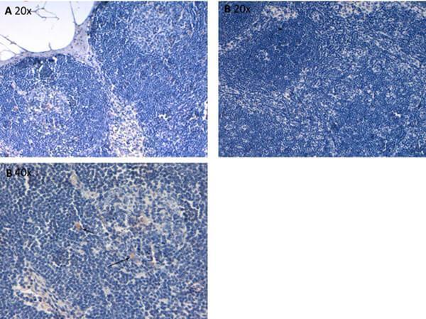
Immunohistochemistry of Rabbit Anti-IL6-Antibody. Tissue: human lymph nodes at pH9. Fixation: formalin fixed paraffin embedded. Antigen retrieval: not required. Primary antibody: Anti-IL-6 antibody at 10 µg/mL for 1 h at RT. Secondary antibody: Peroxidase rabbit secondary antibody at 1:10,000 for 45 min at RT. Localization: IL-6 is cytoplasmic. Staining: IL-6 as precipitated brown signal (B 20x, B 40x) hematoxylin purple nuclear counterstain. Negative Control (A 20x).

Click image to see more details
Western blot using Boster's anti-Human IL6 antibody shows detection of a band ~24 kDa in size corresponding to recombinant human IL6 (arrowhead). Molecular weight markers are also shown. After transfer, the membrane was blocked overnight with 1% BSA in TTBS followed by reaction with primary antibody at a 1:500 dilution. Detection occurred using HRP conjugated anti-Rabbit IgG secondary antibody diluted 1:40,000 in blocking buffer and FemtoMax™ Chemiluminescent substrate . Image was captured using VersaDoc MP 4000 imaging system (Bio-Rad).
Specific Publications For Anti-IL-6 Antibody (A00102-1)
Loading publications
Recommended Resources
Here are featured tools and databases that you might find useful.
- Boster's Pathways Library
- Protein Databases
- Bioscience Research Protocol Resources
- Data Processing & Analysis Software
- Photo Editing Software
- Scientific Literature Resources
- Research Paper Management Tools
- Molecular Biology Software
- Primer Design Tools
- Bioinformatics Tools
- Phylogenetic Tree Analysis
Customer Reviews
Have you used Anti-IL-6 Antibody?
Share your experimental results or join a short interview to earn up to $1,000 in product credits or other rewards.
0 Reviews For Anti-IL-6 Antibody
Customer Q&As
Have a question?
Find answers in Q&As, reviews.
Can't find your answer?
Submit your question
4 Customer Q&As for Anti-IL-6 Antibody
Question
We are currently using anti-IL-6 antibody A00102-1 for human tissue, and we are happy with the IHC results. The species of reactivity given in the datasheet says human. Is it true that the antibody can work on canine tissues as well?
Verified Customer
Verified customer
Asked: 2018-08-10
Answer
The anti-IL-6 antibody (A00102-1) has not been tested for cross reactivity specifically with canine tissues, though there is a good chance of cross reactivity. We have an innovator award program that if you test this antibody and show it works in canine you can get your next antibody for free. Please contact me if I can help you with anything.
Boster Scientific Support
Answered: 2018-08-10
Question
We have observed staining in human left coronary artery. Any tips? Is anti-IL-6 antibody supposed to stain left coronary artery positively?
R. Anderson
Verified customer
Asked: 2017-09-20
Answer
According to literature left coronary artery does express IL6. According to Uniprot.org, IL6 is expressed in left coronary artery, fibroblast, lung, among other tissues. Regarding which tissues have IL6 expression, here are a few articles citing expression in various tissues:
Fibroblast, Pubmed ID: 3758081
Lung, Pubmed ID: 15489334
Boster Scientific Support
Answered: 2017-09-20
Question
Our lab were happy with the WB result of your anti-IL-6 antibody. However we have observed positive staining in lung secreted. using this antibody. Is that expected? Could you tell me where is IL6 supposed to be expressed?
B. Li
Verified customer
Asked: 2016-08-16
Answer
Based on literature, lung does express IL6. Generally IL6 expresses in secreted. Regarding which tissues have IL6 expression, here are a few articles citing expression in various tissues:
Fibroblast, Pubmed ID: 3758081
Lung, Pubmed ID: 15489334
Boster Scientific Support
Answered: 2016-08-16
Question
We ordered your anti-IL-6 antibody for Neu on fibroblast a few months ago. I am using human, and We intend to use the antibody for WB next. We need examining fibroblast as well as lung in our next experiment. Could you please give me some suggestion on which antibody would work the best for WB?
D. Dhar
Verified customer
Asked: 2013-05-24
Answer
I took a look at the website and datasheets of our anti-IL-6 antibody and it seems that A00102-1 has been tested on human in both Neu and WB. Thus A00102-1 should work for your application. Our Boster satisfaction guarantee will cover this product for WB in human even if the specific tissue type has not been validated. We do have a comprehensive range of products for WB detection and you can check out our website bosterbio.com to find out more information about them.
Boster Scientific Support
Answered: 2013-05-24
