Click image to see more details
-

-
-
-
-
+2
Product Info Summary
| SKU: | A00101-3 |
|---|---|
| Size: | 100ug |
| Reactive Species: | Mouse |
| Host: | Rabbit |
| Application: | ELISA, Flow Cytometry, IP, IF, IHC, WB |
Customers Who Bought This Also Bought

Product info
Product Name
Anti-IL1 beta Antibody
SKU/Catalog Number
A00101-3
Size
100ug
Form
Lyophilized
Description
Boster Bio Anti-IL1 beta Antibody (Catalog # A00101-3). Tested in IF, IHC, WB applications. This antibody reacts with Mouse.
Storage & Handling
Store Anti-IL-1 beta antibody at 4°C prior to restoration. For extended storage aliquot contents and freeze at -20°C or below. Avoid cycles of freezing and thawing. Centrifuge product if not completely clear after standing at room temperature. This product is stable for several weeks at 4°C as an undiluted liquid. Dilute only prior to immediate use. Expiration date is six (6) months from date of opening. (Ship at ambient temperature.)
Cite This Product
Anti-IL1 beta Antibody (Boster Biological Technology, Pleasanton CA, USA, Catalog # A00101-3)
Host
Rabbit
Contents
0.02 M Potassium Phosphate, 0.15 M Sodium Chloride, pH 7.2
Clonality
Polyclonal
Isotype
IgG
Immunogen
This antibody was prepared by repeated immunizations with recombinant mouse IL-1ß produced in E.coli. The MW of recombinant mouse IL-1ß was 17 kDa.
Reactive Species
A00101-3 is reactive to IL1B in Mouse
Observed Molecular Weight
42 kDa
Calculated molecular weight
30.9 kDa
Background of IL1B
IL-1 beta (also known as Interleukin-1 beta, IL-1ß and catabolin) is produced by activated macrophages. IL-1 stimulates thymocyte proliferation by inducing IL-2 release, B-cell maturation and proliferation, and fibroblast growth factor activity. IL-1 proteins are involved in the inflammatory response, being identified as endogenous pyrogens, and are reported to stimulate the release of prostaglandin and collagenase from synovial cells. IL-1ß is a monomeric secreted protein that may be released by damaged cells or is secreted by a mechanism differing from that used for other secretory proteins. Anti-IL-1 beta antibody is ideal for investigators involved in Cardiovascular and Immunology research.
Antibody Validation
Boster validates all antibodies on WB, IHC, ICC, Immunofluorescence, and ELISA with known positive control and negative samples to ensure specificity and high affinity, including thorough antibody incubations.
Application & Images
Applications
A00101-3 is guaranteed for ELISA, Flow Cytometry, IP, IF, IHC, WB Boster Guarantee
Recommend Dilution
| Application | Dilution | Species |
|---|---|---|
| ELISA: 1:1 | 000 - 1:5 | 000 |
| WB: 1:500 - 1:2 | 000 | |
| Anti-Mouse IL-1ß has been tested for use in immunohistochemistry | immunoblotting and immunofluorescence. This antibody is useful in ELISA | neutralizations, radioimmunoassays, flow cytometry, and immunoprecipitation. It recognizes the 17,000 MW mature IL-1ß. For immunoblots, typically, IL-1ß is detected from supernatants or lysates of 2 x 10E6 endotoxin-stimulated peripheral blood mononuclear cells (PBMC). PBMC are stimulated for 24 hours with 1% (v/v) serum plus 10 ng/mL E.coli LPS. For immunoprecipitation pre-clearing the preparation with a non-specific Rabbit IgG (p/n 011-001-297) to reduce background is suggested. For immunohistochemistry either paraffin fixation or cryofixation can be used for sample preparation to stain intracellular IL-1ß. For ELISA use HRP Conjugated Anti-Rabbit IgG [H&L] (Goat) (611-1302) for detection. In ELISA formats this antibody is best used as the second antibody in combination with a monoclonal antibody as a capture antibody. This antibody is also useful for neutralization of mouse and rat IL-1ß activity in bioassays. It does not neutralize the biological activity IL-1α. It does not neutralize the biological activity of human or primate IL-1ß. For neutralization, it is recommended to incubate the sample with a dilution of the antibody for at least 4 hours before being tested. A control of similarly diluted normal rabbit IgG is recommended. This antibody can be used for FACS analysis. Caution should be exhibited as the F(c) domain of the rabbit IgG molecule may interact with cells non-specifically. |
Validation Images & Assay Conditions
Click image to see more details
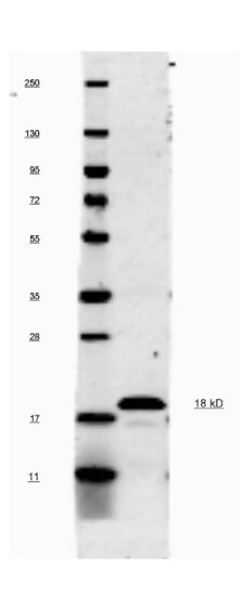
This antibody will recognize 10% of the non-denatured (native) precursor 31,000 MW mouse IL-1ß containing samples but will primarily detect all of the 17,000 MW mature molecule. However, in western blot analysis, the usual procedure of heating the sample in SDS with or without reducing agents will facilitate denaturing of the 31,000 MW IL- 1ß precursor molecule. Denatured IL-1ß will have a 18 kDa band.

Click image to see more details
Immunohistochemistry of Rabbit anti-IL1Beta Antibody in Mouse Embryonic Kidney Tissue: Mouse Embryonic Kidney Fixation: FFPE buffered formalin 10% conc Ag Retrieval: Heat, Citrate pH 6.2. Pressure Cooker Primary antibody: 2ug/ml 1.5 hour @ room T Secondary Ab: MACH 1 HRP POLYMER 1:50 45” RT

Click image to see more details
Immunofluorescence microscopy after staining of mouse carotid artery tissue with anti-Mouse IL-1ß antiserum diluted 1:50. Tissue sections were prepared after cryofixation. Reaction occurred at room temperature for 60' followed by washes and reaction with Rhodamine conjugated Gt-a-Rabbit IgG . Tissue was counterstained with bis-benzimide solution at 0.5 mg/ml in PBS for 15 min at room temperature. Panel A) shows no antibody staining of WT uninjured mouse carotid tissue. Panel B) shows anti-IL-1ß staining of cells after surgical injury of tissue. Panel C) shows no antibody staining of injured carotid tissue from an IL-1ß KO mouse.

Click image to see more details
Production of IL1β occurs through activating NLRP3 inflammasome in an NF-κB-dependent manner. ( A ) Compared with the control, IL1β, NLRP3, caspase-1p20 and ASC all showed increased expression with time, esp in the 48hrs. ( B ) p-p65/p65 ratio increases with time. Control: CAFs were incubated with DMEM and without exposed to the light; Light: CAFs were incubated with DMEM for 5hrs at 37°C and exposed to the light; ALA: CAFs were incubated with ALA (0.5 mM) for 5h at 37°C, but not exposed to the light;12hrs/24hrs/48hrs: CAFs were incubated with ALA (0.5 mM) and exposed to the light. We collected the cell samples in 12hrs/24hrs/48hrs. Notes: *p<0.05; **p<0.01; ***p<0.001.
Index in PubMed under a CC BY license. PMID: 31849516

Click image to see more details
Pseudoephedrine + emodin Inhibited MAPK in LPS-induced acute lung injury in rats. A – D Western blot analysis was performed to detect p-P38, p-ERK1/2 and p-JNK1/2 protein expression. E – I TNF-α, IL-6, IL-1β, IL-10, Arg-1 mRNA expression was determined using Real-time PCR analysis. All data are expressed as mean ± S.D. (n = 3). ## p < 0.01, ### p < 0.001 vs. control group. *p < 0.05, **p < 0.01, ***p < 0.001 vs. LPS alone group. + p < 0.05, ++ p < 0.01, +++ p < 0.001 vs. Combined treatment group (5 + 20 mg/kg)
Index in PubMed under a CC BY license. PMID: 35123524

Click image to see more details
Effects of Pseudoephedrine + emodin on the expression of TNF-α, IL-6, IL-1β, iNOS, IL-10, Arg-1 in LPS‐induced ALI rats. The contents of TNF-α ( A ), IL-6 ( B ), IL-1β ( C ), iNOS ( D ), IL-10 ( E ), Arg-1 ( F ) in the serum and BALF were determined using ELISA. Data were expressed as mean ± S.D. (n = 3). # p < 0.05, ## p < 0.01, ### p < 0.001 vs. control group. *p < 0.05, **p < 0.01, ***p < 0.001 vs. LPS alone group. + p < 0.05, ++ p < 0.01, +++ p < 0.001 vs. combined treatment group (5 + 20 mg/kg)
Index in PubMed under a CC BY license. PMID: 35123524
Specific Publications For Anti-IL1 beta Antibody (A00101-3)
Loading publications
Recommended Resources
Here are featured tools and databases that you might find useful.
- Boster's Pathways Library
- Protein Databases
- Bioscience Research Protocol Resources
- Data Processing & Analysis Software
- Photo Editing Software
- Scientific Literature Resources
- Research Paper Management Tools
- Molecular Biology Software
- Primer Design Tools
- Bioinformatics Tools
- Phylogenetic Tree Analysis
Customer Reviews
Have you used Anti-IL1 beta Antibody?
Share your experimental results or join a short interview to earn up to $1,000 in product credits or other rewards.
0 Reviews For Anti-IL1 beta Antibody
Customer Q&As
Have a question?
Find answers in Q&As, reviews.
Can't find your answer?
Submit your question
15 Customer Q&As for Anti-IL1 beta Antibody
Question
We have seen staining in rat lung. What should we do? Is anti-IL1 beta antibody supposed to stain lung positively?
Verified Customer
Verified customer
Asked: 2019-11-11
Answer
Based on literature lung does express IL1B. Based on Uniprot.org, IL1B is expressed in smooth muscle tissue, leukocyte, histiocytic lymphoma, monocyte, lung, skin, macrophage, among other tissues. Regarding which tissues have IL1B expression, here are a few articles citing expression in various tissues:
Histiocytic lymphoma, Pubmed ID: 3493774
Leukocyte, Pubmed ID: 3490654
Lung, Pubmed ID: 15489334
Macrophage, Pubmed ID: 20148899
Monocyte, Pubmed ID: 2635664, 11991722
Skin, Pubmed ID: 1919436
Boster Scientific Support
Answered: 2019-11-11
Question
I appreciate helping with my inquiry over the phone. Here are the WB image, lot number and protocol we used for lung using anti-IL1 beta antibody A00101-3. Let me know if you need anything else.
Verified Customer
Verified customer
Asked: 2019-09-23
Answer
We appreciate the data. You have provided everything we needed. Our lab team are working to resolve your inquiry as quickly as possible, and we appreciate your patience and understanding! Please let me know if there is anything you need in the meantime.
Boster Scientific Support
Answered: 2019-09-23
Question
We are interested in to test anti-IL1 beta antibody A00101-3 on mouse lung for research purposes, then I may be interested in using anti-IL1 beta antibody A00101-3 for diagnostic purposes as well. Is the antibody suitable for diagnostic purposes?
Verified Customer
Verified customer
Asked: 2019-09-12
Answer
The products we sell, including anti-IL1 beta antibody A00101-3, are only intended for research use. They would not be suitable for use in diagnostic work. If you have the means to develop a product into diagnostic use, and are interested in collaborating with us and develop our product into an IVD product, please contact us for more discussions.
Boster Scientific Support
Answered: 2019-09-12
Question
I see that the anti-IL1 beta antibody A00101-3 works with IHC, what is the protocol used to produce the result images on the product page?
Verified Customer
Verified customer
Asked: 2019-08-08
Answer
You can find protocols for IHC on the "support/technical resources" section of our navigation menu. If you have any further questions, please send an email to support@bosterbio.com
Boster Scientific Support
Answered: 2019-08-08
Question
I was wanting to use your anti-IL1 beta antibody for IHC for mouse lung on frozen tissues, but I want to know if it has been tested for this particular application. Has this antibody been tested and is this antibody a good choice for mouse lung identification?
Verified Customer
Verified customer
Asked: 2019-03-15
Answer
You can see on the product datasheet, A00101-3 anti-IL1 beta antibody has been validated for IP, IF, IHC, WB on mouse, rat tissues. We have an innovator award program that if you test this antibody and show it works in mouse lung in IHC-frozen, you can get your next antibody for free.
Boster Scientific Support
Answered: 2019-03-15
Question
We are currently using anti-IL1 beta antibody A00101-3 for mouse tissue, and we are well pleased with the WB results. The species of reactivity given in the datasheet says mouse, rat. Is it possible that the antibody can work on primate tissues as well?
Verified Customer
Verified customer
Asked: 2018-12-20
Answer
The anti-IL1 beta antibody (A00101-3) has not been tested for cross reactivity specifically with primate tissues, but there is a good chance of cross reactivity. We have an innovator award program that if you test this antibody and show it works in primate you can get your next antibody for free. Please contact me if I can help you with anything.
Boster Scientific Support
Answered: 2018-12-20
Question
I have a question about product A00101-3, anti-IL1 beta antibody. I was wondering if it would be possible to conjugate this antibody with biotin. I would need it to be without BSA or sodium azide. I am planning on using a buffer exchange of sodium azide with PBS only. Would there be problems for me to conjugate the antibody and store it in -20 degrees in small aliquots?
Verified Customer
Verified customer
Asked: 2018-10-05
Answer
We do not recommend storing this antibody with PBS buffer only in -20 degrees. If you want to store it in -20 degrees it is best to add some cryoprotectant like glycerol. If you want carrier free A00101-3 anti-IL1 beta antibody, we can provide it to you in a special formula with trehalose and/or glycerol. These molecules will not interfere with conjugation chemistry and provide a good level of protection for the antibody from degradation. Please be sure to specify this in your purchase order.
Boster Scientific Support
Answered: 2018-10-05
Question
See below the WB image, lot number and protocol we used for lung using anti-IL1 beta antibody A00101-3. Please let me know if you require anything else.
Verified Customer
Verified customer
Asked: 2018-05-04
Answer
Thank you very much for the data. Our lab team are working to resolve this as quickly as possible, and we appreciate your patience and understanding! You have provided everything we needed. Please let me know if there is anything you need in the meantime.
Boster Scientific Support
Answered: 2018-05-04
Question
Is there a BSA free version of anti-IL1 beta antibody A00101-3 available?
Verified Customer
Verified customer
Asked: 2018-01-01
Answer
I appreciate your recent telephone inquiry. I can confirm that some lots of this anti-IL1 beta antibody A00101-3 are BSA free. For now, these lots are available and we can make a BSA free formula for you free of charge. It will take 3 extra days to prepare. If you require this antibody BSA free again in future, please do not hesitate to contact me and I will be pleased to check which lots we have in stock that are BSA free.
Boster Scientific Support
Answered: 2018-01-01
Question
Will A00101-3 anti-IL1 beta antibody work on parafin embedded sections? If so, which fixation method do you recommend we use (PFA, paraformaldehyde, other)?
Verified Customer
Verified customer
Asked: 2017-07-25
Answer
It shows on the product datasheet, A00101-3 anti-IL1 beta antibody as been validated on IHC. It is best to use PFA for fixation because it has better tissue penetration ability. PFA needs to be prepared fresh before use. Long term stored PFA turns into formalin, as the PFA molecules congregate and become formalin.
Boster Scientific Support
Answered: 2017-07-25
Question
Is a blocking peptide available for product anti-IL1 beta antibody (A00101-3)?
H. Patel
Verified customer
Asked: 2017-06-15
Answer
We do provide the blocking peptide for product anti-IL1 beta antibody (A00101-3). If you would like to place an order for it please contact support@bosterbio.com and make a special request.
Boster Scientific Support
Answered: 2017-06-15
Question
Our lab want to know about using your anti-IL1 beta antibody for leukocyte migration studies. Has this antibody been tested with western blotting on carotid artery tissue? We would like to see some validation images before ordering.
C. Zhang
Verified customer
Asked: 2017-02-03
Answer
We appreciate your inquiry. This A00101-3 anti-IL1 beta antibody is validated on carotid artery tissue. It is guaranteed to work for IP, IF, IHC, WB in mouse, rat. Our Boster guarantee will cover your intended experiment even if the sample type has not been be directly tested.
Boster Scientific Support
Answered: 2017-02-03
Question
Will anti-IL1 beta antibody A00101-3 work for IHC with lung?
P. Williams
Verified customer
Asked: 2016-06-03
Answer
According to the expression profile of lung, IL1B is highly expressed in lung. So, it is likely that anti-IL1 beta antibody A00101-3 will work for IHC with lung.
Boster Scientific Support
Answered: 2016-06-03
Question
My colleagues were satisfied with the WB result of your anti-IL1 beta antibody. However we have observed positive staining in histiocytic lymphoma cytoplasm using this antibody. Is that expected? Could you tell me where is IL1B supposed to be expressed?
C. Zhao
Verified customer
Asked: 2013-05-30
Answer
According to literature, histiocytic lymphoma does express IL1B. Generally IL1B expresses in cytoplasm, cytosol. Regarding which tissues have IL1B expression, here are a few articles citing expression in various tissues:
Histiocytic lymphoma, Pubmed ID: 3493774
Leukocyte, Pubmed ID: 3490654
Lung, Pubmed ID: 15489334
Macrophage, Pubmed ID: 20148899
Monocyte, Pubmed ID: 2635664, 11991722
Skin, Pubmed ID: 1919436
Boster Scientific Support
Answered: 2013-05-30
Question
Our lab used your anti-IL1 beta antibody for IP on smooth muscle tissue in the past. I am using mouse, and I plan to use the antibody for IF next. I would like examining smooth muscle tissue as well as histiocytic lymphoma in our next experiment. Could you please give me some suggestion on which antibody would work the best for IF?
J. Banerjee
Verified customer
Asked: 2013-04-23
Answer
I looked at the website and datasheets of our anti-IL1 beta antibody and it seems that A00101-3 has been validated on mouse in both IP and IF. Thus A00101-3 should work for your application. Our Boster satisfaction guarantee will cover this product for IF in mouse even if the specific tissue type has not been validated. We do have a comprehensive range of products for IF detection and you can check out our website bosterbio.com to find out more information about them.
Boster Scientific Support
Answered: 2013-04-23
