
Click image to see more details
-

-
-
-
-
+8
Product Info Summary
| SKU: | M00003 |
|---|---|
| Size: | 100 μl |
| Reactive Species: | Human, Mouse, Rat |
| Host: | Rabbit |
| Application: | Flow Cytometry, IP, IF, IHC, ICC, WB |
Customers Who Bought This Also Bought

Product info
Product Name
Anti-mTOR/Tor Rabbit Monoclonal Antibody
SKU/Catalog Number
M00003
BM4182 is an alternative SKU for this antibody, used in previous lots.
Size
100 μl
Form
Liquid
Description
Boster Bio Anti-mTOR/Tor Rabbit Monoclonal Antibody catalog # M00003. Tested in WB, IHC, ICC/IF, IP, Flow Cytometry applications. This antibody reacts with Human, Mouse, Rat.
Storage & Handling
Store at -20°C for one year. For short term storage and frequent use, store at 4°C for up to one month. Avoid repeated freeze-thaw cycles.
Cite This Product
Anti-mTOR/Tor Rabbit Monoclonal Antibody (Boster Biological Technology, Pleasanton CA, USA, Catalog # M00003)
Host
Rabbit
Contents
Rabbit IgG in stabilizing components, phosphate buffered saline, pH 7.4, 150mM NaCl, 0.02% sodium azide and 50% glycerol.
*This antibody is supplied in a stabilized formulation.
Compatibility with conjugation reactions depends on the chemistry of the conjugation method used.
For conjugation methods that are not compatible with the stabilizing components present in this formulation, a carrier-free antibody format is required.
Clonality
Monoclonal
Clone Number
CBD-13
Isotype
Rabbit IgG
Immunogen
A synthesized peptide derived from human mTOR
Reactive Species
M00003 is reactive to MTOR in Human, Mouse, Rat
Observed Molecular Weight
289 kDa
Calculated molecular weight
288.9 kDa
Antibody Validation
Boster validates all antibodies on WB, IHC, ICC, Immunofluorescence, and ELISA with known positive control and negative samples to ensure specificity and high affinity, including thorough antibody incubations.
Application & Images
Applications
M00003 is guaranteed for Flow Cytometry, IP, IF, IHC, ICC, WB Boster Guarantee
Recommend Dilution
WB 1:500-2000
IHC 1:50-200
ICC/IF 1:50-200
IP 1:30
FC 1:20
Tested application
Suggested blocking solution with 5% non-fat milk or BSA; (*)Recommended protein loading: 20-40 µg per lane
Use TE buffer pH 9.0 for antigen retrieval; (*) citrate buffer pH 6.0 is an alternative.
Validation Images & Assay Conditions
Click image to see more details
Western blot analysis of MTOR using anti-MTOR antibody (M00003).
Electrophoresis was performed on a 5-20% SDS-PAGE gel at 70V (Stacking gel) / 90V (Resolving gel) for 2-3 hours. The sample well of each lane was loaded with 30 ug of sample under reducing conditions.
Lane 1: human 293T whole cell lysates,
Lane 2: human Hela whole cell lysates,
Lane 3: human K562 whole cell lysates,
Lane 4: rat brain tissue lysates,
Lane 5: rat C6 whole cell lysates,
Lane 6: mouse brain tissue lysates,
Lane 7: mouse Neuro-2a whole cell lysates.
After electrophoresis, proteins were transferred to a nitrocellulose membrane at 150 mA for 50-90 minutes. Blocked the membrane with 5% non-fat milk/TBS for 1.5 hour at RT. The membrane was incubated with rabbit anti-MTOR antigen affinity purified monoclonal antibody (Catalog # M00003) at 1:500 overnight at 4°C, then washed with TBS-0.1%Tween 3 times with 5 minutes each and probed with a goat anti-rabbit IgG-HRP secondary antibody at a dilution of 1:500 for 1.5 hour at RT. The signal is developed using an Enhanced Chemiluminescent detection (ECL) kit (Catalog # EK1002) with Tanon 5200 system. A specific band was detected for MTOR at approximately 289 kDa. The expected band size for MTOR is at 289 kDa.

Click image to see more details
Western blot analysis of mTOR/Tor using anti-mTOR/Tor antibody (M00003).
Electrophoresis was performed on a 5-20% SDS-PAGE gel at 80V (Stacking gel) / 120V (Resolving gel) for 2 hours. The sample well of each lane was loaded with 30 ug of sample under reducing conditions.
Lane 1-2: human OCI-LY1-Hmgb1 SRPK1 KOwhole cell lysates.
After electrophoresis, proteins were transferred to a nitrocellulose membrane at 150 mA for 50-90 minutes. Blocked the membrane with 5% non-fat milk/TBS for 1.5 hour at RT. The membrane was incubated with rabbit anti-mTOR/Tor antigen affinity purified polyclonal antibody (M00003) at 1:3000 overnight at 4°C, then washed with TBS-0.1%Tween 3 times with 5 minutes each and probed with a goat anti-rabbit IgG-HRP secondary antibody (Catalog # BA1054) at a dilution of 1:5000 for 1.5 hour at RT. The signal is developed using an ECL Plus Western Blotting Substrate (Catalog # AR1196-200) with Tanon 5200 system. A specific band was detected for mTOR/Tor at approximately 289 kDa. The expected band size for mTOR/Tor is at 289kDa.

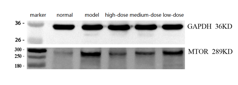
Click image to see more details
Western blot analysis of mTOR/Tor using anti-mTOR/Tor antibody (M00003).
Electrophoresis was performed on a 5-20% SDS-PAGE gel at 80V (Stacking gel) / 120V (Resolving gel) for 2 hours. The sample well of each lane was loaded with 30 ug of sample under reducing conditions.
Lane 1: normal group-Rat skeletal muscle tissue lysates,
Lane 2: model group-Rat skeletal muscle tissue lysates,
Lane 3: high-dose group-Rat skeletal muscle tissue lysates,
Lane 4: medium-dose group-Rat skeletal muscle tissue lysates,
Lane 5: low-dose group-Rat skeletal muscle tissue lysates.
After electrophoresis, proteins were transferred to a nitrocellulose membrane at 150 mA for 50-90 minutes. Blocked the membrane with 5% non-fat milk/TBS for 1.5 hour at RT. The membrane was incubated with rabbit anti-mTOR/Tor antigen affinity purified polyclonal antibody (M00003) at 1:2000 overnight at 4°C, then washed with TBS-0.1%Tween 3 times with 5 minutes each and probed with a goat anti-rabbit IgG-HRP secondary antibody (Catalog # BA1054) at a dilution of 1:10,000 for 1 hour at RT. The signal is developed using an ECL Plus Western Blotting Substrate (Catalog # AR1196-200) with ChemiDoc MP system. A specific band was detected for mTOR/Tor at approximately 289 kDa. The expected band size for mTOR/Tor is at 289kDa.

Click image to see more details
Western blot analysis of MTOR using anti-MTOR antibody (M00003).
Electrophoresis was performed on a 5-20% SDS-PAGE gel at 70V (Stacking gel) / 90V (Resolving gel) for 2-3 hours. The sample well of each lane was loaded with 30 ug of sample under reducing conditions.
Lane 1: mouse 4T1 whole cell lysates,
Lane 2: LPS-stimulated mouse 4T1 whole cell lysates,
Lane 3: Low-dose drug mouse 4T1 whole cell lysates,
Lane 4: Medium-dose drug mouse 4T1 whole cell lysates,
Lane 5: High-dose drug mouse 4T1 whole cell lysates,
Lane 6: Positive control drug mouse 4T1 whole cell lysates.
After electrophoresis, proteins were transferred to a nitrocellulose membrane at 150 mA for 50-90 minutes. Blocked the membrane with 5% non-fat milk/TBS for 1.5 hour at RT. The membrane was incubated with rabbit anti-MTOR antigen affinity purified monoclonal antibody (Catalog # M00003) at 1:10000 overnight at 4°C, then washed with TBS-0.1%Tween 3 times with 5 minutes each and probed with a goat anti-rabbit IgG-HRP secondary antibody at a dilution of 1:10000 for 1 hour at RT. The signal is developed using an Enhanced Chemiluminescent detection (ECL) kit with ChemiDoc MP system. A specific band was detected for MTOR at approximately 289 kDa. The expected band size for MTOR is at 289 kDa.

Click image to see more details
Immunohistochemical analysis of paraffin-embedded rat liver, using mTOR Antibody.

Click image to see more details
PI3K/Akt/mTOR pathway is involved in the cardioprotective effects of LLC. (A) Expression levels of PI3K, p-Akt, Akt, p-mTOR and mTOR in hearts were analyzed. (B) Representative western blots of PI3K (p110α), p-Akt (Ser473), Akt, p-mTOR (Ser2448), mTOR, LC3, Beclin 1 and p62 in the presence or absence of 20 μmol⋅L -1 LY294002 and 40 μg⋅mL -1 LLC. (C) The cell viability was analyzed by MTT assay. (D) The cell injury was detected by LDH measurements. All experiments were repeated at least three times. Data were presented as means ± SD. ∗∗ P < 0.01 vs. Con group, ## P < 0.01 vs. OGD group, & P < 0.05, && P < 0.01 vs. OGD+LLC group.
Index in PubMed under a CC BY license. PMID: 29651246

Click image to see more details
Verification of the effect of Pso on PDLSCs. (A) Representative protein expression bands for mTOR, OCN, RUNX2, ALP, and GADPH. (B–F) : Expression of LC3, PI3K, Rag C, WIPI 1, and Deptor genes. CK, blank control; L, LPS group; P, Pso group; LP, Pso/LPS co-culture group. Data are presented as mean ± SD. * P < 0.05, ** P < 0.01, *** P < 0.001, and **** P < 0.0001 vs. control group.
Index in PubMed under a CC BY license. PMID: 40718710

Click image to see more details
Relationship between ciRNA13761/novel-miR-3880/ ELF2 axis and PI3K/AKT/mTOR/S6K1 pathway. (A) Schematic diagram of animal treatment. C57BL/6 mice were injected with novel-miR-3880 or si ELF2 in an interval of three days and four days alternatively. Samples were harvested at day 22. (B) Immunohistochemistry of mammary gland for p-PI3K, p-AKT, p-mTOR and p-S6K1 in Normal Saline, novel-miR-3880 and si ELF2 groups. (C) Protein phosphorylation level of PI3K, AKT, mTOR and S6K1 in mammary gland. (D,E) Effects of novel-miR-3880 and ELF2 on Bcl2/Bax pathway and protein phosphorylation level of PI3K, AKT, mTOR and S6K1 in MEC. (F) PI3K, AKT, mTOR and S6K1 inhibitors suppressed the phosphorylation of PI3K, AKT, mTOR and S6K1 in MEC. (G–J) The role of novel-miR-3880 and si ELF2 in Bcl2/Bax and protein phosphorylation level of PI3K, AKT, mTOR and S6K1 in MEC with PI3K, AKT, mTOR or S6K1 inhibited. (K) Regulation of ciRNA13761 on Bcl2/Bax and PI3K, AKT, mTOR and S6K1 phosphorylation, and the balance effects of novel-miR-3880. (L) Effects of si DOCK1 on Bcl2/Bax and PI3K, AKT, mTOR and S6K1 phosphorylation.
Index in PubMed under a CC BY license. PMID: 32656203

Click image to see more details
(A) WB results for mTOR, RILP, LC3 and P62. (B) Relative expression levels of mTOR protein. (C) Relative expression levels of RILP protein. (D) The ratio of LC3II to LC3I. (E) Relative expression levels of P62 protein. (*: P < 0.05, **: P < 0.01, ***: P < 0.001, ****: P < 0.0001).
Index in PubMed under a CC BY license. PMID: 39958873

Click image to see more details
Eriocitrin and quercetin are responsible for anti-proliferation by targeting the PI3K protein in PASMCs under hypoxic conditions. ERI, eriocitrin; QCT, quercetin. (A, B) Primitive bands and quantitative evaluation of p-mTOR, mTOR, p-AKT1 (Ser473), and AKT1 with or without PS210 (2 μM) by Western blotting in PASMCs under 3% O 2 . n = 3. All data are represented as the mean ± SD. * p < 0.05 vs. control group, # p < 0.05 vs. 3% O 2 group, and p < 0.05 vs. 3% O 2 + FLA-50 μg/ml group. (C–G) Primitive bands and quantitative densities of p-PI3K and PI3K by Western blots. n = 3. All data are represented as the mean ± SD. * p < 0.05 vs. control group and # p < 0.05 vs. 3% O 2 group. (H–N) BECC, ERI, and QCT treatment increased the stability of PI3K in PASMC protease lysates by the DARTS experiment. (H–K) Primitive Western blots of PI3K. (L–N) Quantitative evaluation of PI3K levels. n = 3. All data are represented as the mean ± SD. * p < 0.05 vs. DMSO group.
Index in PubMed under a CC BY license. PMID: 40385484

Click image to see more details
BECCs regulate PI3K, PDPK1, and mTOR protein levels in HAPH rats. (A–C) Primitive bands of p-PI3K, PI3K, p-PDPK1, PDPK1, p-mTOR, and mTOR by Western blots in lung tissues. (D–F) Quantitative evaluation of p-PI3K, PI3K, p-PDPK1, PDPK1, p-mTOR, and mTOR in lung tissues. n = 5. All data are represented as the mean ± SD. * p < 0.05 vs. control group and # p < 0.05 vs. hypoxia group.
Index in PubMed under a CC BY license. PMID: 40385484

Click image to see more details
Expression of related genes and proteins after the addition of mTOR inhibitor. (A) Expression level of the mTOR gene after adding the inhibitor rapamycin. (B) Change in the expression levels of the Rag C gene. (C–D) Changes in the expression levels of the osteogenesis-related genes ALP and RUNX2. (E) Protein expression levels for of Rag C, Deptor, ALP, and RUNX2. CK, control group; mTOR, mTOR-inhibitor group. (F) Relative expression levels of Rag C, ALP, and RUNX2 proteins. mTOR, mTOR-inhibitor group; Pso, mTOR inhibitor + Pso group. (G) Representative images of PDLSCs with Rag C lentiviral knockdown; green fluorescence indicates the lentiviral vector particles. (H–I) Expression changes in ALP and RUNX2 in PDLSCs with Rag C knockdown. CK, control group; Rag C, PDLSCs (scale bar = 200 μm). Data are presented as mean ± SD. *P < 0.05, **P < 0.01, ***P < 0.001, and ****P < 0.0001 vs. the control group.
Index in PubMed under a CC BY license. PMID: 40718710
Specific Publications For Anti-mTOR/Tor Rabbit Monoclonal Antibody (M00003)
Loading publications
Recommended Resources
Here are featured tools and databases that you might find useful.
- Boster's Pathways Library
- Protein Databases
- Bioscience Research Protocol Resources
- Data Processing & Analysis Software
- Photo Editing Software
- Scientific Literature Resources
- Research Paper Management Tools
- Molecular Biology Software
- Primer Design Tools
- Bioinformatics Tools
- Phylogenetic Tree Analysis
Customer Reviews
Have you used Anti-mTOR/Tor Rabbit Monoclonal Antibody?
Share your experimental results or join a short interview to earn up to $1,000 in product credits or other rewards.
3 Reviews For Anti-mTOR/Tor Rabbit Monoclonal Antibody
MTOR Antibody (M00003) WB on rat skeletal muscle shows clear bands, with higher MTOR in the model and dose-dependent reduction in treatment groups.
Excellent
| SKU | M00003 |
|---|---|
| Application | Western Blot |
| Sample | Rat skeletal muscle tissue |
| Sample Processing Description | Total protein was extracted from rat skeletal muscle tissue: ① normal, ② injury model, ③ high-dose drug treatment, ④ medium-dose drug treatment, ⑤ low-dose drug treatment. |
| Other Reagents | RIPA lysis buffer, Protease inhibitor, Running buffer, Transfer buffer, Blocking buffer |
| Primary Antibody | mTOR/Tor Rabbit Monoclonal Antibody |
| Primary Incubation | 1:2000, overnight at 4 ℃ |
| Secondary Antibody | HRP Conjugated AffiniPure Goat Anti-Rabbit IgG (H+L) (BA1054) |
| Secondary Incubation | 1:10000, 1 h in RT |
| Detection | Substrate: ECL substrate, Image system:ChemiDoc MP |
| Results Summary | The results show clear MTOR bands, with higher levels in the injury model compared to normal, and a dose-dependent decrease in the treatment groups, as expected. |
Guangtian Yu, Ningxia Medical University
Verified customer
Submitted 2026-03-24
The antibody shows a clear target, correct localization, minimal background, and overall good results.
Excellent

| SKU | M00003 |
|---|---|
| Application | Western Blot |
| Sample | mouse 4T1 cells |
| Sample Processing Description | normal 4T1 cells, LPS-stimulated 4T1 cells, cells treated with low, medium, or high doses of the drug, and cells treated with a positive control drug. |
| Other Reagents | RIPA lysis buffer, Protease inhibitor, Electrophoresis buffer, Transfer buffer, Blocking buffer |
| Primary Antibody | Anti-ATF4 Rabbit Monoclonal Antibody |
| Primary Incubation | 1:10000, overnight at 4 ℃ |
| Secondary Antibody | HRP Goat Anti-Rabbit IgG |
| Secondary Incubation | 1:10000, 1 hour in room temperature |
| Detection | Substrate: ECL, Imaging system:ChemiDoc MP |
| Results Summary | With equal loading amounts, the total MTOR protein levels were similar across all groups. However, the levels of phosphorylated MTOR showed significant changes: the blank group had the lowest, the model group the highest, and in the drug-treated groups, the phosphorylation levels decreased sequentially from low, medium, to high dose. The positive control group was comparable to the high-dose group. |
Bolan Guan, Harbin Medical University
Verified customer
Submitted 2025-12-01
Cell samples were lysed by sonication in RIPA buffer containing protease and phosphatase inhibitors, followed by centrifugation for 10 minutes. The supernatant was mixed with loading buffer at a 4:1 ratio and boiled for 10 minutes. Fifteen microliters of
Excellent

| SKU | M00003 |
|---|---|
| Application | Western Blot |
| Sample | human OCI-LY1 cell |
| Sample Processing Description | Cell samples were lysed by sonication in RIPA buffer containing protease and phosphatase inhibitors, followed by centrifugation for 10 minutes. The supernatant was mixed with loading buffer at a 4:1 ratio and boiled for 10 minutes. Fifteen microliters of each sample were loaded per well. |
| Other Reagents | 5% Non-fat milk |
| Primary Antibody | mTOR/Tor Rabbit Monoclonal Antibody |
| Primary Incubation | 1:3000, overnight at 4 ℃ |
| Secondary Antibody | HRP-conjugated Anti-Rabbit IgG Secondary Antibody |
| Secondary Incubation | 1 hour in room temperature |
| Detection | Substrate: ECL reagent, Imaging system:ChemiDoc MP |
| Results Summary | This antibody is highly specific and efficient, with a clean background and no nonspecific bands. The target band has sharp and well-defined edges. |
Maolin Yao, Fujian Medical University
Verified customer
Submitted 2025-11-11
Customer Q&As
Have a question?
Find answers in Q&As, reviews.
Can't find your answer?
Submit your question
17 Customer Q&As for Anti-mTOR/Tor Rabbit Monoclonal Antibody
Question
Would anti-mTOR/Tor Rabbit Monoclonal antibody M00003 work on pig IHC with testis?
Verified Customer
Verified customer
Asked: 2020-02-27
Answer
Our lab technicians have not tested anti-mTOR/Tor Rabbit Monoclonal antibody M00003 on pig. You can run a BLAST between pig and the immunogen sequence of anti-mTOR/Tor Rabbit Monoclonal antibody M00003 to see if they may cross-react. If the sequence homology is close, then you can perform a pilot test. Keep in mind that since we have not validated pig samples, this use of the antibody is not covered by our guarantee. However we have an innovator award program that if you test this antibody and show it works in pig testis in IHC, you can get your next antibody for free.
Boster Scientific Support
Answered: 2020-02-27
Question
I see that the anti-mTOR/Tor Rabbit Monoclonal antibody M00003 works with WB, what is the protocol used to produce the result images on the product page?
Verified Customer
Verified customer
Asked: 2020-01-22
Answer
You can find protocols for WB on the "support/technical resources" section of our navigation menu. If you have any further questions, please send an email to support@bosterbio.com
Boster Scientific Support
Answered: 2020-01-22
Question
Is a blocking peptide available for product anti-mTOR/Tor Rabbit Monoclonal antibody (M00003)?
Verified Customer
Verified customer
Asked: 2019-12-23
Answer
We do provide the blocking peptide for product anti-mTOR/Tor Rabbit Monoclonal antibody (M00003). If you would like to place an order for it please contact support@bosterbio.com and make a special request.
Boster Scientific Support
Answered: 2019-12-23
Question
Do you have a BSA free version of anti-mTOR/Tor Rabbit Monoclonal antibody M00003 available?
Verified Customer
Verified customer
Asked: 2019-11-13
Answer
We appreciate your recent telephone inquiry. I can confirm that some lots of this anti-mTOR/Tor Rabbit Monoclonal antibody M00003 are BSA free. For now, these lots are available and we can make a BSA free formula for you free of charge. It will take 3 extra days to prepare. If you require this antibody BSA free again in future, please do not hesitate to contact me and I will be pleased to check which lots we have in stock that are BSA free.
Boster Scientific Support
Answered: 2019-11-13
Question
I was wanting to use your anti-mTOR/Tor Rabbit Monoclonal antibody for WB for mouse cerebellum on frozen tissues, but I want to know if it has been tested for this particular application. Has this antibody been tested and is this antibody a good choice for mouse cerebellum identification?
Verified Customer
Verified customer
Asked: 2019-10-25
Answer
It shows on the product datasheet, M00003 anti-mTOR/Tor Rabbit Monoclonal antibody has been validated for Flow Cytometry, IP, IF, IHC, ICC, WB on human, mouse, rat tissues. We have an innovator award program that if you test this antibody and show it works in mouse cerebellum in IHC-frozen, you can get your next antibody for free.
Boster Scientific Support
Answered: 2019-10-25
Question
We have seen staining in rat cervix carcinoma. What should we do? Is anti-mTOR/Tor Rabbit Monoclonal antibody supposed to stain cervix carcinoma positively?
Verified Customer
Verified customer
Asked: 2019-09-16
Answer
From what I have seen in literature cervix carcinoma does express MTOR. From what I have seen in Uniprot.org, MTOR is expressed in testis, brain, cerebellum, b-cell, cervix carcinoma, cervix carcinoma erythroleukemia, liver, among other tissues. Regarding which tissues have MTOR expression, here are a few articles citing expression in various tissues:
B-cell, Pubmed ID: 7809080
Brain, Pubmed ID: 8008069
Cerebellum, Pubmed ID: 15489334
Cervix carcinoma, Pubmed ID: 18669648, 18691976, 20068231
Cervix carcinoma, and Erythroleukemia, Pubmed ID: 23186163
Liver, Pubmed ID: 24275569
Boster Scientific Support
Answered: 2019-09-16
Question
Would anti-mTOR/Tor Rabbit Monoclonal antibody M00003 work for WB with cerebellum?
Verified Customer
Verified customer
Asked: 2019-07-26
Answer
According to the expression profile of cerebellum, MTOR is highly expressed in cerebellum. So, it is likely that anti-mTOR/Tor Rabbit Monoclonal antibody M00003 will work for WB with cerebellum.
Boster Scientific Support
Answered: 2019-07-26
Question
you antibody using your anti-mTOR/Tor Rabbit Monoclonal antibody for pip3 activates akt signaling studies. Has this antibody been tested with western blotting on jurkat cell lysate? We would like to see some validation images before ordering.
Verified Customer
Verified customer
Asked: 2019-07-18
Answer
We appreciate your inquiry. This M00003 anti-mTOR/Tor Rabbit Monoclonal antibody is validated on jurkat cell lysate. It is guaranteed to work for Flow Cytometry, IP, IF, IHC, ICC, WB in human, mouse, rat. Our Boster guarantee will cover your intended experiment even if the sample type has not been be directly tested.
Boster Scientific Support
Answered: 2019-07-18
Question
Does M00003 anti-mTOR/Tor Rabbit Monoclonal antibody work on parafin embedded sections? If so, which fixation method do you recommend we use (PFA, paraformaldehyde, other)?
Verified Customer
Verified customer
Asked: 2019-07-11
Answer
As indicated on the product datasheet, M00003 anti-mTOR/Tor Rabbit Monoclonal antibody as been validated on WB. It is best to use PFA for fixation because it has better tissue penetration ability. PFA needs to be prepared fresh before use. Long term stored PFA turns into formalin, as the PFA molecules congregate and become formalin.
Boster Scientific Support
Answered: 2019-07-11
Question
Our team were satisfied with the WB result of your anti-mTOR/Tor Rabbit Monoclonal antibody. However we have observed positive staining in liver endoplasmic reticulum membrane using this antibody. Is that expected? Could you tell me where is MTOR supposed to be expressed?
Verified Customer
Verified customer
Asked: 2019-06-04
Answer
Based on literature, liver does express MTOR. Generally MTOR expresses in endoplasmic reticulum membrane. Regarding which tissues have MTOR expression, here are a few articles citing expression in various tissues:
B-cell, Pubmed ID: 7809080
Brain, Pubmed ID: 8008069
Cerebellum, Pubmed ID: 15489334
Cervix carcinoma, Pubmed ID: 18669648, 18691976, 20068231
Cervix carcinoma, and Erythroleukemia, Pubmed ID: 23186163
Liver, Pubmed ID: 24275569
Boster Scientific Support
Answered: 2019-06-04
Question
I am looking for to test anti-mTOR/Tor Rabbit Monoclonal antibody M00003 on mouse cerebellum for research purposes, then I may be interested in using anti-mTOR/Tor Rabbit Monoclonal antibody M00003 for diagnostic purposes as well. Is the antibody suitable for diagnostic purposes?
N. Brown
Verified customer
Asked: 2019-05-24
Answer
The products we sell, including anti-mTOR/Tor Rabbit Monoclonal antibody M00003, are only intended for research use. They would not be suitable for use in diagnostic work. If you have the means to develop a product into diagnostic use, and are interested in collaborating with us and develop our product into an IVD product, please contact us for more discussions.
Boster Scientific Support
Answered: 2019-05-24
Question
Is this M00003 anti-mTOR/Tor Rabbit Monoclonal antibody reactive to the isotypes of MTOR?
Verified Customer
Verified customer
Asked: 2018-08-15
Answer
The immunogen of M00003 anti-mTOR/Tor Rabbit Monoclonal antibody is A synthesized peptide derived from human mTOR. Could you tell me which isotype you are interested in so I can help see if the immunogen is part of this isotype?
Boster Scientific Support
Answered: 2018-08-15
Question
We are currently using anti-mTOR/Tor Rabbit Monoclonal antibody M00003 for rat tissue, and we are happy with the IP results. The species of reactivity given in the datasheet says human, mouse, rat. Is it likely that the antibody can work on horse tissues as well?
V. Baker
Verified customer
Asked: 2018-04-12
Answer
The anti-mTOR/Tor Rabbit Monoclonal antibody (M00003) has not been tested for cross reactivity specifically with horse tissues, but there is a good chance of cross reactivity. We have an innovator award program that if you test this antibody and show it works in horse you can get your next antibody for free. Please contact me if I can help you with anything.
Boster Scientific Support
Answered: 2018-04-12
Question
We appreciate helping with my inquiry over the phone. Here are the WB image, lot number and protocol we used for cerebellum using anti-mTOR/Tor Rabbit Monoclonal antibody M00003. Let me know if you need anything else.
Verified Customer
Verified customer
Asked: 2017-07-11
Answer
Thank you for the data. You have provided everything we needed. Our lab team are working to resolve your inquiry as quickly as possible, and we appreciate your patience and understanding! Please let me know if there is anything you need in the meantime.
Boster Scientific Support
Answered: 2017-07-11
Question
Our lab used your anti-mTOR/Tor Rabbit Monoclonal antibody for IF on testis in the past. I am using mouse, and We want to use the antibody for ICC next. Our lab want to know about examining testis as well as liver in our next experiment. Could give a recommendation on which antibody would work the best for ICC?
A. Huang
Verified customer
Asked: 2017-01-23
Answer
I have checked the website and datasheets of our anti-mTOR/Tor Rabbit Monoclonal antibody and I see that M00003 has been validated on mouse in both IF and ICC. Thus M00003 should work for your application. Our Boster satisfaction guarantee will cover this product for ICC in mouse even if the specific tissue type has not been validated. We do have a comprehensive range of products for ICC detection and you can check out our website bosterbio.com to find out more information about them.
Boster Scientific Support
Answered: 2017-01-23
Question
Please see the WB image, lot number and protocol we used for cerebellum using anti-mTOR/Tor Rabbit Monoclonal antibody M00003. Please let me know if you require anything else.
L. Collins
Verified customer
Asked: 2016-09-15
Answer
Thank you very much for the data. Our lab team are working to resolve this as quickly as possible, and we appreciate your patience and understanding! You have provided everything we needed. Please let me know if there is anything you need in the meantime.
Boster Scientific Support
Answered: 2016-09-15
Question
Can you help my question with product M00003, anti-mTOR/Tor Rabbit Monoclonal antibody. I was wondering if it would be possible to conjugate this antibody with biotin. I would need it to be without BSA or sodium azide. I am planning on using a buffer exchange of sodium azide with PBS only. Would there be problems for me to conjugate the antibody and store it in -20 degrees in small aliquots?
A. Mangal
Verified customer
Asked: 2013-05-01
Answer
We do not recommend storing this antibody with PBS buffer only in -20 degrees. If you want to store it in -20 degrees it is best to add some cryoprotectant like glycerol. If you want carrier free M00003 anti-mTOR/Tor Rabbit Monoclonal antibody, we can provide it to you in a special formula with trehalose and/or glycerol. These molecules will not interfere with conjugation chemistry and provide a good level of protection for the antibody from degradation. Please be sure to specify this in your purchase order.
Boster Scientific Support
Answered: 2013-05-01
