Click image to see more details
-

-
-
-
-
+2
Product Info Summary
| SKU: | M00759-2 |
|---|---|
| Size: | 100 μg/vial |
| Reactive Species: | Human |
| Host: | Mouse |
| Application: | Flow Cytometry, IF, IHC, ICC, WB |
Customers Who Bought This Also Bought

Product info
Product Name
Anti-TGFBR2 Antibody Picoband® (monoclonal, 2F11)
SKU/Catalog Number
M00759-2
Size
100 μg/vial
Form
Lyophilized
Description
Boster Bio Anti-TGFBR2 Antibody Picoband® (monoclonal, 2F11) catalog # M00759-2. Tested in Flow Cytometry, IF, IHC, ICC, WB applications. This antibody reacts with Human. The brand Picoband indicates this is a premium antibody that guarantees superior quality, high affinity, and strong signals with minimal background in Western blot applications. Only our best-performing antibodies are designated as Picoband, ensuring unmatched performance.
Storage & Handling
Store at -20˚C for one year from date of receipt. After reconstitution, at 4˚C for one month. It can also be aliquotted and stored frozen at -20˚C for six months. Avoid repeated freeze-thaw cycles.
Cite This Product
Anti-TGFBR2 Antibody Picoband® (monoclonal, 2F11) (Boster Biological Technology, Pleasanton CA, USA, Catalog # M00759-2)
Host
Mouse
Contents
Each vial contains 4mg Trehalose, 0.9mg NaCl and 0.2mg Na2HPO4.
Clonality
Monoclonal
Clone Number
2F11
Isotype
Mouse IgG2b
Immunogen
A synthetic peptide corresponding to a sequence at the N-terminus of human TGFBR2, different from the related mouse sequence by five amino acids, and from the related rat sequence by eight amino acids.
Cross-reactivity
No cross-reactivity with other proteins.
Reactive Species
M00759-2 is reactive to TGFBR2 in Human
Observed Molecular Weight
70-85 kDa
Calculated molecular weight
64.6 kDa
Background of TGFBR2
TGFBR2 (transforming growth factor, beta receptor II (70/80kDa)), also known as TGF-beta receptor type-2, TGFR-2, TGF-beta type II receptor, Transforming growth factor-beta receptor type II (TGF-beta receptor type II, TbetaR-II), is a member of the Ser/Thr protein kinase family and the TGFB receptor subfamily. A TGFBR2 cDNA encodes a deduced 565-amino acid protein with a calculated molecular mass of approximately 60 kD in length. The encoded protein is a transmembrane protein that has a protein kinase domain, forms a heterodimeric complex with another receptor protein, and binds TGF-beta. This receptor/ligand complex phosphorylates proteins, which then enter the nucleus and regulate the transcription of a subset of genes related to cell proliferation. Mutations in this gene have been associated with Marfan syndrome, Loeys-Deitz aortic aneurysm syndrome, Osler-Weber-Rendu syndrome, and the development of various types of tumors. Alternatively spliced transcript variants encoding different informs have been characterized.
Antibody Validation
Boster validates all antibodies on WB, IHC, ICC, Immunofluorescence, and ELISA with known positive control and negative samples to ensure specificity and high affinity, including thorough antibody incubations.
Application & Images
Applications
M00759-2 is guaranteed for Flow Cytometry, IF, IHC, ICC, WB Boster Guarantee
Recommend Dilution
| Application | Dilution | Species |
|---|---|---|
| Western blot | 0.25-0.5μg/ml | Human |
| Immunohistochemistry (Paraffin-embedded Section) | 2-5μg/ml | Human |
| Immunocytochemistry/Immunofluorescence | 5μg/ml | Human |
| Flow Cytometry (Fixed) | 1-3μg/1x106 cells | Human |
Tested application
Suggested blocking solution with 5% non-fat milk or BSA; (*)Recommended protein loading: 20-40 µg per lane
Use TE buffer pH 9.0 for antigen retrieval; (*) citrate buffer pH 6.0 is an alternative.
Validation Images & Assay Conditions
Click image to see more details
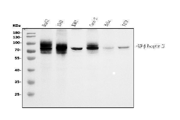
Western blot analysis of TGFBR2 using anti-TGFBR2 antibody (M00759-2).
Electrophoresis was performed on a 5-20% SDS-PAGE gel at 70V (Stacking gel) / 90V (Resolving gel) for 2-3 hours. The sample well of each lane was loaded with 30ug of sample under reducing conditions.
Lane 1: human HepG2 whole cell lysates,
Lane 2: human A549 whole cell lysates,
Lane 3: human K562 whole cell lysates,
Lane 4: human Caco-2 whole cell lysates,
Lane 5: human Hela whole cell lysates,
Lane 6: human T-47D whole cell lysates.
After Electrophoresis, proteins were transferred to a Nitrocellulose membrane at 150mA for 50-90 minutes. Blocked the membrane with 5% Non-fat Milk/ TBS for 1.5 hour at RT. The membrane was incubated with mouse anti-TGFBR2 antigen affinity purified monoclonal antibody (Catalog # M00759-2) at 0.5 μg/mL overnight at 4°C, then washed with TBS-0.1%Tween 3 times with 5 minutes each and probed with a goat anti-mouse IgG-HRP secondary antibody at a dilution of 1:10000 for 1.5 hour at RT. The signal is developed using an Enhanced Chemiluminescent detection (ECL) kit (Catalog # EK1001) with Tanon 5200 system. A specific band was detected for TGFBR2 at approximately 70-85KD. The expected band size for TGFBR2 is at 70-85KD.

Click image to see more details
IHC analysis of TGFBR2 using anti-TGFBR2 antibody (M00759-2).
TGFBR2 was detected in paraffin-embedded section of human placenta tissue. Heat mediated antigen retrieval was performed in EDTA buffer (pH8.0, epitope retrieval solution). The tissue section was blocked with 10% goat serum. The tissue section was then incubated with 2μg/ml mouse anti-TGFBR2 Antibody (M00759-2) overnight at 4°C. Biotinylated goat anti-mouse IgG was used as secondary antibody and incubated for 30 minutes at 37°C. The tissue section was developed using Strepavidin-Biotin-Complex (SABC) (Catalog # SA1021) with DAB as the chromogen.

Click image to see more details
IHC analysis of TGFBR2 using anti-TGFBR2 antibody (M00759-2).
TGFBR2 was detected in paraffin-embedded section of human cervical intraepithelial neoplasia tissue. Heat mediated antigen retrieval was performed in EDTA buffer (pH8.0, epitope retrieval solution). The tissue section was blocked with 10% goat serum. The tissue section was then incubated with 2μg/ml mouse anti-TGFBR2 Antibody (M00759-2) overnight at 4°C. Biotinylated goat anti-mouse IgG was used as secondary antibody and incubated for 30 minutes at 37°C. The tissue section was developed using Strepavidin-Biotin-Complex (SABC) (Catalog # SA1021) with DAB as the chromogen.

Click image to see more details
IHC analysis of TGFBR2 using anti-TGFBR2 antibody (M00759-2).
TGFBR2 was detected in paraffin-embedded section of human esophageal squamous carcinoma tissue. Heat mediated antigen retrieval was performed in EDTA buffer (pH8.0, epitope retrieval solution). The tissue section was blocked with 10% goat serum. The tissue section was then incubated with 2μg/ml mouse anti-TGFBR2 Antibody (M00759-2) overnight at 4°C. Biotinylated goat anti-mouse IgG was used as secondary antibody and incubated for 30 minutes at 37°C. The tissue section was developed using Strepavidin-Biotin-Complex (SABC) (Catalog # SA1021) with DAB as the chromogen.

Click image to see more details
IF analysis of TGFBR2 using anti-TGFBR2 antibody (M00179-1).
TGFBR2 was detected in immunocytochemical section of HepG2 cells. Enzyme antigen retrieval was performed using IHC enzyme antigen retrieval reagent (AR0022) for 15 mins. The cells were blocked with 10% goat serum. And then incubated with 5μg/mL mouse anti-TGFBR2 Antibody (M00179-1) overnight at 4°C. DyLight®488 Conjugated Goat Anti-Mouse IgG (BA1126) was used as secondary antibody at 1:100 dilution and incubated for 30 minutes at 37°C. The section was counterstained with DAPI. Visualize using a fluorescence microscope and filter sets appropriate for the label used.

Click image to see more details
Flow Cytometry analysis of A549 cells using anti-TGFBR2 antibody (M00759-2).
Overlay histogram showing A549 cells stained with M00759-2 (Blue line). The cells were fixed with 4% paraformaldehyde and blocked with 10% normal goat serum. And then incubated with mouse anti-TGFBR2 Antibody (M00759-2, 1μg/1x106 cells) for 30 min at 20°C. DyLight®488 conjugated goat anti-mouse IgG (BA1126, 5-10μg/1x106 cells) was used as secondary antibody for 30 minutes at 20°C. Isotype control antibody (Green line) was mouse IgG (1μg/1x106) used under the same conditions. Unlabelled sample without incubation with primary antibody and secondary antibody (Red line) was used as a blank control.
Specific Publications For Anti-TGFBR2 Antibody Picoband® (monoclonal, 2F11) (M00759-2)
Loading publications
Recommended Resources
Here are featured tools and databases that you might find useful.
- Boster's Pathways Library
- Protein Databases
- Bioscience Research Protocol Resources
- Data Processing & Analysis Software
- Photo Editing Software
- Scientific Literature Resources
- Research Paper Management Tools
- Molecular Biology Software
- Primer Design Tools
- Bioinformatics Tools
- Phylogenetic Tree Analysis
Customer Reviews
Have you used Anti-TGFBR2 Antibody Picoband® (monoclonal, 2F11)?
Share your experimental results or join a short interview to earn up to $1,000 in product credits or other rewards.
0 Reviews For Anti-TGFBR2 Antibody Picoband® (monoclonal, 2F11)
Customer Q&As
Have a question?
Find answers in Q&As, reviews.
Can't find your answer?
Submit your question
1 Customer Q&As for Anti-TGFBR2 Antibody Picoband® (monoclonal, 2F11)
Question
Was M00759-2 derived from a hybridoma?
Verified customer
Asked: 2022-06-09
Answer
Yes, the Anti-TGFBR2 Antibody Picoband™ (Monoclonal, 2F11) (M00759-2) was derived from a hybridoma.
Boster Scientific Support
Answered: 2022-06-10
