Introduction
In IHC experiments, many researchers take for granted the familiar blue stain on every slide – hematoxylin – and may overlook its role. As one expert put it, “How many of us take the time to think about which counterstain would be the best choice?”. Counterstaining can have a big impact on your IHC results. Most cells are colorless and transparent, so without a background stain, even a perfectly developed antibody signal can be hard to place. A good counterstain provides contrast and context, helping antibody-stained cells “stand out more” and pinpointing their exact location in the tissue.
What Are IHC Counterstains and Why Use Them?
Counterstains are dyes applied after the primary IHC staining to colorize cellular features not highlighted by the antibody. In typical IHC, target proteins are visualized via an enzymatic or fluorescent tag: for example, an HRP-linked secondary antibody and DAB substrate produce a brown precipitate at antigen sites. A counterstain then colors other structures (nuclei, cytoplasm, etc.) to create contrast. As one review (Orakpoghenor et al., 2018) explains, the aim is to “provide contrast that enables the primary stain to stand out”. By selectively staining nuclei or cytoplasm, counterstains make it easy to distinguish labeled antigens from the rest of the tissue. In short, counterstains give us the “architecture” of the tissue so we can interpret the specific antibody signal.
Common Chemical Counterstains
Several traditional dyes are used as IHC counterstains, each binding different cellular components and yielding distinct colors:
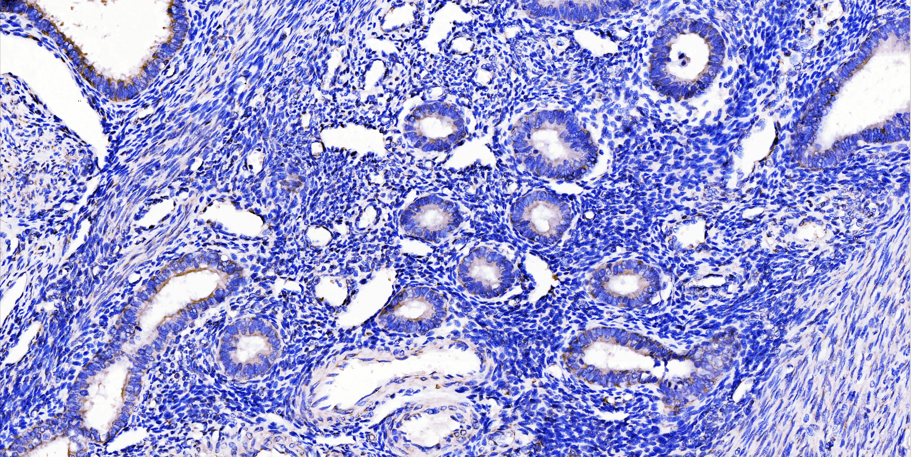
- Hematoxylin (nuclear stain, blue/violet): This is the classic nuclear stain in histology. Oxidized hematoxylin (hematin) forms a positively charged metal–dye complex (usually with aluminum) that binds to negatively charged chromatin (nuclear histones). The result is a blue-purple staining of cell nuclei. Hematoxylin binding involves histone lysine residues, so it colors nuclear proteins rather than DNA directly. The blue-violet nuclei provide excellent contrast to typical IHC colors (brown DAB, red chromogens, or green dyes).
- Eosin (cytoplasmic stain, pink/red): Often paired with hematoxylin in H&E, eosin is an acidic (anionic) dye that stains cytoplasm and extracellular matrix in shades of pink to red. Eosin binds basic (cationic) protein groups, which are abundant in nearly all cells. Because it broadly stains the cytoplasm and stroma, eosin is useful when the target antigen is nuclear (so the nucleus is left unstained/blue). In other words, if your antibody marks a nuclear protein, using eosin as a counterstain colors the rest of the cell without obscuring the nuclear signal. However, because eosin binds so widely, over-staining can flood the tissue with a pink background and diminish contrast.
- Nuclear Fast Red (Kernechtrot, nuclear stain, red): This is an alternate nuclear stain that quickly colors nuclei red (hence the name). Unlike hematoxylin, nuclear fast red targets nucleic acids directly and can achieve strong nuclear staining in about 5 minutes. Its red color provides a different contrast scheme, which can be advantageous if your chromogen is brown or green. For example, red nuclei from nuclear fast red stand out nicely against brown DAB or green HRP products.
- Methyl Green (nuclear stain, green): Methyl green is another DNA-binding dye that stains nuclei green. A typical staining time is around 5 minutes. The green nuclei contrast well with red, blue, or brown targets. One advantage of methyl green (often used with pyronin Y) is that it can help distinguish DNA (green) from RNA (pyronin red) in tissues.
- Other Chromogenic Dyes: Additional options include methylene blue (a basic dye that can stain nuclei and some cytoplasm blue, without needing a mordant), and toluidine blue (a strong basic dye that stains nuclei blue and glycosaminoglycans metachromatically, often appearing pink/red in cartilage). These are less common but useful in special cases.
Each chemical counterstain offers a particular color (blue, red, green, or pink). Selecting one involves matching it to your antigen’s color. For example, if your IHC chromogen is brown (DAB), a blue hematoxylin counterstain is standard because blue/green contrasts well with brown. If your IHC stain is green, a red nuclear counterstain might be preferred. The proper selection of nuclear and tissue counterstains is important to maintain optimal color contrast with your desired IHC stains.
Fluorescent Counterstains for Multiplex IHC
In fluorescence-based IHC, nuclear counterstains are typically fluorescent dyes:
- DAPI (4′,6-diamidino-2-phenylindole): Often called the “gold standard” nuclear stain for fluorescence, DAPI binds strongly to double-stranded DNA and emits blue fluorescence under UV excitation. It has minimal cytoplasmic staining. DAPI is compatible with most other fluorophores (green, red, etc.) because it occupies the blue channel.
- Hoechst 33258/33342: Hoechst dyes also bind A-T-rich regions of DNA and emit blue fluorescence, with behavior very similar to DAPI. Like DAPI, they produce bright blue nuclei and work well with green/red antibody labels.
- Propidium Iodide (PI): PI intercalates into DNA and emits red fluorescence. It can be used for nuclear staining when a red channel is available. Note: PI also binds RNA, so fixed samples should be treated with RNase to avoid RNA staining. PI is often used in live/dead assays (it only penetrates dead cells), but can serve as a nuclear stain in fixed tissues.
- Other Nuclear Dyes: For special situations, there are alternatives. SYTOX Green preferentially stains nuclei in cells with compromised membranes (dead cells). SYBR Green binds double-stranded DNA (with green emission) and is membrane-permeable, so it can be used for live-cell nuclei. These alternatives can be chosen if DAPI/Hoechst (blue) conflicts with other labels. For instance, if you need a blue-emitting antibody, you would use a red or green nuclear stain instead. The key is to avoid spectral overlap between your antibody fluorophores and the nuclear stain.
Choosing Counterstains by Target Location
The choice of counterstain should reflect where your antigen is:
- Nuclear Antigens: If your target protein is in the nucleus (e.g., transcription factors, nuclear phosphoproteins), a heavy nuclear stain might mask the signal. In this case, you can skip nuclear staining or use a cytoplasmic stain like eosin. As one review (https://bitesizebio.com/13467/counterstaining-for-immunohistochemistry-choices-choices/) explains, “eosin can also be used as a counterstain when an antibody localized to the nucleus is used”. Eosin will stain the cytoplasm and extracellular matrix pink, leaving the nucleus (and your target) more visible. In summary: use a non-nuclear dye (pink or similar) to provide background contrast while keeping nuclei clear.
- Cytoplasmic/Membrane Antigens: If the antigen is in the cytoplasm or on the cell membrane, a nuclear counterstain is ideal. A standard choice is hematoxylin (blue nuclei). The blue nuclei provide context for the cell structures, while your target (often brown or red) is highlighted against them. Alternatives like nuclear fast red (red nuclei) or methyl green (green nuclei) can also be used, especially if you want different color schemes.
- Multiplex Staining: In multi-color IHC, think in terms of color contrast. For example, if your marker antibody is conjugated to a green fluorophore, using blue DAPI or Hoechst for nuclei works well. But if your antibody is blue (e.g., Alexa Fluor 405), you would switch to a red or green nuclear stain instead. In practice, ensure that the nuclear stain’s color is distinct from all your antibody signals.
Color Compatibility Table (examples):
- DAB (brown) signal → blue (hematoxylin) nuclei.
- A red chromogen (e.g. Fast Red AP) → blue or green nuclei.
- A green chromogen or Alexa Fluor 488 → red nuclei (NFR) or blue (DAPI).
- Fluorescent Alexa 594 (red) → blue (DAPI) nuclei.
- Fluorescent Alexa 488 (green) → red (PI) or blue (DAPI) nuclei.
Always verify that colors contrast well under the microscope.
Common Problems in IHC Counterstaining
Despite the simplicity of counterstains, various issues can arise in IHC:
- Overstaining with Hematoxylin: If the nuclei turn too dark blue, they can obscure nuclear targets or overall morphology. The CAP troubleshooting guide notes that too-dark hematoxylin “masks positive staining of nuclei”. The solution is to shorten the hematoxylin staining time or use a more dilute solution.
- Underdifferentiated Hematoxylin: Conversely, if hematoxylin staining (or differentiation in acid alcohol) is too short, nuclei appear faint and pale, providing little contrast. You may need to stain longer or adjust pH/acid steps.
- Uneven Staining: Irregular counterstain intensity across a slide can result from inconsistent section thickness, incomplete deparaffinization, or automated stainer issues. Ensure uniform section thickness, fully wet the tissue throughout processing, and verify automated program timing (machine dips often require longer exposure than manual dips).
- Eosin (or Other Dye) Background: Because eosin binds proteins widely, too much eosin can flood the tissue with pink/red, making it hard to see fine details. To fix this, reduce the eosin concentration or time, or perform a brief acid wash to differentiate the excess stain.
- Fluorescent Dye Photobleaching: Dyes like DAPI/PI fade under prolonged light. Always minimize exposure to excitation light and use anti-fade mounting media. Re-image as soon as possible after staining.
- Spectral Overlap (Crosstalk): In multiplex IF, DAPI and Hoechst fluoresce blue; if your antibody also emits blue, you’ll get bleed-through. Use dyes with well-separated emission spectra, and ensure proper filter settings. For instance, if using a blue-emitting label, switch to a red nuclear dye (PI) or a green (SYTOX Green) to prevent overlap.
- Autofluorescence: Some tissues (e.g., lipofuscin in liver, collagen) naturally fluoresce, sometimes in blue/green channels. If background is high, consider quenching steps (e.g., Sudan black treatment) or shifting to far-red probes.
- Incomplete Washing: Poor washing after counterstaining can leave excess dye. For example, DAPI residue will appear as a diffuse blue haze. Thorough PBS/TBS washes after staining remove unbound dye and reduce background.
- Mounting Medium Issues: Certain mountants can affect dye stability or refractive index. Use anti-fade media for fluorescence and ensure the medium is compatible with your counterstain.
- Automation Variability: On automated stainers, “time” and “dipping” are not directly equivalent to manual steps. As one histology expert noted, machines often move slides slowly, requiring longer exposures to match manual results. If using an autostainer, calibrate your counterstaining times by comparing machine vs manual stains.
Counterstains in Fluorescent IHC: Special Considerations
Unlike chromogenic IHC, fluorescent IHC (IF) relies on fluorophores to emit light signals. Nuclear counterstains must therefore be compatible with the fluorophores in use:
- DAPI and Hoechst are the most common nuclear counterstains in IF. They bind DNA and emit blue fluorescence when excited by UV or violet light.
- These dyes should not overlap with other fluorophores in your panel. For example, avoid pairing DAPI with blue-emitting labels like Alexa Fluor 405.
Note that chromogenic dyes like hematoxylin are not suitable for use in fluorescent IHC. Use dye types appropriate for the detection system.
Conclusion
Counterstains are the unsung heroes of IHC, providing essential context so that antibody signals “stand out” against the background. By choosing the right dye (blue, red, green, or fluorescent) for your experiment, considering where your antigen is and what colors you’ve used, you can dramatically improve the interpretability of your slides. We have reviewed the definitions, mechanisms, and examples of common counterstains (hematoxylin, eosin, nuclear fast red, methyl green, etc.), including fluorescent alternatives (DAPI, Hoechst, PI), and discussed how each provides contrast to different IHC stain colors. We also covered practical guidelines (e.g., use cytoplasmic dyes when staining nuclear targets) and listed common pitfalls with tips to troubleshoot them. Following these principles will help ensure crisp, informative IHC images.