This website uses cookies to ensure you get the best experience on our website.
- Table of Contents

DAPI, or 4',6-Diamidino-2-Phenylindole, Dihydrochloride, is a commonly used fluorescent dye that binds to double-stranded DNA (dsDNA).
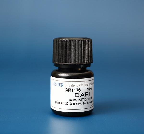
DAPI binds to and ‘stains’ double-stranded DNA, preferably binding to A-T-rich regions in DNA. DAPI stain is excited by ultraviolet (UV) light, with its largest excitation wavelength at ~360nm, and it produces a vibrant blue color with its largest emission wavelength at ~460nm when bound to DNA. Due to its fluorescent properties and rich blue color, it is readily used for visualization in fluorescence microscopy and other assays. Because it can pass through the cell membrane and stain DNA, DAPI is a useful dye for nuclear quantification and has been utilized in numerous assays, such as live or fixed cell staining, cell viability assays, flow cytometry, cell cycle analysis, mycoplasma contamination detection and in fluorescence microscopy. Because of its wide range of applications, Boster offers an affordable DAPI stain solution (Catalog# AR1176) that has been validated and cited in several publications.
Cell cycle analysis & flow cytometry
Since DAPI binds to DNA, it can be used to determine the relative amount of DNA in cells for cell cycle analysis. Cells currently in the G2 phase of mitosis will have twice the amount of DNA as cells in the G1 phase of mitosis, which will be reflected in the amount of fluorescence from DAPI in each cell. Apoptotic cells will have less DNA than a single cell, since DNA is being degraded, and will be reflected in the decreased amount of fluorescence. Cells processed with DAPI can then be analyzed with flow cytometry using a UV laser at ~360nm to determine the fluorescence per cell. Relative fluorescence will correspond with the different stages of the cell cycle. Live or fixed cells incubated with DAPI can be processed via flow cytometry, though live cells have a lower efficiency of DAPI staining.
DAPI live cell staining
DAPI has the benefit of staining both live and fixed cells. In order to stain live cells, though, the concentration of DAPI must be increased. Common DAPI live cell staining protocols generally involve incubating cells with DAPI stain for approximately 10 minutes, then visualizing cells under a fluorescent microscope.
Mycoplasma detection
Mycoplasma is a type of bacteria without a cell wall that contaminates cell culture. Mycoplasma cannot be visualized by simply looking at cells through the microscope, but this contamination is often suspected due to changes in cell growth, viability, and other alterations in cell lines. One simple method of detecting mycoplasma presence in cell culture is by staining cells with DAPI. Cells without contamination will only show fluorescent DAPI staining within the nucleus, but cells with mycoplasma contamination will also have DAPI staining outside of the nucleus. This simple visualization with DAPI, performed on a regular basis, can help ensure your cell culture remains free of mycoplasma.
DAPI in fluorescence microscopy
DAPI is commonly used in fluorescence microscopy due to its ease of use and bright blue color. Utilizing DAPI-stained cells in fluorescence microscopy enables researchers to visualize the nucleus. Common protocols call for either live or fixed cells. Fixed cells are typically incubated in DAPI for 5-10 minutes, then rinsed in PBS or TBS-T multiple times to remove excess stain. Once mounted, antifade reagents can be added. Samples can then be visualized under a fluorescence microscope with a blue/cyan filter for DAPI. To note, the excitation wavelength for DAPI stain is 360nm. For more information, Boster offers a simple protocol for staining fixed cells, tissues, adherent, and suspension cells using our DAPI stain.
DAPI as a counterstain
Due to DAPI’s vibrant blue fluorescence and relatively low background ‘noise’ in labeling, it is frequently used as a counterstain in assays requiring the use of multiple fluorescent probes. Its specificity as a nuclear stain also helps DAPI to be used in multicolor fluorescence staining assays.
DAPI is one of many fluorescent dyes that are often used in cell staining. Other frequently used stains include Hoechst 33342 (blue) and Propidium iodide (PI, red). PI is a fluorescent dye that binds DNA and RNA, but it cannot pass the plasma membrane, so it can only stain apoptotic, dead, or permeabilized cells. Hoechst 33342 stain is a DNA stain that can pass the plasma membrane, like DAPI, and binds to A-T rich regions in DNA. Some non-fluorescent stains include Trypan Blue and H&E (Haematoxylin–eosin). Trypan blue is a simple-to-use blue dye that cannot pass through the plasma membrane of healthy, live cells; hence it is most often used in cell counting to stain dead cells and in determining cell viability in mouse tissue. H&E staining is a well-known histopathology stain, in which hematoxylin (H) stains the nucleus and eosin (E) stains the cytoplasm of fixed cells. In comparison with other stains, DAPI has the advantage of being able to be used in live and fixed cells, it only stains DNA (unlike PI which stains RNA too), and it provides a vibrant, rich blue that can be easily visualized in fluorescent microscopy, imaging, flow cytometry, and numerous assays.