This website uses cookies to ensure you get the best experience on our website.


- Table of Contents


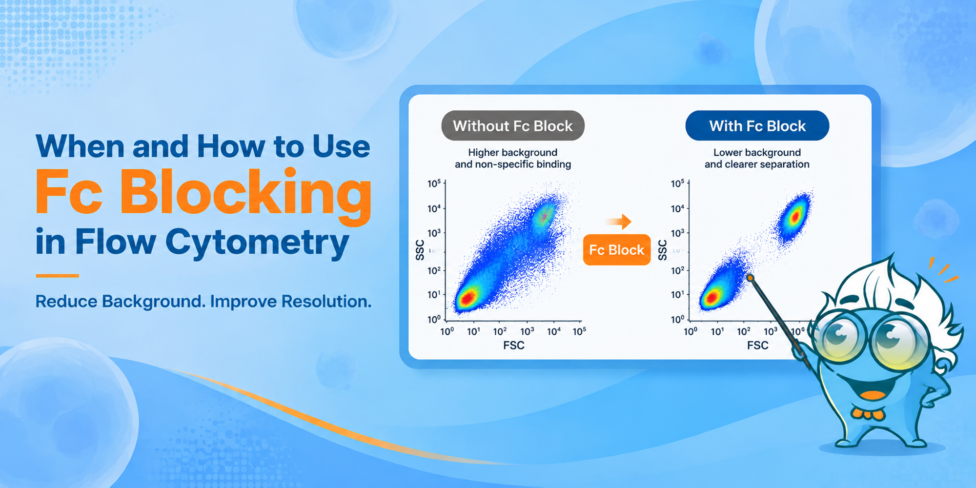
High background in flow cytometry is not always an antibody-specificity problem. Cells that express Fc receptors can capture antibodies through the Fc region, adding fluorescence that is unrelated to the target antigen. The result may be a higher negative population, poorer separation, or an apparently positive population that is difficult to interpret.
Fc blocking in flow cytometry is used to reduce that Fc receptor-mediated component before antibody staining. The more useful questions are when the step is likely to matter, how inadequate blocking appears in the data, and whether the blocker can interfere with a marker in the panel.
Those answers depend on the sample. Fc receptor expression varies with lineage and activation state, and blocker chemistry and antibody isotype also matter. For an unfamiliar preparation, a blocked-versus-unblocked comparison is often the quickest way to see whether Fc-mediated binding is affecting the assay.
Most flow cytometry antibodies contain an antigen-binding Fab region and an Fc region. The Fab region recognizes the target antigen; the Fc region can also interact with Fc receptors on leukocytes. When that second interaction contributes to staining, the measured fluorescence is no longer explained by antigen binding alone. [1,5,6]
Fc blocking reduces this unwanted binding by occupying or inhibiting Fc receptors before the staining antibodies are added. Depending on the system, the blocker may be a receptor-specific antibody, purified immunoglobulin, serum-derived immunoglobulin, or a commercial Fc receptor-blocking formulation. [1,5,6]
Fc-mediated binding can raise background, shift negative populations, and make dim or rare populations harder to resolve. In complex samples, it can compound other artifacts such as dead cells and doublets, particularly when profiling tumor-infiltrating immune cells. Fc blocking addresses the Fc-mediated component; viability and singlet controls still need to be handled separately. [1,2]
The need for Fc blocking follows the cells in the tube more than the name of the assay. Vendor protocols flag monocytes, macrophages, neutrophils, B cells, NK cells, and some T-cell subsets because they can express Fc gamma receptors. But the amount of nonspecific binding is not the same across all of these populations or across antibody isotypes. [1,5,6]
Fc receptor-rich myeloid cells deserve the most attention. Lymphocyte-only assays are more variable and should be judged in the context of the panel and sample.
Sample type is a useful shortcut, but cell composition is what matters. A tumor digest rich in macrophages, for example, is a different Fc-blocking problem from a purified T-cell preparation.
| Sample | Fc Blocking Consideration | Why |
|---|---|---|
| Whole blood | Strongly consider | Contains multiple Fc receptor-expressing leukocyte populations, including monocytes and granulocytes. |
| PBMCs | Often useful | Monocytes are retained, but granulocytes are largely absent after standard density separation; importance depends on the populations being analyzed. |
| Bone marrow | Often useful in mixed/myeloid-rich samples | Contains diverse developing and mature immune populations, including myeloid cells. |
| Spleen | Depends on populations analyzed | Mixed immune-cell composition can include substantial Fc receptor-positive populations. |
| Dissociated tumor | Strongly consider when myeloid-rich | Tumor-infiltrating myeloid cells can be particularly sensitive to Fc-mediated artifacts. [2] |
| Inflamed tissue | Often useful when FcR-rich | Inflammatory infiltrates can increase the proportion and activation state of Fc receptor-expressing leukocytes. |
| Purified monocytes/macrophages | Strongly consider | Directly enriched for populations in which Fc-mediated binding can be prominent. [1] |
| Purified conventional T cells | Usually lower priority | Fc-mediated background is commonly less prominent, but activation state and panel composition still matter. |
| Fc receptor-negative cultured cells | Usually low priority if truly FcR-negative | A blocker may add little if the cells do not express relevant Fc receptors and no other Fc-interacting component is present. |
For IgG-based staining, CD64 (Fc gamma RI), CD32 (Fc gamma RII), and CD16 (Fc gamma RIII) are the receptors most often encountered in this context. Their expression varies by lineage, differentiation state, and activation. BD and Thermo Fisher guidance discusses CD16/CD32-associated Fc-mediated binding across B cells, NK cells, granulocytes, monocytes, and macrophages. [5,6]
| Fc receptor | Commonly relevant populations | Note |
|---|---|---|
| CD64 / Fc gamma RI | Especially important in myeloid populations such as monocytes/macrophages | High-affinity Fc gamma receptor; blocker compatibility depends on reagent design. |
| CD32 / Fc gamma RII | B cells, monocytes, macrophages, granulocytes and other FcR-positive cells | A major target of common mouse anti-CD16/CD32 blocking antibodies. [6] |
| CD16 / Fc gamma RIII | NK cells, neutrophils, macrophage/monocyte subsets |