This website uses cookies to ensure you get the best experience on our website.


- Table of Contents
A clean loading control band can still normalize the wrong thing. If the control is saturated, treatment-sensitive, or mismatched to the sample fraction, it can make a blot look corrected while quietly distorting the target result. In Western blotting, a reliable internal loading control should remain proportional to the amount of sample loaded and independent of changes in the protein of interest.
To choose a reliable Western blot loading control, define what variation you need to control: total protein loading, transfer efficiency, fraction recovery, or densitometry normalization. Selecting appropriate loading controls antibodies can help ensure the chosen reference is suitable for the type of variation being assessed. The control should be stable under the treatment, appropriate for the sample fraction, detected within the linear range, and processed under the same workflow as the target. GAPDH, beta-actin, and tubulin can work for routine total lysates, but they are not universal references. Across different experimental conditions, the selected reference should reflect technical variation without changing alongside protein expression or overall protein abundance.
A loading control is not just a band under the target. It is the reference used to argue that target differences are biological rather than technical. If that reference is unstable or overloaded, normalization can make weak data look stronger than it is. For a broader product starting point, Boster's Loading Control Antibodies page is useful, but the real decision still depends on the experiment. A sound Western blot analysis therefore requires evidence that the internal loading control is suitable for the sample type and treatment.
Most Western blot normalization relies on one assumption: the loading control changes because of technical variation, not because of the biology being tested. This means the internal loading control should remain stable even when the protein of interest and related protein levels respond to treatment.
That assumption is easy to violate. If a treatment lowers both the target protein and GAPDH, normalizing the target to GAPDH may underestimate the real target decrease. If the target is unchanged but beta-actin changes because the treatment affects cytoskeletal organization, normalization may create a false target difference. If the loading control is saturated, every lane can look equal even when loading differences remain. This concern is especially important when the control is a cytoskeletal protein or when the experiment examines signaling proteins that can alter cell structure or metabolism.
The question is not simply, “Do I have a loading control?” The better question is whether that control is allowed to behave as a reference in this experiment.
Different blots need different reference logic. In routine total lysate experiments, the main concern is usually lane-to-lane loading and transfer variation. A common housekeeping protein may be enough if it is stable and not saturated. Before loading, a protein assay should be performed against a standard curve so that comparable amounts of sample enter each lane.
Fractionated samples are different. A nuclear fraction needs a nuclear reference. A mitochondrial fraction needs a mitochondrial reference. A membrane-enriched sample should not be normalized blindly to a soluble cytoplasmic protein. Equal total protein loading does not always mean equal recovery of the compartment you care about. The internal loading control should match the subcellular localization of the fraction and should represent recovery of the compartment containing the target.
This is where mismatched controls become dangerous. Strong GAPDH signal in a nuclear fraction may be a contamination warning, not a loading control. A cytosolic control in a membrane prep may not reflect membrane protein recovery. Boster's Western Blot Antibody Selection Guide gives a useful overview of compartment-based antibody choices, but the key is to match the control to the technical question. For nuclear proteins, lamin B1 can serve as a nuclear reference when its stability has been validated. However, lamin B1 may change during apoptosis, senescence, or nuclear envelope disruption. For mitochondrial samples, a marker associated with the mitochondrial membrane may better reflect organelle recovery than a total lysate control.
GAPDH, beta-actin, and tubulin are popular because they are abundant and easy to detect. That convenience is also why they can mislead.
GAPDH is tied to glycolysis and can shift in metabolism, hypoxia, stress, and cell-state experiments. Beta-actin becomes risky when the study affects migration, differentiation, morphology, apoptosis, or the cytoskeleton. Tubulin needs caution in microtubule-drug, cell-cycle, neuronal, or differentiation models. In these settings, altered protein levels may reflect biology rather than loading variation.
Nuclear controls such as Histone H3, Lamin, or TBP are better suited to nuclear fractions, but they still need context. Apoptosis, chromatin remodeling, or nuclear envelope disruption can change what looks like a stable marker. Cell-cycle synchronization can also influence histone abundance during DNA replication. In studies focused on DNA replication, validate Histone H3 and other nuclear references before using them for normalization.
A housekeeping protein becomes a bad control the moment the experiment starts regulating it. Post-translational modifications can also change antibody recognition or apparent band migration, even when the amount of the reference protein has not changed.
One of the most common loading-control failures is not absence. It is saturation.
Housekeeping proteins are often so abundant that their bands become too strong before the target is detected well. A clean, dark GAPDH or beta-actin band may look reassuring, but if the signal has plateaued, it no longer reflects protein amount. A saturated loading control cannot correct loading differences. It only makes different lanes look more equal than they are. For quantitative Western blot analysis, the internal loading control and the target must both remain within a response range where signal intensity tracks protein levels.
A simple warning sign is that shorter exposure or lower sample loa...

How to separate heat-related effects from contact problems, over-transfer, and membrane-selection errors.
You start a 90-minute wet transfer with cold buffer and...

A practical old-versus-new lot comparison that moves from experimental setup to a documented pass, conditional pass, or fail decision.
ANTIBODY LOT BRIDGING SERIES
Western Blot Worked Example — Use this article to see the cross-application requalification framework applied to one semi-quantitative W


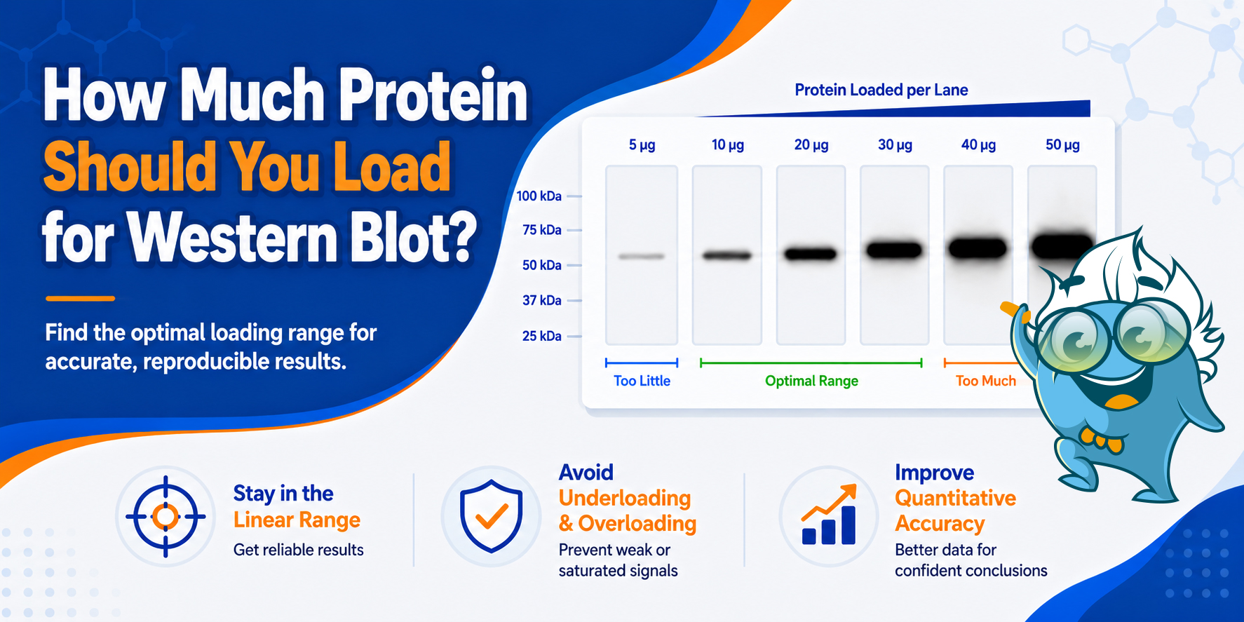
For many western blots, 20–30 μg of total lysate per lane is a practical starting point—but the right amount depends on target abundance, sample type, and detection sensitivity. Learn how to identify the usable linear range and avoid saturation.
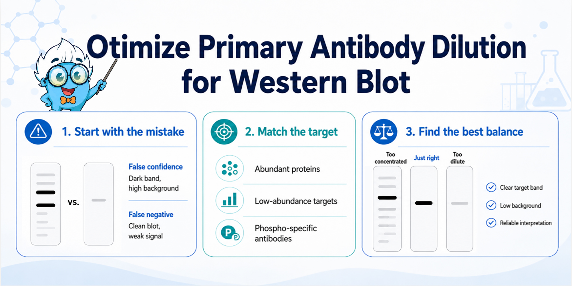
More primary antibody is not a shortcut to a better Western blot. A stronger band only helps if the rest of the membrane stays clean enough to trust.
To optimize primary antibody dilution for Western blot, start from the datasheet range and test a small dilution series. Choose the condition with the best signal-to-background balance, not simply the darkest band. Too much primary antibody can raise...
High background after secondary antibody incubation in Western blot does not always mean the blot needs to be re-run. First, decide whether the background is caused by excess secondary antibody, insufficient TBST washing, blocking mismatch, ECL overexposure, or non-specific signal carried over from the primary antibody step. If the target band is still visible and the membrane is not saturated or dried, shorter exposure or extra washes may still rescue the blot. If the background appears in a secondary-only control, follows non-specific bands, or overwhelms every exposure, the next run needs adjusted conditions.
For broader workflow issues, see Boster Bio's Western Blot Troubleshooting guide. This article focuses on background that appears or becomes obvious after secondary antibody incubation and detection.
A blot that looks clean after transfer can still turn into a gray, hazy membrane after secondary antibody and ECL. That does not always mean the gel, transfer, or sample preparation failed. It often means the detection layer has amplified something that was already weakly present.
Start by looking at the pattern. If a short exposure looks clean but a long exposure looks dirty, the problem may be exposure range, not blot failure. If the whole membrane has a smooth haze, check secondary antibody concentration and washing.
In many Western blotting workflows, membrane type can also influence background. A PVDF membrane and nitrocellulose membrane may respond differently to blocking and washing conditions, and some laboratories observe varying levels of non-target signal depending on the membrane selected. Similar considerations apply when working with nitrocellulose membranes from different manufacturers.
If the background appears in a secondary-only control, the issue is not target-specific primary antibody binding. If the membrane has speckles, think about precipitated antibody, contaminated trays, dried membrane spots, or old ECL working solution.
Extra bands are different from whole-membrane haze. If the background follows bands or lanes, the problem may involve primary antibody cross-reactivity, sample loading, or the antibody pair, not just the secondary incubation step.
Review whether the unexpected bands overlap with the expected protein of interest or appear independently of the anticipated molecular weight. Comparing the target signal with a suitable loading control can help determine whether the pattern reflects true biology or technical variation.
The first decision is not "Which reagent is bad?" It is: can this membrane still be interpreted, or has the background become part of the result?
The secondary antibody is supposed to amplify the primary antibody signal. That is why HRP-conjugated secondary antibodies and ECL detection are useful. But the same amplification can turn weak background into a visible problem.
If the secondary antibody is too concentrated, it can bind weakly across the membrane and create a gray haze.
Optimizing antibody dilution is often one of the simplest ways to reduce excess background. Appropriate dilution factors should be determined empirically because antibody performance varies across applications and protein targets.
If washing is incomplete, unbound HRP-labeled secondary antibody can remain on the membrane and light up during ECL.
This effect can be particularly noticeable when using HRP-conjugated secondary antibodies or a secondary antibody conjugated to HRP in highly sensitive detection workflows.
If the primary antibody already produced weak non-specific binding, the secondary antibody may amplify it into extra bands or broad background.
This is why background after secondary incubation does not automatically prove the primary antibody is innocent. It also does not mean you should change the primary antibody first. Check whether the background appears only on long exposure, whether a secondary-only control is clean, and whether the pattern is diffuse, speckled, lane-specific, or band-specific.
For general secondary antibody selection principles, Boster's secondary antibody guide can be a useful reference.
Secondary antibodies recognize specific immunoglobulin molecules and immunoglobulin proteins, including defined antibody chains present in the host species used to generate the primary antibody.
A secondary-only control is useful when the blot becomes dirty after secondary incubation, but it is easy to overread.
If the secondary-only control shows background, the problem is not target-specific primary binding. It may come from the secondary antibody, membrane background, blocking condition, wash stringency, or detection reagent. If the signal appears as distinct bands rather than diffuse background, also consider sample-derived immunoglobulins, IgG heavy/light chains in immunoprecipitated samples, or contamination from trays, buffers, or membrane handling.
The choice of blocking buffer can significantly influence background levels. Common blocking reagents include Bovine Serum Albumin and milk-based formulations, although optimal conditions depend on the assay and antibody pair.
When the secondary-only control is positive, lowering the secondary antibody concentration, improving TBST washing, changing the blocking condition, or checking the ECL reagent may matter more than changing the primary antibody.
If the secondary-only control is clean, that does not prove the secondary antibody has no role. The secondary antibody may still be amplifying weak non-specific primary binding. The control is best used to ask one narrow question: is there primary-independent background? It is not proof that the primary antibody is specific.
Some secondary-stage background can still be reduced on the same membrane. Other problems can only be fixed in the next run.
If the target band is visible and the membrane is not dried or saturated, start with a shorter exposure. Long exposure can make low-level membrane background look worse than it is. If the short exposure gives a clean target band, the blot may still be usable.
If the membrane has not dried, additional fresh TBST washes may help reduce residual secondary antibody.
This is most useful when the background is diffuse haze rather than strong non-specific bands. For more on Tween 20 in blotting buffers, see Boster's guide to Tween 20 in Western blotting.
If the background is speckled, replace or filter the antibody or ECL working solution and clean the incubation tray. Speckles often come from precipitate, contaminated containers, dried droplets, or uneven reagent contact rather than true antibody binding.
If the membrane is still in good condition, stripping and re-probing may be possible.
Researchers performing quantitative western blots often verify signal quality before stripping by reviewing exposure linearity and evaluating a total protein stain or total protein staining approach.
But if the membrane is already saturated, unevenly blocked, or dominated by non-specific bands, re-running with adjusted conditions is usually cleaner. Boster's Western blot stripping buffer protocol can help with re-probing decisions.
Some problems should not be fixed by trying harder on the same blot. Re-run with adjusted conditions if every exposure is saturated, if the target band cannot be separated from background, or if the membrane dried during incubation. Re-running is also usually better when the background follows lanes or bands, because that pattern often points to antibody specificity, sample loading, or primary antibody conditions rather than free secondary antibody left on the membrane.
Next-run changes may include using a lower secondary antibody concentration, shortening secondary incubation, improving TBST wash stringency, changing the blocker, or reducing primary antibody concentration if extra bands are present.
Additional optimization may include reviewing primary antibody dilution buffer composition, reassessing dilution factors, and ensuring that incubation solutions are prepared using high-quality solutions and reagents, including reverse osmosis deionized water when appropriate.
If the issue is exposure-related, capture a shorter exposure or use a less sensitive ECL substrate.
Instruments used for image acquisition can also contribute to interpretation differences. An appropriate imaging system and well-calibrated detection system help distinguish true target signal from low-level background.
For exposure context, see Boster's ECL Western blot substrate article and Western blot quantification guide.
Do not make all changes at once. If you change primary dilution, secondary dilution, blocker, wash time, and ECL exposure together, you may fix the blot without knowing which condition mattered.
| What you see | Same-blot action | Next-run change |
|---|---|---|
| Clean short exposure, dirty long exposure | Use shorter exposure | Reduce exposure time or ECL sensitivity |
| Whole-membrane haze | Extra fresh TBST washes | Lower secondary antibody; improve wash stringency |
| Secondary-only control is positive | Confirm background occurs without primary antibody | Lower secondary, improve washes, adjust blocker, check ECL, or review sample/contamination |
| Secondary-only control is clean but blot is dirty | Check primary-driven binding | Lower primary or review antibody specificity |
| Speckled background | Replace/filter reagent; clean tray | Use fresh antibody/ECL; avoid drying |
| Saturated bands and background | Re-image shorter if possible | Reload less or lower antibody/detection intensity |
Do not immediately reduce the primary antibody if the secondary-only control is dirty. Do not keep increasing wash time if the membrane is fragile or already drying. Do not judge background only from the longest exposure. Do not treat every dirty membrane as a blocking failure. And do not strip every blot just because the first image looks bad.
The fastest fix is not always changing the antibody. First identify which layer created the background.
The secondary antibody and HRP/ECL detection system can amplify weak background. Common causes include excess secondary antibody, incomplete washing, blocker mismatch, long exposure, or non-specific primary signal that becomes visible after detection.
Sometimes. Extra fresh TBST washes can reduce leftover unbound secondary antibody and lower diffuse haze. They will not fix saturated exposure, severe primary-driven bands, or an antibody pair that is not specific enough.
It suggests the background can occur without target-specific primary antibody binding. The source may be secondary antibody, membrane/blocking condition, wash stringency, or detection reagent.
Not always. If the target band is visible and a shorter exposure is interpretable, the blot may still be usable. Re-run if the membrane is saturated, dried, uneven, or if background overwhelms every exposure.
High background after secondary antibody incubation is not always a failed blot. It is often an amplification problem. The secondary antibody and ECL system may be revealing excess secondary antibody, incomplete washing, blocker mismatch, overexposure, or weak non-specific binding from the primary step.
Before re-running, decide whether the membrane can still be interpreted. If the target band survives shorter exposure or extra washes, you may still use the blot. If the background is saturated...

Before you move on to blocking, make sure the membrane is worth taking forward.
A Western blot can start failing long before the antibodies ever touch the membrane. If transfer is incomplete, uneven, or locally disrupted, you may not realize it until much later—after blocking, primary incubation, washes, secondary, and detection. By then, transfer problems are harder to separate from antibody or detection problems, and the blot has already cost you time.
That is why one of the most useful QC steps in the workflow happens immediately after transfer and before blocking.
At that point, you are not trying to finish the analysis. You are trying to answer one practical question: Is this membrane good enough to keep going—or am I about to waste the blot?
Check transfer quality right after transfer and before blocking.
For many routine blots, Ponceau S is enough for a first-pass check. It can show whether protein reached the membrane, whether lanes look broadly even, and whether there are obvious local defects.
If you need a clearer lane-level readout—or already expect total protein signal to matter later—a total protein stain may be the better choice. A loading control can still be useful later, but it should not be your first transfer QC step.

A practical guide to choosing the right membrane based on workflow, reprobing needs, and downstream use.
Western blot problems are often blamed on antibodies, transfer conditions, or blocking. But in many experiments, the first preventable mistake happens even earlier: membrane selection.
If you are deciding between PVDF and nitrocellulose for Western blot, the best choice depends on what the blot needs to do after transfer. As a practical rule, PVDF is often the better choice for stronger protein retention, reprobing, and more demanding downstream workflows, while nitrocellulose is often the more practical choice for simpler, routine Western blotting.
A membrane that fits one workflow well may be less suitable for another. The right choice affects how the blot behaves during detection, whether it holds up for reprobing, and how easy the overall workflow is to manage.
This article focuses on that bench-level decision. Rather than repeating general membrane definitions, it is designed to help you choose between PVDF and nitrocellulose based on workflow, detection needs, and downstream use.
If your workflow involves reprobing, more demanding target detection, or stronger emphasis on membrane durability, PVDF is often the stronger fit. It is commonly chosen when researchers want a membrane that can hold up well beyond a simple one-time readout, including workflows that use loading control antibodies for normalization...