This website uses cookies to ensure you get the best experience on our website.


- Table of Contents
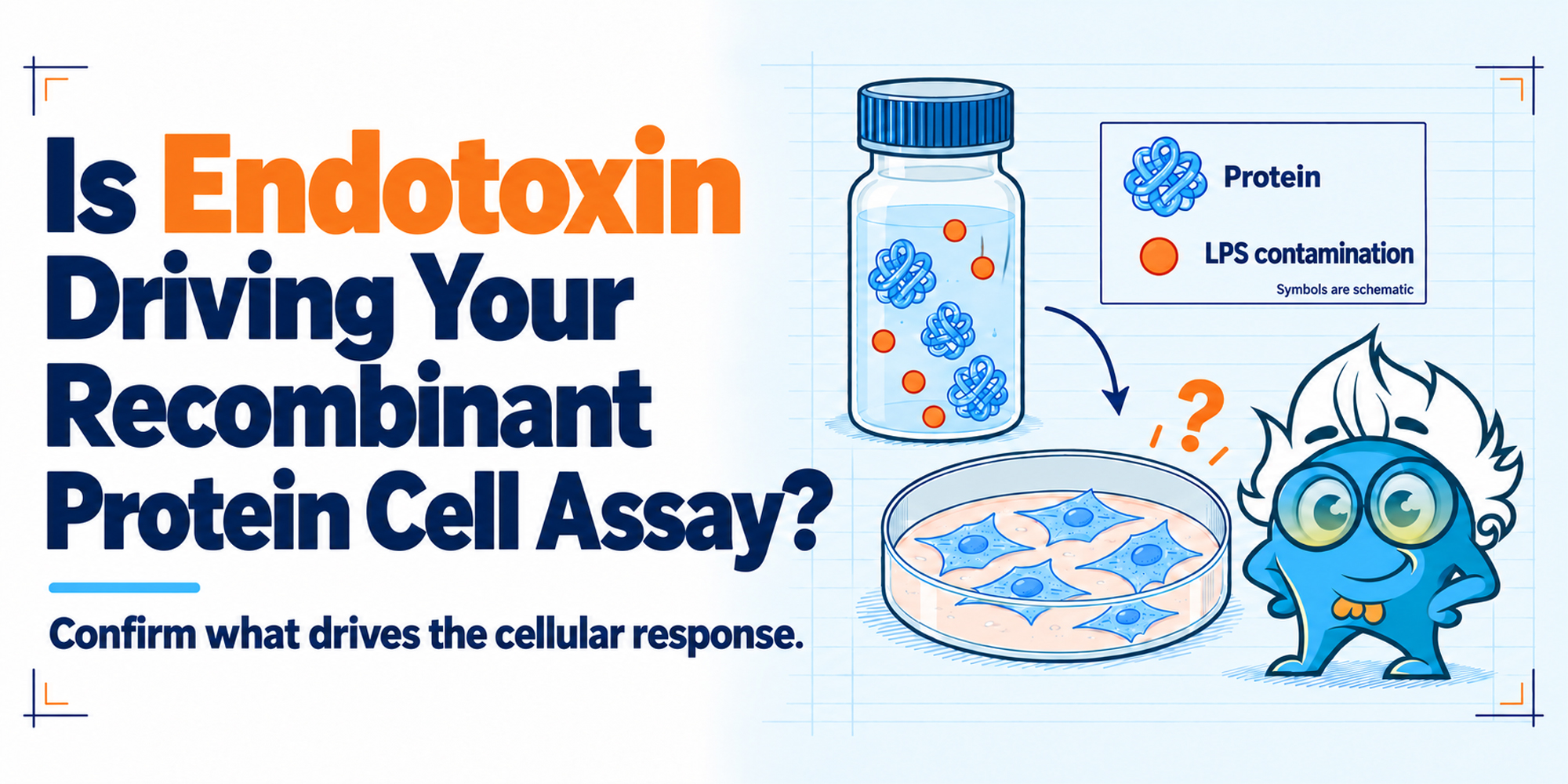
A recombinant protein changes your cells.
NF-κB increases. IL-6 or TNF-α rises. Cell viability drops. A differentiation marker shifts. The response may even increase neatly as the protein concentration goes up.
That is often taken as evidence that the recombinant protein is biologically active. In a cell-based assay, however, the observation and the cause are not always the same thing.
Residual endotoxin, particularly lipopolysaccharide (LPS) associated with Gram-negative bacterial expression systems such as E. coli, can trigger many of the same cellular readouts used to evaluate recombinant protein activity. Because expression host and downstream purification both influence the final preparation, Boster's Recombinant Proteins resource provides a useful overview of recombinant protein formats and workflows.
In sensitive models, even a relatively small amount of LPS can complicate interpretation.
So the practical question is not simply whether endotoxin is present. It is whether the amount delivered with the recombinant protein at the actual working concentration is enough to affect the cells.
That distinction should shape the control design from the beginning.
The biologically active lipid A region of LPS can activate Toll-like receptor 4 (TLR4) signaling in responsive cells. Monocytes, macrophages, dendritic cells, and other models with functional TLR4/CD14/MD-2 signaling are especially relevant.
Depending on the system, endotoxin exposure can alter NF-κB activity, cytokine secretion, surface markers, proliferation, viability, metabolism, or differentiation-related phenotypes.
Those are also common endpoints in recombinant protein bioactivity experiments.
If a protein preparation increases IL-6 secretion in macrophages, that result does not identify which component of the preparation caused the response. The readout only shows that the cells reacted.
This is why endotoxin becomes particularly important when a recombinant protein produces an inflammatory or stress-related phenotype that overlaps with known LPS responses.
This issue is especially relevant in pathway-based functional assays. Boster's Reporter Cell Lines for Pathway Activity Assays includes NF-κB, TLR/NF-κB, STAT, and related reporter systems that illustrate how pathway activation can become the assay endpoint itself.
A datasheet may report an endotoxin specification such as <1 EU/µg or <0.1 EU/µg. Those numbers are useful, but they only become experimentally meaningful once they are connected to the amount of protein added to the cells.
If a recombinant protein contains 0.1 EU/µg and is used at 5 µg/mL, the corresponding endotoxin exposure is approximately 0.5 EU/mL. At 20 µg/mL protein, the same preparation would introduce approximately 2 EU/mL.
That is why 'low endotoxin' cannot be interpreted independently of working concentration.
The same lot may be suitable for one assay and problematic in another simply because the dose, cell type, exposure time, or endpoint is different.
Cell sensitivity also matters. A transformed line with weak TLR4 signaling may tolerate an endotoxin concentration that produces a clear response in primary monocytes. An acute reporter assay may behave differently from a multi-day differentiation experiment.
A small LPS titration in the actual cell model is often more informative than adopting a generic endotoxin threshold from another application.
A clean concentration-response relationship is reassuring, but it does not by itself establish protein-specific activity.
| Recombinant protein | Cellular response |
|---|---|
| 1 µg/mL | Low |
| 5 µg/mL | Moderate |
| 10 µg/mL | Strong |
If the preparation carries a relatively constant amount of endotoxin per microgram of protein, increasing the protein dose also increases the endotoxin dose.
The dose-response may still be real, but the curve alone cannot tell you which component is driving it.
This becomes especially important when the observed phenotype resembles a typical LPS response or appears predominantly in endotoxin-sensitive cells.
Lot-to-lot variation can provide another clue. If two preparations of the same recombinant protein produce very different inflammatory responses, the difference may come from protein quality, aggregation, formulation, or endotoxin burden rather than sequence alone.
None of these patterns proves contamination. They simply justify a more deliberate control strategy.
A good cell assay should make the competing explanations experimentally separable.
The same principle applies broadly across assay design: controls should isolate the likely failure mode rather than simply provide a generic positive and negative reference. Boster's guide to How to Design Positive and Negative Controls provides additional examples of this control logic across common protein assays.
A buffer-only vehicle control should match the recombinant protein formulation as closely as possible.
Protein preparations may contain glycerol, salts, carrier proteins, reducing agents, detergents, or stabilizers. Any of these can affect cells independently of the target protein.
That control addresses formulation effects, but it does not address endotoxin carried with the protein.
Suppose endotoxin testing indicates that the protein treatment introduces about 0.2 EU/mL into the culture. A vehicle-treated condition containing approximately the same endotoxin exposure can show whether that amount alone changes the readout.
This is more informative than relying only on a very high-dose LPS positive control.
A strong LPS control still has value. It confirms that the cells can respond to LPS and shows what an LPS-driven phenotype looks like in that assay.
The most useful design often includes both a clear LPS positive control and an LPS concentration close to the endotoxin exposure introduced with the recombinant protein.
The first confirms responsiveness. The second asks whether the actual contamination level is biologically relevant.
If practical, testing the same protein preparation before and after a validated endotoxin-reduction step can be highly informative.
If the endotoxin level decreases substantially while the cellular phenotype remains similar, that supports a protein-dependent effect.
If the phenotype falls together with endotoxin reduction, LPS becomes a more plausible contributor.
This comparison is only interpretable if the cleanup step itself is characterized. Additional purification can change protein recovery, concentration, aggregation, oligomeric state, or buffer composition.
A lower cellular response after cleanup is not automatically evidence that endotoxin was responsible unless protein integrity and dose are also controlled.
Polymyxin B is often used because it binds the lipid A region of LPS and can reduce LPS-mediated activity.
A practical design can include Protein, Protein + polymyxin B, LPS, and LPS + polymyxin B.
If polymyxin B reduces both the LPS control and the protein-associated response, endotoxin involvement becomes more plausible.
If the LPS control is suppressed but the recombinant protein response remains, the result supports the possibility that the protein effect is not primarily driven by polymyxin-sensitive LPS.
The interpretation should remain cautious. Polymyxin B does not neutralize every LPS preparation equally, and at some concentrations it can affect cells independently.
It works best as part of a larger pattern of evidence.
Another useful question is whether the cellular effect depends on the structural integrity of the recombinant protein.
Depending on the target, a controlled heat-denaturation step, protease treatment, or another validated protein-inactivation approach can help.
| Treatment | Protein-dependent activity | Endotoxin-driven activity |
|---|---|---|
| Untreated protein | Response | Response |
| Protein + polymyxin B | May remain | May decrease |
| Denatured protein | Often decreases | May remain |
| Protease-treated protein | Often decreases | May remain |
| LPS + polymyxin B | - | Should decrease if the control is working |
This is not an absolute decision table. Some proteins are heat stable, and added treatment steps can introduce new variables.
The purpose is to determine whether the phenotype continues to follow the intact recombinant protein.
Where possible, pathway-level controls can strengthen the interpretation. A validated TLR4-blocking approach, a TLR4-deficient model, or comparison with a cell system lacking strong LPS responsiveness can help distinguish LPS-mediated signaling from the protein's intended pathway.
If viability or cytotoxicity is the primary endpoint, assay chemistry should also be considered separately from endotoxin effects. Boster's What Is the CCK-8 Assay? reviews how WST-8-based measurements reflect cellular metabolic activity and viability.
Orthogonal evidence is strongest when the expected protein function is measured through a relevant biological endpoint rather than inferred from purity alone. Boster recombinant protein pages may include recombinant protein bioactivity data based on functional cell assays, providing an example of how biological activity can be demonstrated independently of simple concentration or purity measurements.
For an initial cell-based qualification experiment, the following conditions cover most of the important questions:
| Condition | What it helps determine |
|---|---|
| Untreated cells | Baseline behavior |
| Vehicle control | Formulation effects |
| Recombinant protein | Observed treatment phenotype |
| LPS positive control | Whether the cell model responds to LPS |
| Endotoxin-matched LPS control | Whether the estimated endotoxin exposure could explain the phenotype |
| Protein + polymyxin B | Whether LPS contributes to the protein-associated response |
| LPS + polymyxin B | Whether polymyxin B is effective in this assay |
| Endotoxin-reduced protein | Whether activity survives endotoxin reduction |
| Denatured or protease-treated protein | Whether the response depends on intact protein |
| Orthogonal functional assay | Whether the expected protein biology can be confirmed independently |
Not every routine experiment needs every condition.
The broader point is that initial lot qualification should establish what the assay is actually responding to before a streamlined design is used for routine work.
Common endotoxin testing approaches include LAL-based assays and recombinant Factor C methods.
For cell-assay work, the choice of method matters less than three practical questions: Is the method sensitive enough around the endotoxin level that affects the cells? Does the protein formulation interfere with the endotoxin measurement? Was the tested material representative of the preparation actually used in the cell experiment?
The second question is easy to overlook.
Concentrated protein, detergents, salts, chelating agents, reducing agents, color, turbidity, and other formulation components can inhibit or enhance an endotoxin assay.
A low result is only useful if the method performs correctly in that sample matrix.
Two checks are especially helpful.
Dilution linearity asks whether the calculated endotoxin result remains reasonably consistent as the sample is diluted away from matrix interference.
Spike recovery asks whether a known amount of endotoxin added to the sample can actually be measured.
If a known spike cannot be recovered, a very low endotoxin result may reflect assay inhibition rather than a genuinely clean preparation.
This is why analytical endotoxin testing and biological controls should be used together. One estimates how much endotoxin is present; the other asks whether that amount explains the phenotype.
A recombinant protein is used at 5 µg/mL in a macrophage assay.
Measured endotoxin content: 0.1 EU/µg.
The treatment therefore introduces approximately 0.5 EU/mL into the culture.
The cells show increased NF-κB activity, TNF-α, and IL-6.
The high response is reproducible.
A 0.5 EU/mL LPS control also produces a measurable cytokine response.
Adding polymyxin B substantially reduces both the LPS response and the response associated with the recombinant protein.
An endotoxin-reduced preparation of the same protein produces much weaker cytokine induction.
Heat treatment of the protein does not eliminate most of the inflammatory phenotype.
Taken together, those results make endotoxin a plausible major contributor.
Now consider a second experiment.
The same recombinant protein produces a strong response, but 0.5 EU/mL LPS has little effect; polymyxin B suppresses the LPS control but not the protein response; endotoxin reduction does not substantially change the activity; disrupting the protein removes the response; and an independent receptor-specific assay confirms activity.
That pattern is much more consistent with genuine recombinant protein function.
The value comes from several results pointing in the same direction rather than from any single control.
For E. coli-expressed proteins, endotoxin risk begins with the expression system itself. Cell disruption releases bacterial membrane components into the preparation, and downstream purification must separate the target protein from them.
Contamination can also be introduced later through water, buffers, chromatography hardware, consumables, storage containers, or handling.
This means expression system changes the starting risk but does not define the final endotoxin status.
The same principle applies in the opposite direction: proteins produced in mammalian or insect systems should not automatically be treated as endotoxin-free. The preparation that reaches the cell assay reflects the entire production and handling workflow.
The choice of expression host and purification workflow affects not only protein yield and folding but also the contaminants that must be controlled. Boster's Recombinant Protein Production guide provides a broader overview of expression systems and purification strategies.
Several approaches can be used for endotoxin reduction, including anion-exchange chromatography, endotoxin-binding resins, polymyxin-based affinity methods, and detergent-based approaches such as Triton X-114.
The best method depends on the protein.
For cell assays, a successful cleanup step should preserve enough protein, maintain biological activity, avoid problematic carryover, and actually reduce the endotoxin burden when re-tested.
A lower EU value is not useful if the cleanup step also alters the protein in a way that changes the assay.
For projects that require tighter control over expression system, purification, buffer exchange, or endotoxin testing, Boster's Custom Recombinant Protein Expression Service includes multiple expression platforms together with downstream purification and QC options.
A low endotoxin specification is useful quality information, but it should not be treated as a universal guarantee of suitability for every cell assay.
The practical sequence is straightforward:
What is the endotoxin content per unit of protein?
How much protein is being added?
What endotoxin concentration does that introduce into the culture?
Does the cell model respond at that concentration?
Only then does the specification become meaningful for the experiment.
This is particularly important when recombinant proteins are used at relatively high concentrations or in primary immune-cell systems.
Endotoxin is difficult to ignore in recombinant protein cell assays because it can produce data that look entirely plausible.
The response may be reproducible. It may increase with protein concentration. The phenotype may even fit a biologically interesting story.
That still does not establish causality.
A stronger interpretation comes from calculating the actual endotoxin exposure, measuring how sensitive the cell system is to that exposure, and using controls that separate protein-dependent activity from LPS-dependent activity.
Measure endotoxin, but do not stop at the specification.
Use LPS at a relevant concentration. Compare untreated and endotoxin-reduced material when needed. Use polymyxin B or pathway controls where appropriate. Check whether the phenotype still requires an intact protein and whether the expected biology can be reproduced with an independent readout.
If the response continues to follow the recombinant protein after those alternatives are challenged, the biological conclusion becomes much more convincing.
For researchers who need support with expression-system selection, purification, characterization, or endotoxin QC, Boster's Recombinant Protein Expression resources provide an overview of available workflows and service options.







