This website uses cookies to ensure you get the best experience on our website.


- Table of Contents
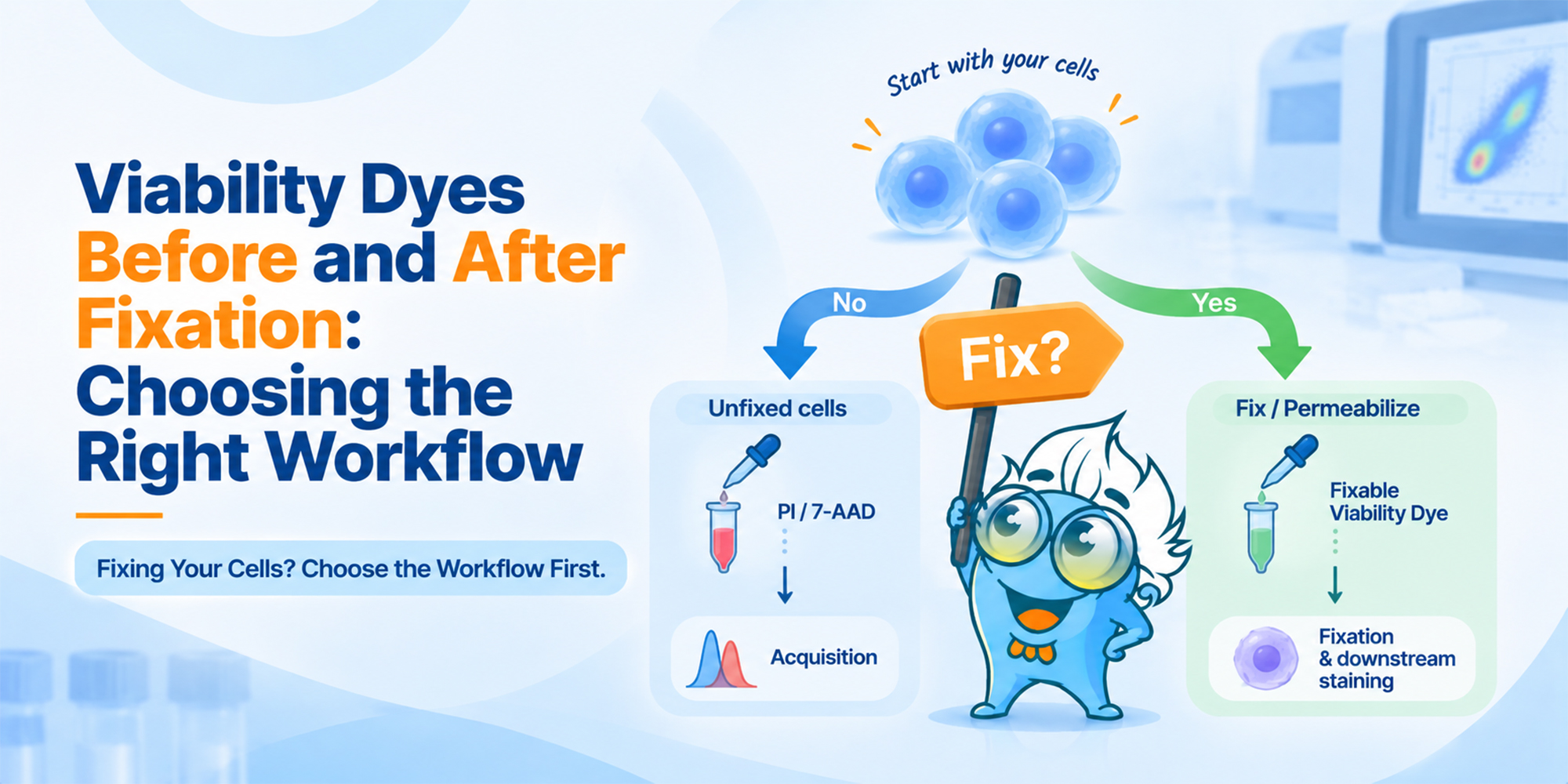
The first question is not which viability dye is brightest. It is whether the live/dead information has to survive fixation and permeabilization.
Choosing a viability dye for flow cytometry is often treated as a fluorophore-selection problem. In practice, it is usually a workflow decision first. If the cells will remain unfixed until acquisition, a membrane-impermeant dye such as PI or 7-AAD may be enough. If the cells will be fixed or permeabilized, viability usually needs to be recorded beforehand with a compatible fixable dye.
That distinction also clears up a common wording trap: a dye being “fixable” does not mean it should be added after the cells have already been fixed. It means the live/dead distinction established before fixation can remain detectable afterward.
Fast answer: If fixation or permeabilization will occur before acquisition, plan the viability stain before that step.
| Experimental workflow | Typical viability approach | When viability is established |
|---|---|---|
| Surface staining; acquire live | PI, 7-AAD, or another compatible nonfixable dye | Near the end of staining, before acquisition |
| Surface staining; fix before acquisition | Fixable viability dye | Before fixation |
| Surface + intracellular staining | Fixable viability dye | Before fixation/permeabilization |
| Intracellular cytokine or transcription-factor panel | Fixable viability dye | Before fixation/permeabilization |
| Phospho-flow | Fixable dye only if compatible with the timing and fixation method | Before fixation, using an assay-validated workflow |
| Sample already fixed without a viability label | Original membrane-integrity viability usually cannot be reconstructed | Not recoverable with a standard post-fix exclusion stain |
For a routine live-cell surface panel, membrane-impermeant DNA dyes provide a simple live/dead gate because intact cells exclude the dye while membrane-compromised cells take it up.
Typical sequence: Prepare cells → surface staining → wash → PI/7-AAD → acquire
PI and 7-AAD are commonly added near acquisition rather than early in the workflow. Flow cytometry guidelines note that these dyes can enter viable cells over time, so incubation and acquisition timing should be standardized. When setting up a routine surface-staining workflow, Boster's Flow Cytometry Protocol provides the overall staining sequence; if the starting suspension is variable or debris-rich, the Flow Cytometry Sample Preparation guide is the better place to troubleshoot upstream sample quality.
Once fixation or permeabilization changes membrane permeability, a dye that depends on membrane exclusion can no longer report the cell's original pre-fixation state in the same way. For intracellular panels, the usual solution is a fixable amine-reactive viability dye applied before fixation.
Common sequence: Prepare cells → fixable viability dye → surface staining as appropriate → fixation/permeabilization → intracellular staining → acquire
Exact ordering can vary with the antibody panel and manufacturer instructions, but the key point is stable: the viability information must be encoded while live and dead cells still differ in membrane integrity. For combined surface and intracellular panels, Boster's fixation and permeabilization optimization guide is useful when validating the next steps, because fixation can also change scatter, autofluorescence, epitopes, and fluorophore performance.
PI and 7-AAD work because viable cells with intact plasma membranes largely exclude them. When membrane integrity is lost, the dyes gain access to nucleic acids and the dead population becomes fluorescent. The measurement therefore depends on membrane integrity still having its biological meaning at the moment of staining.
Fixable viability dyes use a different strategy. Amine-reactive dyes label accessible protein amines. Live cells expose relatively few amines at the outer surface and remain dim; in dead cells, the compromised membrane allows much more dye to enter and react with intracellular proteins. Because that labeling is covalent, the difference can remain detectable after fixation and permeabilization. This principle is described in published flow cytometry guidelines and in a protocol paper on amine-reactive viability dyes.
Terminology check: “Fixable” describes retention of the label after fixation. It does not mean “fix first, then stain for the original viability state.”
An amine-reactive dye cannot distinguish between an amine on a cell and an amine on free protein in the staining solution. If serum or substantial BSA is present during the viability-labeling step, the reagent can be consumed by extracellular protein and reduce separation between live and dead cells.
A common approach is to wash the cells into PBS or another manufacturer-recommended protein-free buffer, perform the fixable viability stain, wash away unbound reagent, and then return to the appropriate antibody-staining buffer. The exact buffer and incubation conditions should follow the specific dye instructions.
If cells were fixed without a pre-fix viability label, adding PI, 7-AAD, or a fixable amine dye afterward does not generally reconstruct which cells had intact membranes before fixation. You may still measure DNA content or other death-related biology, but that is a different readout from pre-fix live/dead discrimination.
Annexin V is often mentioned alongside viability dyes, but it answers a different biological question. It detects phosphatidylserine exposure and is commonly paired with a membrane-integrity dye when apoptosis staging is the endpoint. If the goal is only to exclude dead cells from an immunophenotyping analysis, that extra layer is usually unnecessary; when apoptosis populations do matter, Boster's Flow Cytometry Gating Strategies guide shows the downstream gating context for these populations.
Phospho-flow is the clearest example. Phosphorylation can change rapidly after stimulation, so fixation may need to happen immediately. In that setting, do not insert a generic viability-staining step simply because it is standard elsewhere. Use a viability reagent and order that have been validated for the phospho-flow workflow, or prioritize preservation of the signaling state if the assay design requires it.
Dead and dying cells often move in FSC/SSC space, but scatter is not a reliable substitute for a dedicated viability marker. Nonviable cells can contribute increased autofluorescence and nonspecific reagent binding while still overlapping the scatter region of cells you would otherwise keep. Published guidelines therefore recommend active dead-cell exclusion when accurate analysis depends on a clean viable population.
Once the workflow has determined whether the dye must be fixable, the second decision is spectral. The viability dye still occupies fluorescence space, and its spillover can affect nearby markers. Avoid assigning the viability dye to a detector that compromises a low-abundance or biologically critical marker, and include the viability reagent in the compensation or spectral-unmixing plan.
The fixation method matters here as well. Some fluorophores and tandem dyes tolerate formaldehyde or methanol better than others. Validate the complete panel under the same fixation/permeabilization conditions used for the samples rather than assuming live-cell performance will carry over unchanged.
Do not choose a viability dye in isolation. First decide whether the live/dead information has to survive fixation. That decision usually determines the dye class and the order of the workflow.







