This website uses cookies to ensure you get the best experience on our website.
- Table of Contents
13 Q&As
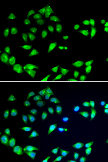
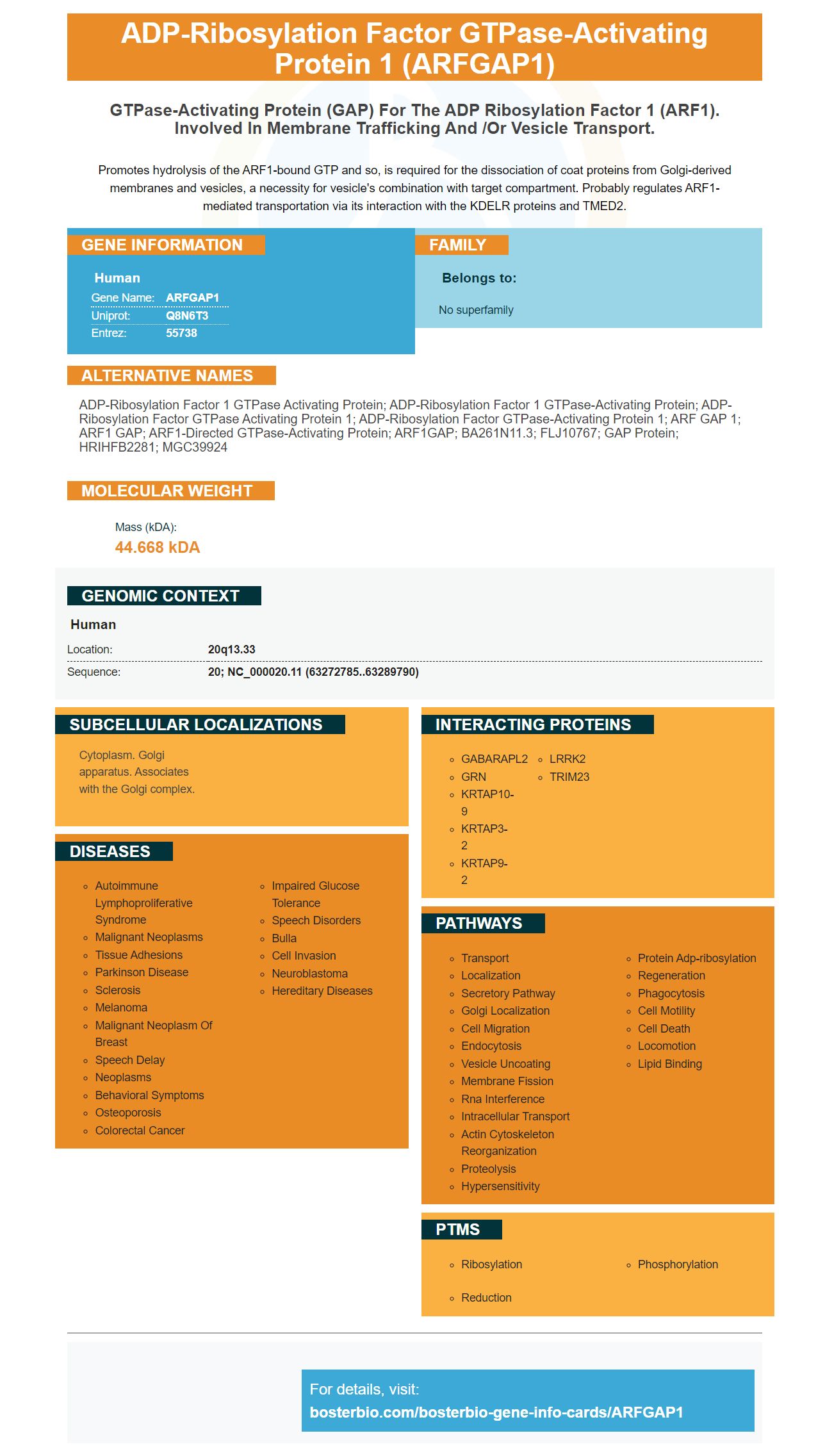
Facts about ADP-ribosylation factor GTPase-activating protein 1.
Promotes hydrolysis of the ARF1-bound GTP and so, is required for the dissociation of coat proteins from Golgi-derived membranes and vesicles, a necessity for vesicle's combination with target compartment. Probably regulates ARF1-mediated transportation via its interaction with the KDELR proteins and TMED2.
Human | |
---|---|
Gene Name: | ARFGAP1 |
Uniprot: | Q8N6T3 |
Entrez: | 55738 |
Belongs to: |
---|
No superfamily |
ADP-ribosylation factor 1 GTPase activating protein; ADP-ribosylation factor 1 GTPase-activating protein; ADP-ribosylation factor GTPase activating protein 1; ADP-ribosylation factor GTPase-activating protein 1; ARF GAP 1; ARF1 GAP; ARF1-directed GTPase-activating protein; ARF1GAP; bA261N11.3; FLJ10767; GAP protein; HRIHFB2281; MGC39924
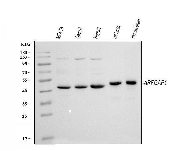
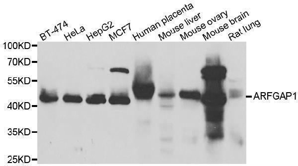
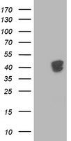
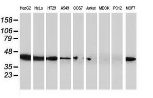
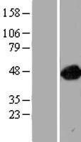
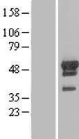
Mass (kDA):
44.668 kDA
Human | |
---|---|
Location: | 20q13.33 |
Sequence: | 20; NC_000020.11 (63272785..63289790) |
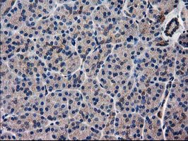
Cytoplasm. Golgi apparatus. Associates with the Golgi complex.
If you are looking to find an anti-GFAP anti-body, you have come to the right place. We have information about Boster Bio as well as Anti-Bax(S4), Anti–MT1-MMP and more. We're available to answer your questions regarding optimizing your research. Keep reading to find out more about these products, and how they could benefit your research.
It is critical to choose the correct antibodies for your ELISA design. The best way to do this is by using a concentrated antibody. It is also important to use a blocking buffer. It is crucial to use the correct buffer. Boster Bio offers optimization tips and guidelines to help you design the perfect ELISA. These guides will answer all your questions about your ELISA.
DCLOAO derivatives increased early apoptotic rates in mice, while topotecan showed a proapoptotic action. The resulting protein expression levels of Bax and Caspase-3 were increased. These compounds are much more toxic than topotecan. It is important to use anti–Bax(S4) Boster Bia with caution.
Recent research has shown that MT1 and MMP can be used to transfect HT1080 human fibrosarcoma cell lines. The researchers used immunofluorescence imaging to determine the level of MT1–MMP expression. This allowed them to detect membrane deposits on the cells and detect MT1–MMP at the cell surface. To quantify the activity of this protease, 5 x 5 images of the cells were taken in two places on each cover slip, and the total intensity of the MT1-MMP-positive spots was quantified using an analysis program for the Olympus ScanR microscope. All images were taken under the same conditions and settings, as described above.
This antibody was generated by using synthetic peptides to recognize MT1-MMP. It binds to both the MT1–MMP subunit and its pro-enzyme version. It recognizes both the 55- and 34-kDa processed forms of the enzyme. This antibody did not detect MT2-MMP. The data for this anti MT1–MMP boster biologic product are not yet available.
MT-1, MMP is a type metalloproteinase derived form a murine cell. It regulates extracellular micromolecules and is developmentally regulated. MT-1 -MMP secreted mesenchymal fibroblasts. Membrane-associated metalloproteinase activates it. MMP-2 is inhibited when STP-2 is present.
This protein is often lost due to metastatic cancers. It acts as a gatekeeper of VAMP3-recycling. It reduces the proliferative and invasion potential of cancer cells by removing it from their cells. This increases the risk of death from metastasis. The role of matrix-metalloproteinases is critical in the progression and control of cancer.
The recombinant MT1–MMP protein was obtained using soluble GST from E. coli. It was then combined with CNBr-activated Spharose B. Cells were plated on a Labtek slide at 3.2x10⁴ cells in each well. Incubation was performed for 1 min in glycine buffer containing 15% glycerol. After incubation, the specific antigen was extracted from the Nitrocellulose.
PMID: 14702039 by Ota T., et al. Complete sequencing and characterization of 21,243 full-length human cDNAs.
PMID: 11780052 by Deloukas P., et al. The DNA sequence and comparative analysis of human chromosome 20.