This website uses cookies to ensure you get the best experience on our website.
- Table of Contents
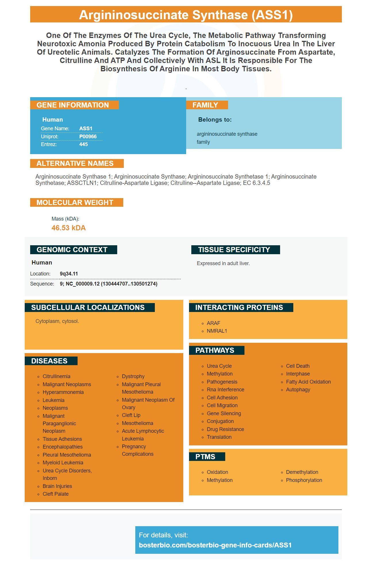
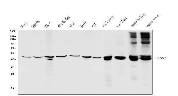
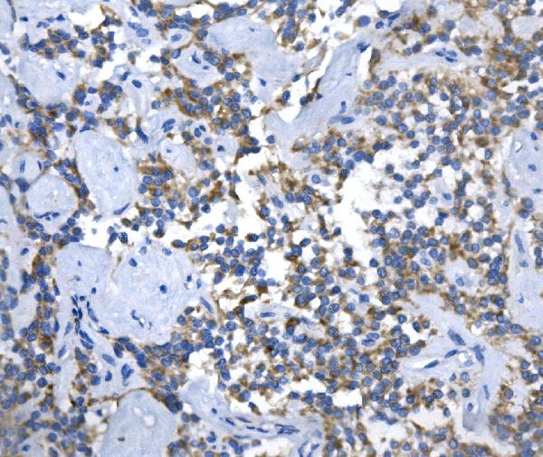
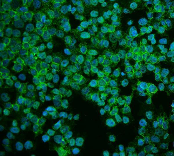
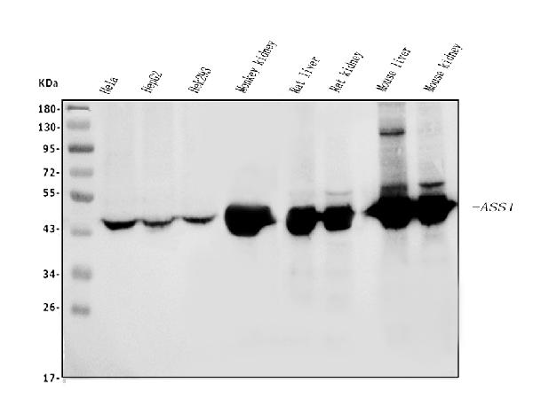
Facts about Argininosuccinate synthase.
.
Human | |
---|---|
Gene Name: | ASS1 |
Uniprot: | P00966 |
Entrez: | 445 |
Belongs to: |
---|
argininosuccinate synthase family |
argininosuccinate synthase 1; argininosuccinate synthase; argininosuccinate synthetase 1; argininosuccinate synthetase; ASSCTLN1; citrulline-aspartate ligase; Citrulline--aspartate ligase; EC 6.3.4.5
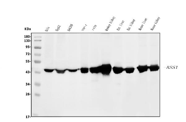
Mass (kDA):
46.53 kDA
Human | |
---|---|
Location: | 9q34.11 |
Sequence: | 9; NC_000009.12 (130444707..130501274) |
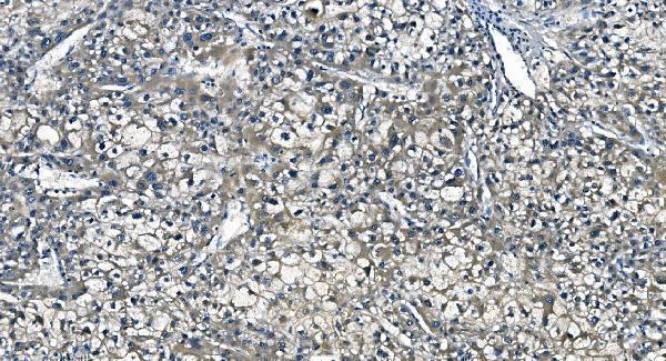
Expressed in adult liver.
Cytoplasm, cytosol.
Boster Bio has an Anti-ASS1 Picoband(tm), Antibody, catalog number A02212-1. This antibody reacts to Humans as well as Rats, Dogs, and Mice. The product contains Trehalose and Na2HPO4 in addition to the recombinant human as well as mouse ASS1 protein in positions S3-S365. You can purchase a blocking peptide for this antibody at an amount based on the length of the immunogen.
The Anti-ASS1 marker in BoSTER Bio is a recombinant protein that has an amino acid sequence that is S3-S365. It is versatile and can be used in a variety of areas of research. This antibody reacts with range of species including mice, humans, and rats. You can also purchase this antibody with blocking peptides for a tiny amount, based on length and cost.
The ASS mRNA expression in pancreatic cancer cells of humans and breast cancer cell lines has been studied. Western Blotting has demonstrated that ASS protein expression in the cell cytoplasms of BxPC-3 pancreatic cancer cells was comparable to the mRNA level. In contrast, immunohistochemical analyses of SW1990 pancreatic cancer cells failed to reveal significant ASS protein expression. For these reasons, the Anti-ASS1 Marker in Boster Bio is a valuable tool for performing tumor immunohistochemistry.
ADI decreased the viability of ASS deficient and ASS-expressing lines. However, it had no effect on cell expansion. The ADI inhibitory level of 50% (IC50) was 1 mU/mL in PANC-1 cells. This implies that ADI is extremely effective in detect the presence of ASS in cancer cells. Additionally, ADI inhibits the production of certain proteins like p-gph-receptors and the NF-kB.
The Anti-CPS1 Marker found in Boston Bio is an antibody with a catalog id of A01320-1. It is suitable for IP, IF and IHC applications. It reacts with Human, Mouse and Rat. Numerous studies have been conducted using the boster bio CPS1 antibody for various purposes including immunohistochemistry as well as cell cultivation. It is safe to use in a variety of applications and is effective in blocking the CPS1 activity in various cancer cells.
It is important to recognize that the lower expression of CPS1-IT1 was associated with poor overall and disease free survival. CPS1-IT1 expression in tumors can be used as a molecular indicator to help identify patients at a high risk for treatment failure. This is a promising technique in the diagnosis and treatment for cancer patients. There is still much to learn about CPS1's functions in this disease.
The physiological significance of the interaction between CPS1 & SIRT5 is obvious. CPS1 activity is controlled by sirt5 in mice. The sirt5 KO mice failed to induce the deacetylation of CPS1 by starvation. The prolonged starvation process causes CPS1 protein deacetylation in wild type mice. The CPS1 activity of SIRT5 mice was extremely small or insignificant.
In addition to binding to Hsp90 CPS1-IT1 also interacted Hsp90 and, as a result, decreased the binding affinity between Hsp90 and HIF-1a. These interactions controlled the expression of EMT-related protein. However, CPS1-IT1 cells did not block HIF-1a activity. Thus, anti-CPS1 in Boster Bio has numerous clinical applications.
For positive and negative control, we used liver mitochondria matrix extracts from SIRT5 knockout mice as well as wild type littermates. After that, we applied an the anti-CPS1 marker in western blots to determine CPS1 activity. The results were presented as a percentage of the wild-type value at the time zero. Error bars indicate the standard deviations for three experiments.
Boster Bio's anti-CPS1 Monoclonal antibodies react with mice, human and rat. The product has been thoroughly tested to ensure specificity and is not hazardous. The Boster Bio Anti-CPS1 Monoclonal Antibody is identified under the catalog number A01320. It is recommended for use in the laboratory. The Boster Bio Anti-CPS1 antibody is able to be purchased without BSA. It has a specificity target of 99%.
It is the market leader in high-quality primary antibodies because of its expertise in the creation of high-affinity antibodies. It has spent the past two decades perfecting its methods and techniques and has been cited in over 29,000 scientific publications. Its products have undergone rigorous validation processes in ELISA, Flow Cytometry, Western Blotting, and Immunohistochemistry.
This monoclonal antibody made by Boster Bio can be utilized in a variety of situations. It is able to bind ASS1 and has been validated through Western Blotting, IHC-P, and Flow Cytometry. It reacts with Rat, Mouse, and Human. Scientists who use this product should contact Boster Bio if they are working on a specific sample or application.
ADI treatment inhibited the growth of cells in 21/25 (84%) MMC cell lines. However only four of the 21 (33 percent) cells from MM expressed constitutive ASS. Cells with constitutive ASS expression showed a markedly higher IC 50 value. Additionally the metastatic and primary MM samples showed similar responses to ADI treatment.
The antibody targets the arginine deminase (ASS), which is a protein implicated in a myriad of metabolic pathways as well as the production of creatine. It was also used to treat tumors sensitive to arginine deprivation. ASS is essential to create endogenous arginine. Researchers analyzed OTC expression in 25 malignant cell lines that are primary and normal fibroblasts.
PMID: 6194510 by Bock H.-G.O., et al. Sequence for human argininosuccinate synthetase cDNA.
PMID: 6321498 by Freytag S.O., et al. Molecular structures of human argininosuccinate synthetase pseudogenes. Evolutionary and mechanistic implications.