This website uses cookies to ensure you get the best experience on our website.
- Table of Contents
1 Citations 17 Q&As
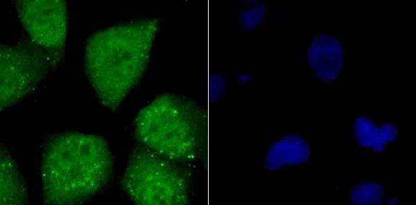
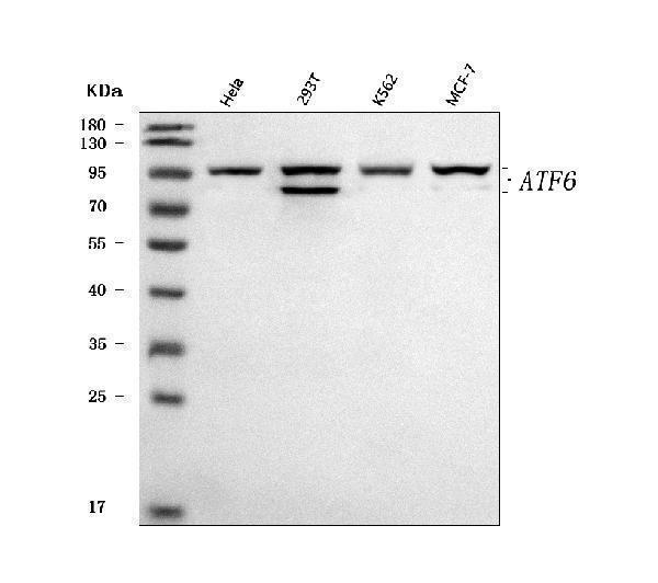
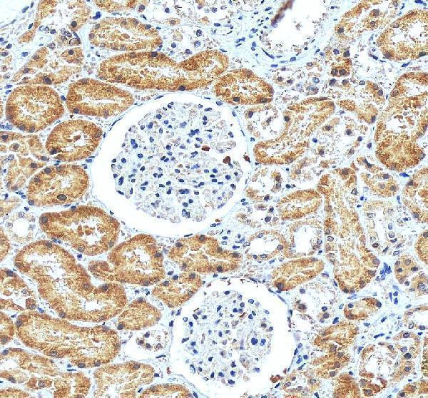
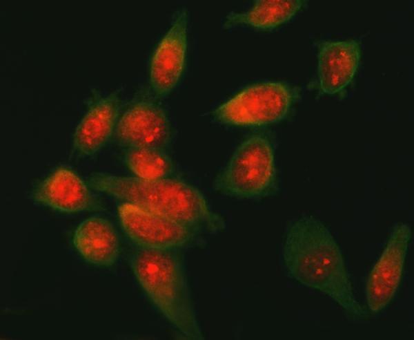
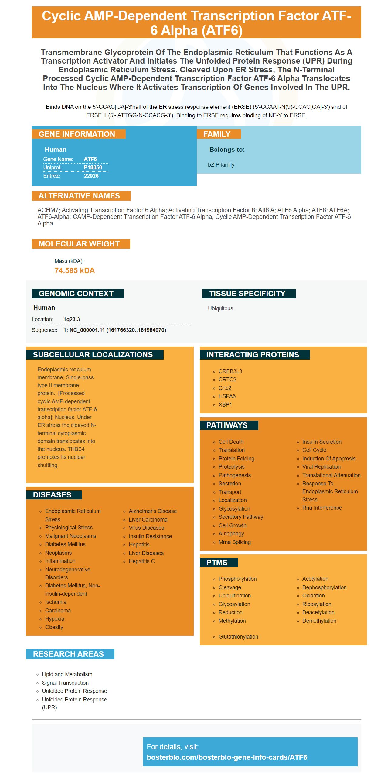
Facts about Cyclic AMP-dependent transcription factor ATF-6 alpha.
Binds DNA on the 5'-CCAC[GA]-3'half of the ER stress response element (ERSE) (5'-CCAAT-N(9)-CCAC[GA]-3') and of ERSE II (5'- ATTGG-N-CCACG-3'). Binding to ERSE requires binding of NF-Y to ERSE.
Human | |
---|---|
Gene Name: | ATF6 |
Uniprot: | P18850 |
Entrez: | 22926 |
Belongs to: |
---|
bZIP family |
ACHM7; Activating transcription factor 6 alpha; activating transcription factor 6; atf6 a; ATF6 alpha; ATF6; ATF6A; ATF6-alpha; cAMP-dependent transcription factor ATF-6 alpha; cyclic AMP-dependent transcription factor ATF-6 alpha
Mass (kDA):
74.585 kDA
Human | |
---|---|
Location: | 1q23.3 |
Sequence: | 1; NC_000001.11 (161766320..161964070) |
Ubiquitous.
Endoplasmic reticulum membrane; Single-pass type II membrane protein.; [Processed cyclic AMP-dependent transcription factor ATF-6 alpha]: Nucleus. Under ER stress the cleaved N-terminal cytoplasmic domain translocates into the nucleus. THBS4 promotes its nuclear shuttling.
There are a variety of uses for the ATF6 marker. This article will focus on the most significant. Continue reading to find out more about the role of ATF6 in the UPR and ocular development and hepatic Steatosis. To further your knowledge, you can make use of this boster bio: The Best Uses of the ATF6 Marker to enhance your understanding of these topics. For more details, go through the entire article.
There are numerous branches to the UPR pathway, including the IRE1 or ATF6 branches. Both are thought to be prosurvivaland protect cardiomyocytes from damage due to ischemic. The IRE1 pathway is believed to be involved in the splicing process of the Xbp1 transcript, which results in a frame shift in the codon region and the production of the 54 kDa protein.
The transcription factor ATF6 regulates gene transcription. The transmembrane protein, known as ATF6 is activated when misfolded proteins build up within the ER. ATF6 activates a gene that is responsible for ER proteins, which in turn increases the rate of folding proteins in the ER. Activation of ATF6 gene during UPR is an important aspect of the ER proteostasis system.
Activation of ATF6 is a new pathway involved in regulating the antioxidant capacity of cells. When cells are exposed ROS and antioxidant levels in the cell decrease. Protein levels of SOD, catalase and GR also decrease. These results suggest that the ATF6 marker during the UPR pathway is a key regulator of cellular death. Also, increased levels ATF4 protein and caspase-4 protein are indicative of an apoptotic state.
The cleavage and activation of ATF6 markers in an ER-mediated URPR procedure is tied to the ATF6 protein. Activation of ATF6 marker during the UPR process is achieved by S1P/S2P and then imported into the nucleus via the THBS4.
ATF6 isn't the only signaling pathway that is important in the ER stress response. IRE1 as well as CHOP are also crucial. They are responsible for regulating the levels of ER stress as well as the ability of cells to live. The ER stress response requires the coordinated activation of these signaling pathways. Scientists can examine the UPR of the ER by detecting activation of IRE1 and ATF6.
Activation UPRER causes a global decrease in protein synthesis , and the degradation of mRNA related to ER membrane. This activation could clear the ER proteome of its somatic signature , and allow for the reprogramming of the ER to a pluripotent state. The UPRER may be temporary and function as a buffer for cells when reprogramming is taking place. It is not known how this signaling mechanism works.
The ROS caused by UPRs was also observed in human lens. UPR-induced ROS produced a decrease of antioxidant levels and an increase in the apoptotic signaling protein which, in turn, caused cataract formation. In the long run this could lead to the development of age-related cataracts. This study has shown that the UPR is crucial in the development of cataracts and could play a role in the treatment and prevention of this condition.
This study suggests that activating XBP1s can significantly improve reprogramming efficiency. However, activation does not directly correlate with the rate of iPSC programming. It is more related to the rate of activation of the UPRER. These factors will determine how effective the reprogramming process is.
It is not known what role Activation of ATF6 plays in ovarian development. However, it is known to play a role in apoptosis as well as contributing to the death of photoreceptor cells. The ER stress response, in turn, is known to play an important part in the development of cone and rod cells. This response is tightly controlled and may aid in vascular renewal and the regulation of repairative angiogenesis.
The activation of ATF6 throughout ocular development has been linked with eye disease. Recent studies suggest that the ATF6 protein is involved in the early differentiation of stem cells. ATF6 signaling promotes the differentiation of mesodermal stem cells lines. In addition, it is involved in the loss of pluripotency and Achromatospia. However further research is required to better understand the role played by ATF6 in the development of ocular vision.
In the eye, ATF6 is expressed in all retinal layers. It is also present in cones as well as the inner and outer segments of photoreceptor cells. Additionally, ATF6 has a role in the processing of visual information. These processes may be mediated by activation of ATF6 during the development of the eye. This study indicates that the ATF6 protein is a critical component of retinal ganglion cells, but more research is needed.
An ATF6 deletion in Pakistan has been linked to an eye disease that is severe, called ACHM. This is due to the loss of exons 2 and 3 , which are present in ATF6. The patient was diagnosed with nystagmus in infancy, impaired vision, and photophobia. A fundus exam revealed that the patient had an overcrowded optic disc in both eyes. Fundus autofluorescence imaging showed mild macular atrophy. Spectral domain optical coherent imaging (SDOCT) showed focal disruption in inner segment of the ellipsoid.
Although activation of ATF6 is not yet understood in Achromatopsia but it is believed that ATF6 could play a role in the pathology of the condition through disrupting the normal function of cone photoreceptor cell. Cone photoreceptor cell development depends on the proper folding of OS proteins. This could be affected by the absence of fully developed ER.
ATF6 is found in all pigment epitheliums of retina in mice. There are two forms of ATF6A: ATF6A, and ATF6B. Both are expressed in the cones and function as functional components of the phototransduction cascade. It is crucial to understand that ATF6a as well as ACHM have been linked to both ATF6b and the adult retina.
Mutations in ATF6 has been linked to rod-cone photoreceptor degeneration in patients with ACHM. Similarly, ATF6 mutations in other ACHM genes have been shown to contribute to ACHM. ATF6a is found throughout the retina, with its strongest expression in photoreceptor-ganglion cells. It is also expressed in peripheral cones.
Recent research has proven that ATF6 overexpression in the liver increases fat acid oxidation, and protects from hepatic steatosis in insulin resistant mice. While the function of ATF6 isn't clear, strategies to activate ATF6 can improve liver function and offer an alternative treatment option for patients suffering from hepatic steatosis.
ATF6 activation inhibits lipogenesis by inhibiting transcription SREBP-2. It also inhibits the activity of PPAR-g transcription factors. The activation of ATF6 in hepatic steatosis can increase the burden of unfolded proteins in the ER. The activation of ATF6 reduces the synthesis of fatty acids in the liver in hepatic statosis.
Steatosis activation of ATF6 causes liver damage, as it hinders the synthesis of FFA. It also raises the levels of ROS in the liver. Apoptosis and inflammation are a result of these processes. The activation of ATF6 in hepatic inflammation is linked to the progression of NASH to NAFL.
ATF6 activation triggers autophagy, inflammation and apoptosis within the liver. Increased levels of ROS result in the apoptosis and death of hepatic cells, which further contributes to NAFLD progression. Therefore, the role of ATF6 in hepatic steatosis must be clarified.
Activation of ATF6 in steatosis, as well as other inflammatory bowl diseases may be a connection. Steatosis and damage to the liver may be caused by inflammation and ER stress. This inflammation may exacerbate the effects of ATF6 on the hepatic steatosis. These results highlight the importance of ER stress and hepatic steatosis in the liver.
PMID: 9271374 by Zhu C., et al. Interaction of ATF6 and serum response factor.
PMID: 9837962 by Yoshida H., et al. Identification of the cis-acting endoplasmic reticulum stress response element responsible for transcriptional induction of mammalian glucose-regulated proteins; involvement of basic-leucine zipper transcription factors.
*More publications can be found for each product on its corresponding product page