This website uses cookies to ensure you get the best experience on our website.
- Table of Contents
Facts about Caldesmon.
In muscle cells, inhibits the actomyosin ATPase by binding to F-actin. This inhibition is attenuated by calcium-calmodulin and is potentiated by tropomyosin.
Human | |
---|---|
Gene Name: | CALD1 |
Uniprot: | Q05682 |
Entrez: | 800 |
Belongs to: |
---|
caldesmon family |
CAD; CALD1; caldesmon 1; Caldesmon; CDM; CDMH-CAD; HCAD; LCAD; L-CAD; MGC21352; NAG22
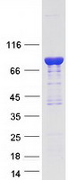
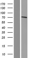
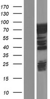
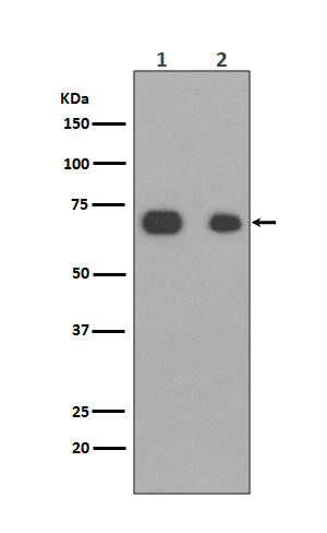
Mass (kDA):
93.231 kDA
Human | |
---|---|
Location: | 7q33 |
Sequence: | 7; NC_000007.14 (134779629..134970729) |
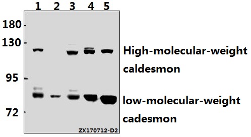
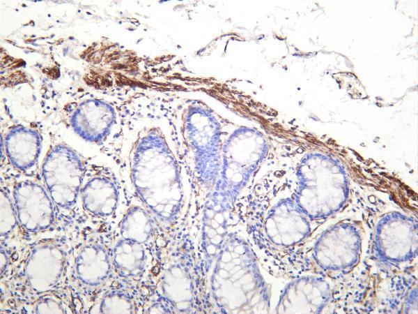
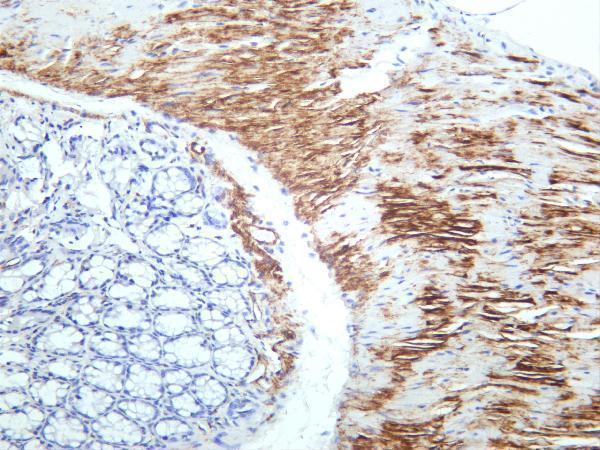
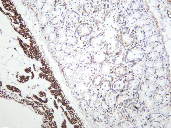
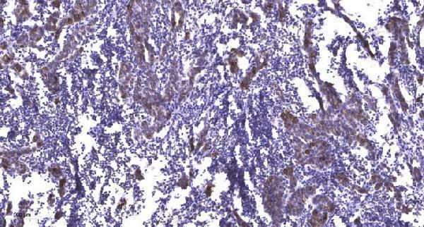
High-molecular-weight caldesmon (isoform 1) is predominantly expressed in smooth muscles, whereas low-molecular- weight caldesmon (isoforms 2, 3, 4 and 5) are widely distributed in non-muscle tissues and cells. Not expressed in skeletal muscle or heart.
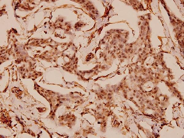
Cytoplasm, cytoskeleton. Cytoplasm, myofibril. Cytoplasm, cytoskeleton, stress fiber. On thin filaments in smooth muscle and on stress fibers in fibroblasts (nonmuscle).
Boster Bio's CALD1 marker can be used for a variety of purposes and is versatile. This protein is part of the CALD superfamily and is extensively utilized in biomedical research. It can be analyzed using an anti-DDK affinity columns and conventional chromatography steps to analyze it. It is used by researchers from all over the world.
Analyzing the reliability of the CALD1 gene expression marker was conducted. It was found to be in line with markers of TAM and M2 macrophages as well as neutrophils, and immune cells. It also showed a correlation with markers of Treg and T cell exhaustion. However this marker comes with limitations. More research is needed to confirm its reliability. There are no alternatives to CALD1.
In this study, researchers showed that the expression of CALD1 is positively associated with the proportion of M2 macrophages and M0 macrophages. However, the expression of the gene was not correlated with the percentage of CD8 T cells, resting NK cells, and dendritic cells. The number of plasma cells was also negatively associated with CALD1. However, CALD1's expression is strongly associated with the grade of glioma.
The CALD1 marker showed promising results. This suggests that the gene may be involved with chemokine signaling, TAM recruitment and/or polarization. It also has the potential to function as biomarker and possibly a therapeutic target in the stage III/IV of pMMR CRC. These results, while not yet validated fully, prove that CALD1 could be a biomarker.
The CALD1 gene encodes the protein Atypical polypoid Adenomyoma. The gene has been associated with Integrin Pathway, Smooth Muscle Contraction, and Gene Ontology annotations for myosin binding and actin binding. LSP1 is an important paralog to CALD1.
The CALD1 gene is a possible molecular marker that could be related to BC prognosis. This gene can promote malignant progression by upregulating the expression of PD-L1 through the JAK/STAT signaling pathway. Researchers have recently discovered that CALD1 is associated with a low overall survival rate and a lower rate of survival for patients who are disease-free. This gene promotes tumor cell proliferation and inhibits the process of apoptosis.
CALD1 expression is highly associated with TAM monocyte, TAM and M2 macrophage markers. The gene is also linked with T cell exhaustion and Tregs. However, this relationship isn't fully understood. Further research is required to understand the role played by CALD1 in the pathogenesis and progression of disease. Researchers are currently studying its role in the process of cancer diagnosis. However, CALD1's benefits may outweigh its cost.
A recent study suggests that CALD1 is a prognostic marker in pMMR stage III/IV. Its expression correlates with M2 macrophage infiltrations in this subtype. Additionally, CALD1 is associated with the TGF-b signaling pathway and angiogenesis gene sets. This gene is positively associated with immune and stromal ESTIMATE scores. Additionally, CALD1 promotes the proliferation and invasion of CRC cells.
Researchers from China have discovered that CALD1 gene expression strongly is associated with T cell activity. Because CALD1 is an overlap gene, and does not have other genes, this result was possible. This allows CALD1 to detect T cells that are activated under conditions of inflammation. However, this association is not fully established. Further research is needed. In the meantime, CALD1 remains a promising biomarker for the treatment of cancer.
PMID: 1885618 by Novy R.E., et al. Characterization of cDNA clones encoding a human fibroblast caldesmon isoform and analysis of caldesmon expression in normal and transformed cells.
PMID: 1555769 by Humphrey M.B., et al. Cloning of cDNAs encoding human caldesmons.