This website uses cookies to ensure you get the best experience on our website.
- Table of Contents
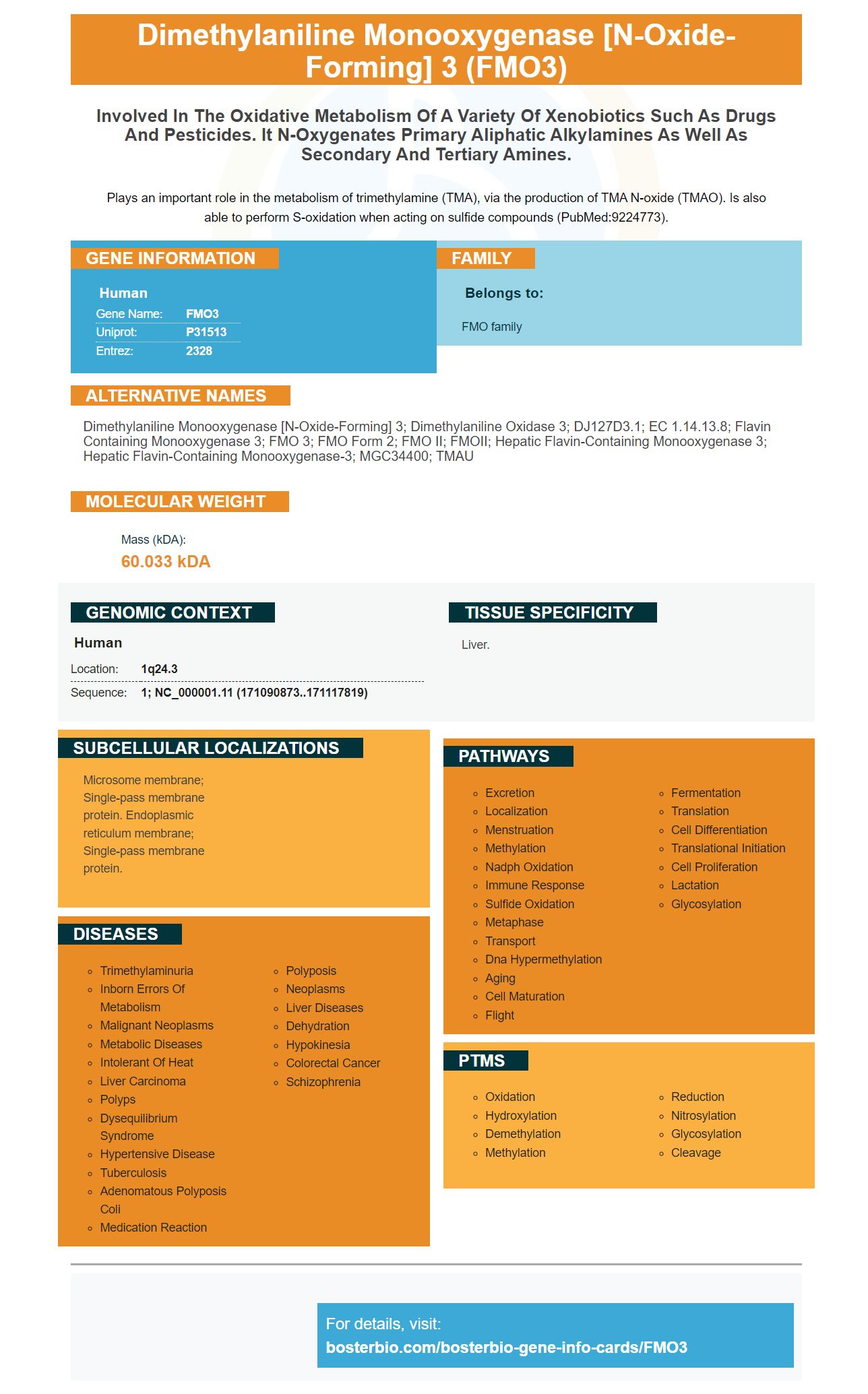
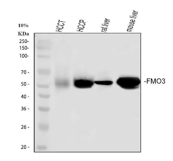
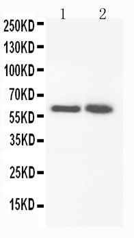
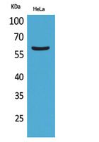
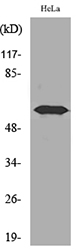
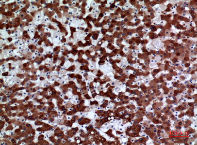
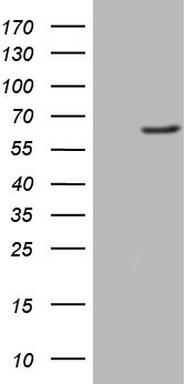
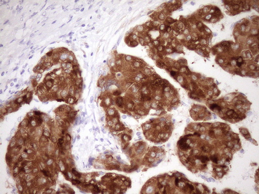
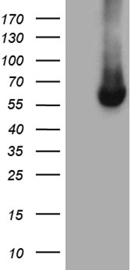
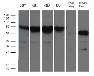
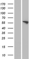
Facts about Dimethylaniline monooxygenase [N-oxide-forming] 3.
Plays an important role in the metabolism of trimethylamine (TMA), via the production of TMA N-oxide (TMAO). Is also able to perform S-oxidation when acting on sulfide compounds (PubMed:9224773).
Human | |
---|---|
Gene Name: | FMO3 |
Uniprot: | P31513 |
Entrez: | 2328 |
Belongs to: |
---|
FMO family |
dimethylaniline monooxygenase [N-oxide-forming] 3; Dimethylaniline oxidase 3; dJ127D3.1; EC 1.14.13.8; flavin containing monooxygenase 3; FMO 3; FMO form 2; FMO II; FMOII; Hepatic flavin-containing monooxygenase 3; hepatic flavin-containing monooxygenase-3; MGC34400; TMAU
Mass (kDA):
60.033 kDA
Human | |
---|---|
Location: | 1q24.3 |
Sequence: | 1; NC_000001.11 (171090873..171117819) |
Liver.
Microsome membrane; Single-pass membrane protein. Endoplasmic reticulum membrane; Single-pass membrane protein.
The most important tools used to determine the affinity of secondary antibodies are fluorescence without one (FMO) controls, as well as high-affinity prima antibodies. This article will discuss the use of these controls, optimization, and troubleshooting. This article will help you optimize your Boster Bio experiments and improve the results. You're bound to encounter difficulties during your experiments. While proper controls can remove numerous errors, it's often necessary to refer to a troubleshooting manual to determine the source of your experiment's difficulties.
Controls using Fluorescence Minus One are commonly used for multicolour experiments. They consist of cells stained with all fluorochromes, with the exception of one. FMO controls are essentially gate controls that permit researchers to effectively to gate. To determine whether FMO is necessary for a specific study Consider a sample which contains the same fluorochromes in the sample. Then, select an FMO control for each staining stage.
Fluorescence Minus One (FMO) controls are a vital part of multicolor flow cytometry panels. They assist in setting the gate by reducing fluorescence spread which is a problem following compensation and cross-laser excitation. Fluorescence Minus One controls (FMO) are a great solution as they incorporate all fluorochromes within one panel. They enable accurate identification of the fluorescence spread.
FMOs provide more accurate controls than unstained samples as they take into account spillover spread. However, FMO controls cannot account for low basal expression levels. Autofluorescence and secondary marker expression levels can also affect FMO controls. FMO controls must be made up of the same kinds of cells as the experimental specimens. You cannot substitute them with irrelevant cell lines or beads.
In Immunohistochemistry, you can use antibodies to detect antigens in cells. It is possible to make these antibodies more specific by concentration. In addition you can enhance your immunohistochemistry process when you select the appropriate method for preparation of your sample. ELISA optimization is a process that involves many steps that include sample fixation, embedding, antigen retrieval and the concentration of antibodies. The guide to Immunohistochemistry optimization gives technical advice and troubleshooting tips. In addition to the guide, Boster offers a comprehensive technical resource library that includes a blog with details about the disease.
Most likely, you've heard of the FMO3 marker. FMO is an enzyme that is membrane-associated and is found in all kinds of secretory cells, including lung. Furthermore, it is expressed by both genders. So, what exactly is it and how can you fix it? Here are some suggestions to help.
In order to identify the clonal antibody sequences high-affinity antibody have the FMO3 marker. This marker is derived from a gene within the Fab fragment. It allows reconstruction of the lineages of antibodies by inferring germ line progenitor sequences. The sequences of these progenitors could differ from the sequences of their ancestral lineage that is not altered. Mutations in the VL and VH gene segments are easy to identify but the VLJLJH junctional sequencing isn't. It is also important to know the insertions and deletions of Fab fragments in the process of maturing affinity.
To identify antigens with potential First, we obtained full-length sequences of amino acids in rat FMO1 (and -MO4) from the National Center for Biotechnology Information. These protein peptides then were studied using GeneRunner. After identifying the peptides, we conducted homology analysis to confirm their specificity. Utilizing the FMO3 marker, we found that the antigens recognized FMO3 molecules and were capable of recognizing it.
The FMO3 sequence of the rat is different from the mouse FMO3 proteins. This study also identified the FMO4 antibody in rat tissues and identified the immunochemical location of the rat FMO4 proteins in human and mouse tissues. This study demonstrates that FMOs are well-positioned within the tissues of rat. However, further research is needed to understand the role they play in FMO-mediated reactions.
The FMO3 marker allows for the identification of peptides with high affinity in biochemical tests. It is also a great indicator for high-affinity protein detection in cell culture and immunoblotting. It is accurate and sensitive, and is therefore highly specific. These antibodies are particularly useful in research on non-model organisms or plants. They are also very sensitive and can be used to detect proteins that are targets without using any other antibodies.
PMID: 1542660 by Lomri N., et al. Molecular cloning of the flavin-containing monooxygenase (form II) cDNA from adult human liver.
PMID: 8654418 by Dolphin C.T., et al. Differential developmental and tissue-specific regulation of expression of the genes encoding three members of the flavin- containing monooxygenase family of man, FMO1, FMO3 and FM04.