This website uses cookies to ensure you get the best experience on our website.
- Table of Contents
1 Citations 11 Q&As
2 Citations 1 Q&As
3 Citations
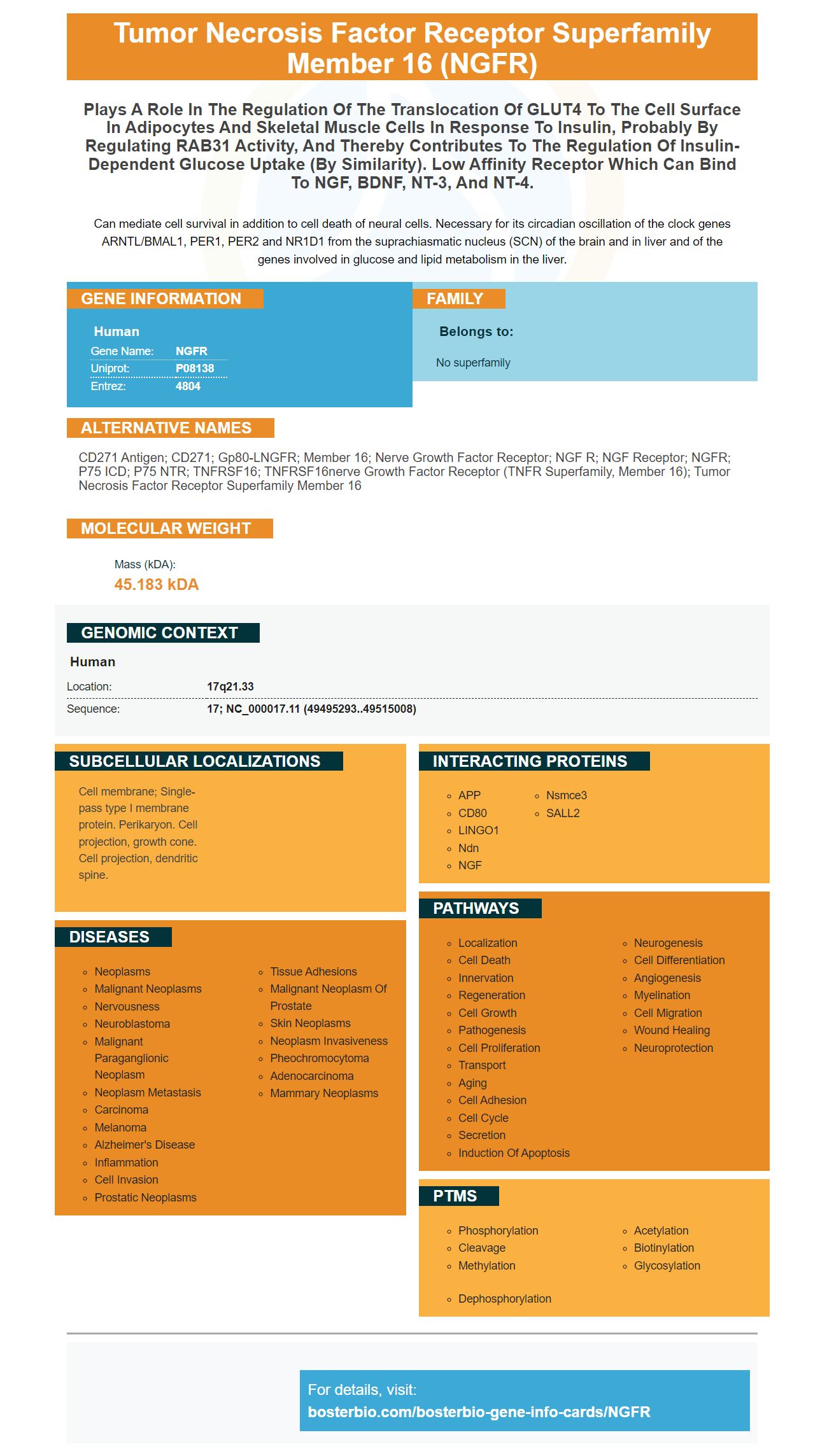
Facts about Tumor necrosis factor receptor superfamily member 16.
Can mediate cell survival in addition to cell death of neural cells. Necessary for its circadian oscillation of the clock genes ARNTL/BMAL1, PER1, PER2 and NR1D1 from the suprachiasmatic nucleus (SCN) of the brain and in liver and of the genes involved in glucose and lipid metabolism in the liver.
Human | |
---|---|
Gene Name: | NGFR |
Uniprot: | P08138 |
Entrez: | 4804 |
Belongs to: |
---|
No superfamily |
CD271 antigen; CD271; Gp80-LNGFR; member 16; nerve growth factor receptor; NGF R; NGF receptor; NGFR; p75 ICD; p75 NTR; TNFRSF16; TNFRSF16nerve growth factor receptor (TNFR superfamily, member 16); tumor necrosis factor receptor superfamily member 16
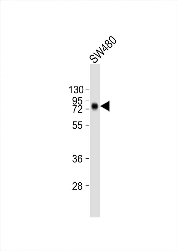
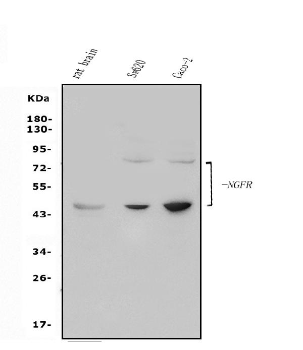
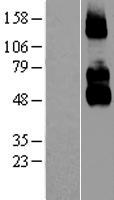
Mass (kDA):
45.183 kDA
Human | |
---|---|
Location: | 17q21.33 |
Sequence: | 17; NC_000017.11 (49495293..49515008) |
Cell membrane; Single-pass type I membrane protein. Perikaryon. Cell projection, growth cone. Cell projection, dendritic spine.
Some of the immunohistochemical markers belonging to the NGFR family are malignant melanomas and substance P-mediated abortion. However, it has more applications than simply identifying cancerous cells. This article will discuss some of its greatest uses. They include: Identifying mesenchymal-derived stem cells. It is the main goal of research to identify mesenchymal stem cell.
NGFR is one transmembrane, a 75 kDa transmembrane orphan receptor that is found in the central and peripheral nervous system. It is often associated with the TrkA receptor and is involved in a variety of neurogenic processes, such as neurite growth, differentiation, and synaptic plasticity. Its role in human cancer is not known. However, research has shown that high levels of NGFR expression are linked to malignant melanoma. This is the case for MICs or metastasis.
The expression of NGFR isn't limited to neural cells, and can also be found in hair follicles, basal Keratinocytes and the outer sheaths of hair strands. Because it isn't restricted to neural cells, NGFR should be used in conjunction with other immunohistochemical markers such as S100. It is also a good option in the case of melanomas that are desmoplastic.
It can be difficult to identify desmoplastic melanoma, particularly when there are small samples from biopsy or clinical settings that are unusual or unusual locations. In these instances, immunohistochemistry can be crucial to make an precise diagnosis. The immunohistochemical staining of S-100 protein is a reliable and accurate marker for melanocytes in these cases. NGFR, which is a marker for Schwannian differentiation, has also proved to be effective in confirmation of the diagnosis of desmoplastic melanoma.
Recently, a study of the 67-year old man showed that his tumors were characterized by strong expression of CD68 and did not have brown pigment. The malignancy was distinguished from a fibrohistiocytic cancer by analysing the immunohistochemical stain of CD163, S100 and Melan A. The cancerous cells displayed strong nuclear SOX10 expression. The Hepatic Stellate cell types were different from malignant melanomas.
The NGFR expression in melanomas of the cutaneous primary was associated with a variety of parameters such as stage of metastasis to lymph nodes, and invasiveness. The relationship between NGFR expression and the stage of clinical progression of the disease was not significant, but the presence of Ki-67 and Kindlin-1 staining in melanoma of the cutaneous origin may be predictive of poor prognosis.
Three researchers recently investigated whether NGFR was an effective immunohistchemical marker to detect melanomas that are malignant. The results showed that NGFR expression was high in myoepithelial cells, but not in luminal or stromal cells. NGFR expression was also observed in tumors that had not metastasized to lymph nodes at the time of diagnosis.
Malignant melanomas are able to express NGFR in a manner that is similar to normal breast and DCIS. Double immunolabelling allows the examination of p63/NGFR fusion. A slide was microwaled in citrate buffer (pH 6.0) and then allowed to cool to an ambient temperature. After this the slides were incubated in Envision+peroxidase blocking solution for five minutes. The slides were then rinsed with water before being transferred to TBS.
NGFR is an immunohistochemical indicator that can be used to detect malignant melanomas. The antibodies used to detect NGFR are typically highly specific. They are not intended to be used to diagnose. They should be part of an immunohistochemical panel. The results of this panel must be interpreted in context with other studies to determine whether NGFR is a valuable immunohistochemical marker for malignant melanomas.
In addition to its function in uterine weight, NGFR could also be an immunohistochemical marker of hormone-triggered or substance-mediated abortion. The findings could be relevant in studies of spontaneous abortions as well as adenomyosis-related hysterauxesis. It is therefore crucial to find women with high levels of NGFR.
Nerve growth factor (NGF) is a crucial neurotrophic protein, helps protect cells from certain types of death. The hormone triggers neurite outgrowth and neuronal differentiation by activating the NGF receptor tyrosine Kinase. Different kinds of cells express the NGF receptor, including keratinocytes and endothelium as well as glial cells.
NGFR was used to track and diagnose the first stages of abortion triggered by stress or by substance-P. High-resolution immunohistochemistry has also been used to identify the protein in different tissues. Its location is essential for the diagnosis and treatment of abortion caused by substance P. NGFR expression is easily detectable in these tumors and it has been found to be a significant indicator of abortion mediated by substance P.
In addition to its role in the pathogenesis of adenomyosis, it is also a useful immunohistochemical indicator to aid in the diagnosis of abortion mediated by substance P and other fetal deaths triggered by stress. Researchers are now able identify the expression of NGFR in human uteri in the form of a high-resolution image.
In addition it has been found to be a sensitive marker for the treatment of uterine pregnancy-related disorders. Additionally, NGFR expression has been discovered to be higher in ovoma, compared to normal-ovaries. In addition, NGFR has been shown to decrease the risk of substance-induced uterine cramps.
It has also been demonstrated to block EMT and cell migration in MDAMB-231 cells. This is in line with the results of earlier studies that discovered a significant correlation between PEDF and EGFR. The increased NGFR in the uterine tissue facilitated tube development and prevented Apoptosis. Propranol and PEDF also increased the PEDF/VEGF relationship, and had a stronger suppressive impact on HemEC.
Neurotrophic factor/receptor expression is also an important determinant of uterine function in women suffering from adolescent disorder. In addition to the NGFR PACAP and VIP are associated with impaired urinary bladder function. These findings may suggest that NGF and PACAP play a role in controlling bladder function.
Furthermore, NGFR expression in the lungs of mice that have CCSP-NGF transgenics has been associated with a reduction in micrometastasis. In addition, these mice have deficits in tachykinin-containing sensory nerve fibers and impaired nociception. However the lungs of NGFR mice that have been knocked out are not significantly altered when compared to wild-type mouse.
Because they express NGFR gene mesenchymal stem cell can be distinguished from fibroblasts. There are a variety of biomarkers to identify mesenchymal stem cells but the NGFR genes are the most important. The NGFR gene has been proven to be expressed in a variety of mesenchymal forms, including mesenchymal cell fibroblasts and mesenchymal mesen cells.
NGFR expression in the mesenchymal stem cell stroma can help identify stem cells and differentiate them into cartilage, bone and fat. These cells can be isolated from various types of adult tissues through density-gradient fractionation and immunohistochemistry. They are also known as plastic-adhered cells, and can be used to determine mesenchymal stem cell populations.
CK5 is a valuable indicator to differentiate epidermal as well as mesenchymal stem cells. This protein is present in multiple epithelia, including the ovary as well as the tubal epithelia. NGFR is an immunohistochemical marker which can be used to determine mesenchymal stem cells.
Recent research has revealed a new class of extracellular signaling molecule that can help sustain and promote mesenchymal-derived cells. Recent studies have revealed a new group of mesenchymal stem cells which express a series of genes related to self-renewal and growth. For example the oct-4 gene is known to stimulate mesenchymal cell self-renewal. These cells also contain rex-1 and.
Other mesenchymal stem cell markers cells include NGFR, NKFR, GCTM-2, and CD133. They are useful for identifying mesenchymal stem cells since they have high proliferative capacity. Additionally, the NGFR also is a prominent expression in adult bone marrow. If NGFR expression is high in a tumor cell stem cells may develop into osteoblasts or muscles, or fat.
Although the NGFR gene is a crucial biomarker for identifying mesenchymal stem cell populations, its precise function is not known. As an added bonus, NGFR also produces SSEA-3 and SSEA-4, which are expressed in undifferentiated primate ESC. SSEA-3 and SSEA-4 are expressed in both human embryonic germ cells as well as the teratocarcinoma, teratocarcinoma.
MSCs can be identified by isolating a homogeneous community using cell surface markers that target antibody-targeted cells. MSCs display natural variation in the cell surface markers, which makes it difficult to identify them in live. Antibodies that target specific cell surface markers can be useful in identifying mesenchymal and other stem cells.
The in vitro and in vivo differentiation capacity of MSCs from donors that are different are similar, however the capacity for healing in vivo might differ. The secretome of MSCs has been extensively documented and a variety of molecules have been examined for their potential. Recombinant bioactive substances are a crucial element of medical regenerative therapy. However they are still in the experimental phase because it is difficult to identify the dose that is clinically effective based on in vitro results.
PMID: 3022937 by Johnson D., et al. Expression and structure of the human NGF receptor.
PMID: 2850481 by Sehgal A., et al. A constitutive promoter directs expression of the nerve growth factor receptor gene.
*More publications can be found for each product on its corresponding product page