This website uses cookies to ensure you get the best experience on our website.
- Table of Contents
2 Citations
Facts about Podoplanin.
Through MSN or EZR interaction promotes epithelial-mesenchymal transition (EMT) leading to ERZ phosphorylation and activating RHOA activation leading to cell migration growth and invasiveness (PubMed:17046996, PubMed:21376833). Interaction with CD44 promotes directional cell migration in epithelial and tumor cells (PubMed:20962267).
Human | |
---|---|
Gene Name: | PDPN |
Uniprot: | Q86YL7 |
Entrez: | 10630 |
Belongs to: |
---|
podoplanin family |
36-KD; Aggrus; Gp36; Gp38; GP40; HT1A-1; hT1alpha-1; hT1alpha-2; lung type I cell membrane associated glycoprotein; lung type-I cell membrane-associated glycoprotein (T1A-2); OTS 8; OTS8; PA2.26 antigen; PDPN; Podoplanin; RANDAM-2; T1A; T1A-2; T1-alpha
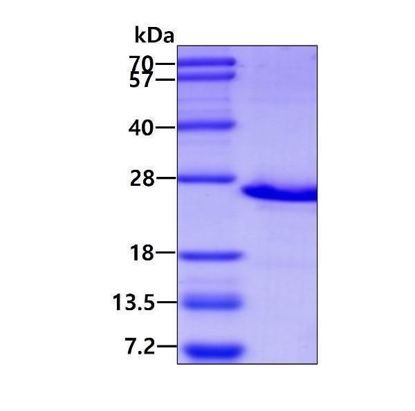
Mass (kDA):
16.698 kDA
Human | |
---|---|
Location: | 1p36.21 |
Sequence: | 1; NC_000001.11 (13583757..13617957) |
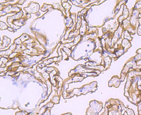
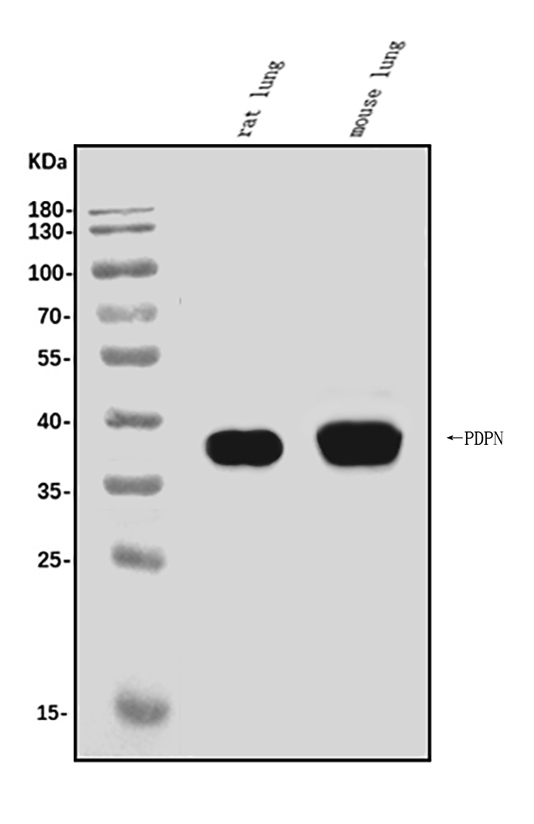
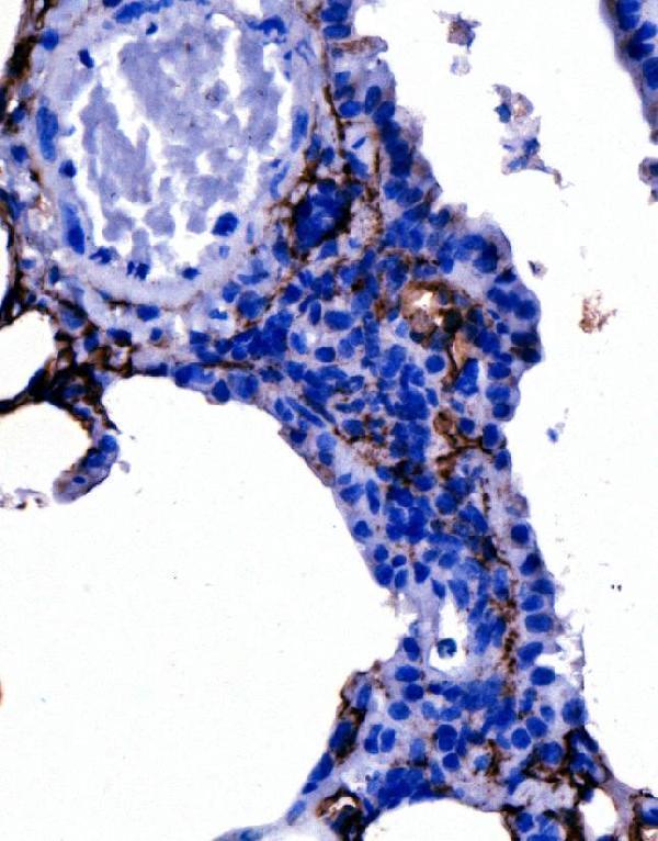
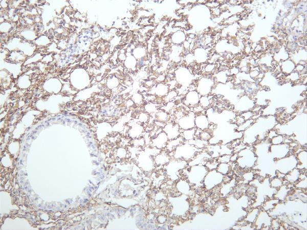
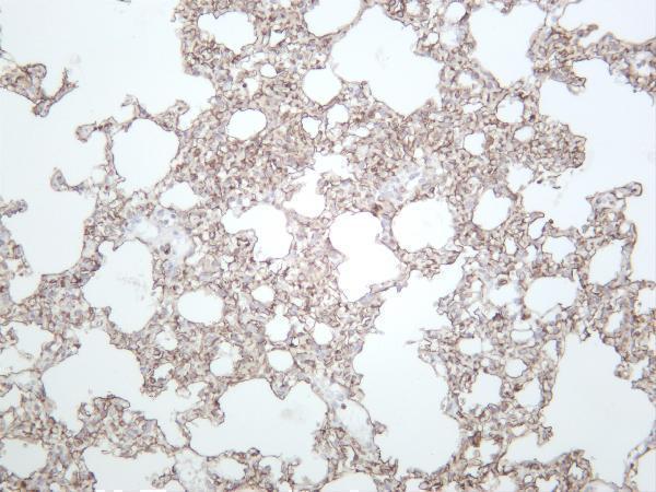
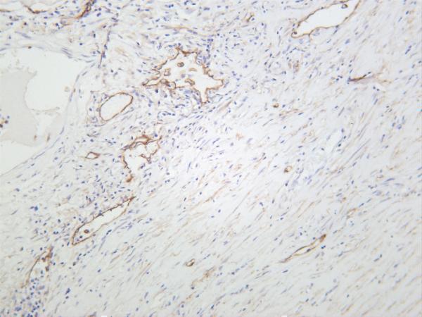
Highly expressed in placenta, lung, skeletal muscle and brain. Weakly expressed in brain, kidney and liver. In placenta, expressed on the apical plasma membrane of endothelium. In lung, expressed in alveolar epithelium. Up-regulated in colorectal tumors and expressed in 25% of early oral squamous cell carcinomas.
[Podoplanin]: Membrane; Single-pass type I membrane protein. Cell projection, lamellipodium membrane; Single-pass type I membrane protein. Cell projection, filopodium membrane; Single-pass type I membrane protein. Cell projection, microvillus membrane; Single-pass type I membrane protein. Cell projection, ruffle membrane; Single-pass type I membrane protein. Membrane raft. Apical cell membrane. Basolateral cell membrane. Cell projection, invadopodium. Localized to actin-rich microvilli and plasma membrane projections such as filopodia, lamellipodia and ruffles (By similarity). Association to t
The PDPN marker, a novel immunogen that can detect lymphatic endothelium that is present in cancer tissues. This protein can be used to detect papillary thyroid carcinoma invasion. Recent studies have proven that PDPN can be reversed through Western blot. But the question is "What are the most beneficial uses for PDPN?"
PDPN is located in the lymphatic vessels' endothelium and aids in the migration of dendritic cells to the LN. Additionally, it blocks retrograde blood flow into the lymphatic system. It also hinders the transfer of postnatal blood to the lymphatic system. This protein is involved in lymphatic angiogenesis, however it could also perform other functions.
While the role of PDPN in the lymphatic system is not clear yet, it has been linked with a number important biological processes. PDPN is a marker of lymphangiogenesis, lymphatic endothelium and retrograde blood flow from veins to the lymphatic system. However, this hasn't been examined in adult stages.
PDPN is also known as podoplanin is a mucin-type O–glycoprotein, is expressed in a variety cell types, including platelets and endothelial cells. The protein binds to VEGF, which is a vascular growth factor and FGFR (a growth factor for fibroblasts).
It is now recognized that lymphatic vessels can be vessels for tumor cells and serve a variety of other purposes. Endothelial lymphocytes can express chemotactic substances that stimulate tumor cell mobilization and alter immune system function in the peripheral. Further, lymphatic endothelial cells are involved in the process of tumor metastasis.
It is a unique marker of the lymphatic endothelium. It is expressed in bone marrow progenitor cells that is able to be recruited from the blood circulation. In addition they can be implanted into lymphatic vessels that are associated with tumors. In the case of cancer the high density of M-LECPs is associated with metastasis of the lymph nodes.
The growth of papillary thyroid cancer is affected by the PDPN gene. The overexpression of PDPN can increase the expression of E/R/M pathways while silencing PDPN reduced these pathways. The gene is also involved in the EMT pathway. Thus, silencing PDPN may be a reliable indicator for the development of papillary thyroid carcinoma. However, more research is required to determine if this is the case.
PDPN is a transmembrane proteins that is specific to the lymphatic system is a transmembrane glycoprotein. It has been shown to be present in a variety human cancers, including papillary as well as thyroid cancer. Studies have shown that PDPN may stimulate cell migration and invasion, which could be a factor in the development of tumors. This study was designed to determine if the PDPN gene is an important marker in papillary thyroid carcinoma invasion.
This study was conducted on three papillary thyroid cancer cell lines that were derived from patients who have positive diagnoses of PDPN. Dr. Nobuo Sato developed the cell lines and they are genetically distinct. BcPAP cells carry the V600E mutation in the BRAF gene. They also carry the RET/PTC1 reverse-rearrangement. The cells were created in a humidified environment of 5 % CO2 and the number of viable cells was determined using Trypane Blue stain.
In addition to PDPN being a marker for the development of papillary tumors It could also be a marker of cell migration and spread. According to one study, the expression of PDPN in papillary thyroid cancer cells contributes to the structure of cytoskeleton components. This result translates to altered cell spreading and cellular protrusion in TPC1 cells and BcPAP cells.
PDPN is an O-glycoprotein of the mucin type which is expressed on many cell types. Its interaction with FRCs activates the platelet aggregation-activating protein CLEC-2. PDPN enhances lymphatic vessel development and maintenance by interfacing with leukocytes through transcriptional activation through Prox1.
PDPN interacts with numerous transmembrane and intracellular proteins mediating effects on cell adhesion and migration. Binding to MMP CCL21 results in increased cell migration. Binding to ERMs causes rearrangement of the actin cytoskeleton. PDPN is associated with MMP-9 in the lymphatic endothelial cell.
PDPN was determined using 11 frozen IDC fragments and paraffin-embedded IDC block blocks. The number of cells was determined through an optical microscope, immunohistochemical staining, and semiquantitative as well as immunereactive scales. To see the cells, the samples were stained with 0.5% of crystal violet. For quantitative analysis, stained cells were examined using ImageJ.
PDPN is a protein of great importance that is needed by many organs, and serves many functions. It is essential to regulate the formation of platelets by CLEC-2. It also is involved in cell proliferation and differentiation. Its physiological effects are specific to the tissue and could be influenced by different binding partners and protein interactions. In addition, there are several possible physiological functions for PDPN.
PDPN is mucinlike transmembrane glycoprotein. It is found in numerous tissues during the process of ontogeny as well as in adult animals. It is involved in the development of the heart. It plays a crucial role in the development of the lymphatic system of the skin and other organs. Tumor cell studies have also revealed that tumor cells upregulate PDPN during the epithelial-mesenchymal transition. Tumor cells that are upregulated in expression of PDPN are highly invading and mobile.
Boster Bio is an antibody manufacturer that specialises in IHC-optimized monoclonal antibodies and picogram sensitivity ELISA kits, as well as other products. The company's product line includes more than 12,000 tested antibodies that have been validated using WB, IHC, and Flow. These antibodies are quantitatively tested against 250 untransfected cells and tissues to determine their specificity and affinity. Boster Bio validates all its antibodies against a known, recombinant proteins to ensure accuracy.
The Boster Quality Guarantee includes every product sold. It ensures that the product will function in the manner described. If you'd like you can also go for custom services or BeNeLux delivery. To buy Boster products or to inquire about Boster products, contact Gentaur. Boster Bio Quality Guarantee backs these lab supplies. The products are available on the internet and from retailers worldwide. Alternatively, contact Boster directly for further information. After you've placed your order, you will be able to track it from beginning to finish.
Boster Bio Anti–AKT AKT1 Antibody reacts to Human and Mouse. It is stored at 20°C. It is suitable for ELISA, Flow Cytometry and IHC. After multiple immunizations with AKT1 and AKT2 proteins, the Boster Bio Anti–AKT Pan Reactive Antibody was produced in rabbits. The antibody is available in PBS and is available in a single dose vial.
Boster Bio PDPN is an IHC optimized polyclonal antibody which can be used in immunofluorescence (IHC). The antibody has been validated against a panel of 250 tissues and non-transfected cell lines. The antibody is highly specific and has excellent affinity. The Boster Bio PDPN can also be purchased in ELISA or WB/IHC formats.
The PDPN gene can be located in plasma membrane protrusions that are high in actin. PDPN is associated with E/R/M proteins, and was found to enhance cell motility when PDPN is depleted in an environment. Additionally it was discovered that PDPN activation triggers cytoskeletal rearrangement, such as redistribution of ezrin to protrusions at the surface. PDPN might be involved in migration.
PDPN has been evaluated in cancer models for mice as well as in cancer cell culture. Its expression promotes cellular invasion. This effect is inhibited when TIMP2 an inhibitor of natural MMPs, has been added to the cells. PDPN is found in lymphatic endothelial cells with MMP-9. PDPN has been confirmed using immunofluorescence, which allows it to be utilized in a variety of clinical situations.
To confirm Boster Bio PDPN on immunofluorescence The Boster Bio PDPN marker was tested in three separate experiments. The primary antibody was PDPN specific, and the secondary antibodies DyLight(tm) 549-conjugated. Indirect immunomagnetic enrichment (IIM) of CD34+ cells in rabbits was not as efficient as it is in human samples. The positive rabbit fraction also contained low levels of CD34 mRNA.
The LpMab-12 is used to detect glycopeptide hPDPN. This is a posttranslational modification of hPDPN, which enhances its association with CLEC-2 and activates platelet aggregation. To confirm PDPN-targeting treatments, it could be beneficial to combine epitope-specific antibodies with GpMabs.
PMID: 9651190 by Ma T., et al. Evidence against a role of mouse, rat, and two cloned human T1alpha isoforms as a water channel or a regulator of aquaporin-type water channels.
PMID: 10393083 by Zimmer G., et al. Cloning and characterization of gp36, a human mucin-type glycoprotein preferentially expressed in vascular endothelium.
*More publications can be found for each product on its corresponding product page