This website uses cookies to ensure you get the best experience on our website.
- Table of Contents
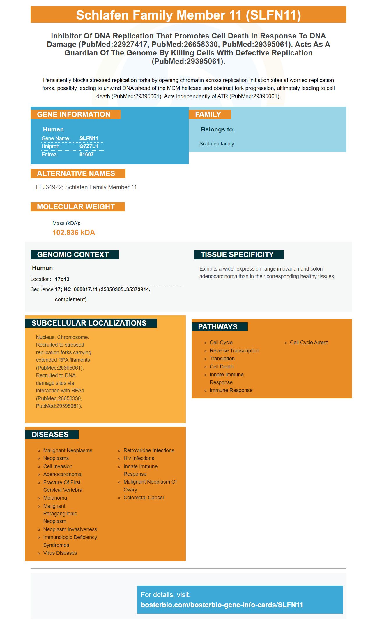
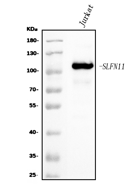
Facts about Schlafen family member 11.
Persistently blocks stressed replication forks by opening chromatin across replication initiation sites at worried replication forks, possibly leading to unwind DNA ahead of the MCM helicase and obstruct fork progression, ultimately leading to cell death (PubMed:29395061). Acts independently of ATR (PubMed:29395061).
Human | |
---|---|
Gene Name: | SLFN11 |
Uniprot: | Q7Z7L1 |
Entrez: | 91607 |
Belongs to: |
---|
Schlafen family |
FLJ34922; schlafen family member 11
Mass (kDA):
102.836 kDA
Human | |
---|---|
Location: | 17q12 |
Sequence: | 17; NC_000017.11 (35350305..35373914, complement) |
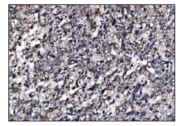
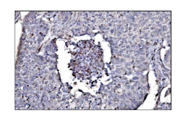
Exhibits a wider expression range in ovarian and colon adenocarcinoma than in their corresponding healthy tissues.
Nucleus. Chromosome. Recruited to stressed replication forks carrying extended RPA filaments (PubMed:29395061). Recruited to DNA damage sites via interaction with RPA1 (PubMed:26658330, PubMed:29395061).
A team of researchers recently published a study that examined SLFN11 expression within human cancer cell lines. The researchers wanted to determine the role of SLFN11 on tumor response to DNA-damaging drug. The researchers used four different lines of human cancer cells that had the SLFN11 genetic deleted experimentally. They found that the cancer cells regained their responsiveness to the cancer drugs olaparib and talazoparib when treated with ATR inhibitors. This research highlights how important SLFN11 expression is as a biomarker.
A transient transfection of CD47-cells using an SLFN11 expression gene vector increased SLFN mRNA transcription, but a significant decrease was observed in cell viability after 20 Gy irradiation. Cells transfected with a control plasmid, however, were viable and showed no changes in SLFN11 mRNA. This suggests radiotherapy targeting the SLFN11 gene promoter might be beneficial in improving radiation sensitivity.
CD47 mRNA expression was found to be negatively correlated with SLFN11 mRNA transcription, suggesting that there may have been a relationship. Using a panel of cancer cell lines, the promoter methylation levels of SLFN11 were negatively correlated with the expression of CD47 mRNA. Further analysis revealed that SLFN11 mRNA expression correlates negatively with CD47 mRNA.
SLFN11 knockdown caused GC cell death to be reduced and increased cleaved cleaved CASSE-3 and Bax expressions within human GC cell lines. SLFN11 knockdown also increased GC cells' sensitivity to cisplatin. This study demonstrates SLFN11's therapeutic effectiveness in treating cancer cells. Why is SLFN11 so important? It can help patients with cancer get the drugs that they need to live a longer and more fulfilling life.
The results of this study show that SLFN11 doesn't inhibit tumor growth. However, they don't rule out the possibility of SLFN11 being an early marker for cancer treatment sensitivity. This is especially true in patients with metastatic diseases. Patients with cancer who have SLFN11 in the primary tumor also have elevated SLFN11 concentrations. The frequency distributions for SLFN11 in cancer cell line cells are similar to those found in other tissues. A H-score above 31 is considered a good cutoff for dividing PDX cancer models in high and low-SLFN11 areas.
To determine whether SLFN11 is an important marker for cancer therapy, we used a combination of two selective HDAC inhibitors, rocilinostat and entinostat, in order to induce SLFN11 mRNA in human cancer cell lines. Quantitative realtime PCR was used in order to measure SLFN11 mRNA levels. These values were then normalized to those from untreated cells. We also used Western blotting as a loading control to measure protein levels.
SLFN11 is also expressed in several types of cancer cells, including colon, liver, prostate, endometrial, and bladder. SLFN11 has also been shown to have a positive impact on glioma cell growth. However, the effects SLFN11 has on cancer cells is still not fully understood. This is why more research is needed in order to understand the effects of this protein on cancer.
RNA from drug-treated cells was extracted with the RNeasy Mini RNA kit by Qiagen. Complementary DNA was created using the SuperScript(tm), II Reverse Transcriptase Kit from Invitrogen. We then used specific RNAi primers, derived from a previous study (5). To measure relative expression of SLFN11 and FLI-1 compared to GAPDH, we employed the 2(-DDCt) method.
Numerous studies have shown that DNA-damaging drug treatment can alter gene expression in cells. The latest study evaluated 100 genes essential for survival. While gene expression responses of DNA-damaging drugs don't correlate with survival, some genes are affected. To identify the essential genes for surviving DNA damage, a comprehensive analysis of gene expression in response to a wide variety of DNA-damaging drugs is necessary.
Despite being important, gene expression profiling does not necessarily reveal the specific mechanism through which DNA-damaging chemicals affect cells. Endogenous levels of DNA repair proteins are sufficient for cells to resist lesions. In fact, only a few changes are needed to protect cells' viability. The UV irradiation doesn't affect the highly conserved DNA-repair pathways.
Despite the number of genes that have seen mutations due to DNA-damaging agent, very few of these changes have been reported in yeast. This study nonetheless reveals a cluster consisting of 9 DNA damage genes. These genes include homologous Recombination and ribonucleotide-reductase Subunit genes. This cluster was induced by four agents, each with a factor greater than 2.
SigG is induced by a variety of DNA-damaging agents. However, it does nothing to regulate the RecA-independent response. Instead, it controls another wave of gene expression that isn't responsible for DNA repair. Hence, the gene expression of DNA-damaging drugs may not be regulated by SigG. Therefore, SigG may be a good indicator of whether or not a cell is capable of repairing itself.
The cell-cycle-regulated genes expressed by mec1 were not significantly affected by ionizing radiation or MMS. The mutant cells did no arrest the cell cycle but did show a decrease of expression after MMS treatment and ionizing radio. The cell cycle-regulated genes are also affected by DNA-damaging drugs. Moreover, mec1 mutant cells showed no significant changes in gene expression.
Additionally, miRNAs are a key target of DNA-damaging drugs and have the potential to influence their response. The molecules that target miRNAs are more effective at preventing cancer cells from repairing DNA damaged DNA. These drugs may also increase the sensitivity of breast carcinoma cells to chemotherapy and radiation treatments. It is important to note that miRNAs are regulated by a host of biological factors, including oxidative stress and vitamin E.
Our goal was the study of the relationship between SLFN11 and patient response to DNA-damaging drugs at HGSOC. We first examined HGSOC's sensitivity to DDAs. Second, we examined whether SLFN11 methylation status was associated to improved survival in HGSOC. Finally, we examined whether SLFN11 expression is associated with the number of tumor-infiltrating lymphocytes (TILs).
The results showed SLFN11 overexpression increases cancer cell sensitivity against DNA-damaging agents. Moreover, tumors containing high levels of SLFN11 showed enhanced DNA-damaging sensitivity. Therefore, the underlying molecular mechanisms responsible for SLFN11 biology and its association with tumor responsiveness are still unclear. More studies are needed to improve the clinical efficacy of these compounds.
This study demonstrated the correlation between SLFN11 response and expression in four isogenic cell types. SLFN11 has the ability to sensitize cells against PARP inhibitors, olaparibs and talazoparib. It also sensitizes cells to PARP inhibitors under the S-phase checkpoint induced by ATR activation. A correlation between SLFN11 sensitivity and DNA-damaging drugs could help guide targeted therapy development and improve patient outcomes.
SLFN11 is not expressed in cancer cells, despite its role in genome maintenance. SLFN11 inhibits may be used to release SLFN11 and increase the sensitivity of patients with SLFN11 tumors. Moreover, the researchers evaluated SLFN11 expression in cancer cell line databases and patient samples treated with romidepsin. They also used isogenic cell line to study the impact of HDAC inhibitors on HDAC inactivated tumor cells.
We found a strong correlation between SLFN11 methylation levels and DNA-damaging drugs sensitivity. Our results also revealed that platinum-derived compound resistance is associated with reduced SLFN11 levels in HCT116 cells. We also found a correlation between SLFN11 methylation status, poor patient prognosis, and poor SLFN11 expression in HCT-116 cells. In clinical cases as well as in cancer models, SLFN11 methylation has been associated with sensitivity to PARP inhibitors.
Our results suggest that SLFN11 is induced in part by HDAC inhibitors of class I. HDAC inhibitor romidepsin activates SLFN11 in cancer cell lines and patients. We believe that SLFN11 may play a part in the response to DDA inhibiters. There are other mechanisms that could be used to treat cancer cells.
A study of children showed that SLFN11 proteins strongly correlate with patient response for TAL and IRN. The drug's sensitivity also drops significantly if SLFN11 has been removed. This suggests that SLFN11 may be a tumor-specific molecular marker and that tumors that express high levels of SLFN11 are highly responsive to these drugs.
SLFN11 is a dual function biomarker that detects the immunological and intrinsic dispositions of cancer cells. The heatmap's bubbles indicate pathways that are significantly enriched. The bubble size corresponds to q value. The color indicates direction and concentration. The heatmap shows log-folded changes in immune-related transcripts. In the case of cisplatin and carboplatin, SLFN11 is highly expressed and significantly increases sensitivity to these drugs.
PMID: 20956525 by Katsoulidis E., et al. Role of interferon {alpha} (IFN{alpha})-inducible Schlafen-5 in regulation of anchorage-independent growth and invasion of malignant melanoma cells.
PMID: 23000900 by Li M., et al. Codon-usage-based inhibition of HIV protein synthesis by human schlafen 11.