This website uses cookies to ensure you get the best experience on our website.
- Table of Contents

5 Citations 6 Q&As
17 Q&As


Facts about Tumor protein 63.

Isoform 2 activates RIPK4 transcription. May be required in conjunction with TP73/p73 for initiation of p53/TP53 dependent apoptosis in response to genotoxic insults and the presence of activated oncogenes.
| Human | |
|---|---|
| Gene Name: | TP63 |
| Uniprot: | Q9H3D4 |
| Entrez: | 8626 |

| Belongs to: |
|---|
| p53 family |

amplified in squamous cell carcinoma; Chronic ulcerative stomatitis protein; CUSP; EEC3; EEC3B(p51B); Keratinocyte transcription factor KET; KET; KETRHS; LMS; OFC8; OFC8SHFM4TP73LNBP; p40; p51; p51tumor protein p53-like; p53CP; p63; p63AIS; p73H; p73L; p73LB(p51A); SHFM4; TP53CP; TP53L; TP63; TP73L; Transformation-related protein 63; tumor protein 63; tumor protein p53-competing protein; tumor protein p63 deltaN isoform delta; tumor protein p63; Tumor protein p73-like
Mass (kDA):
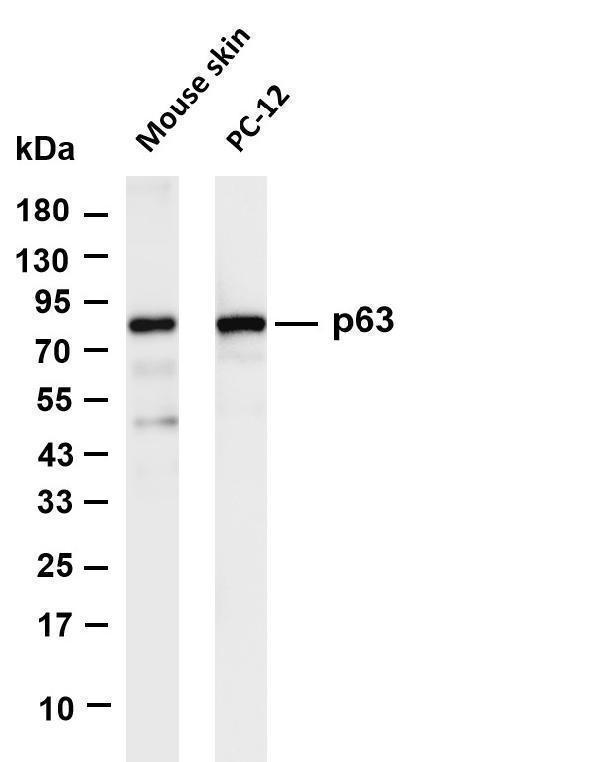
76.785 kDA

| Human | |
|---|---|
| Location: | 3q28 |
| Sequence: | 3; NC_000003.12 (189596746..189897279) |
Widely expressed, notably in heart, kidney, placenta, prostate, skeletal muscle, testis and thymus, although the precise isoform varies according to tissue type. Progenitor cell layers of skin, breast, eye and prostate express high levels of DeltaN-type isoforms. Isoform 10 is predominantly expressed in skin squamous cell carcinomas, but not in normal skin tissues.
Nucleus.


PMID: 9703973 by Senoo M., et al. A second p53-related protein, p73L, with high homology to p73.
PMID: 9799841 by Augustin M., et al. Cloning and chromosomal mapping of the human p53-related KET gene to chromosome 3q27 and its murine homolog Ket to mouse chromosome 16.
*More publications can be found for each product on its corresponding product page