This website uses cookies to ensure you get the best experience on our website.
- Table of Contents

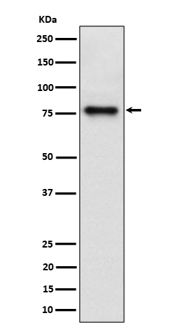
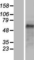
Facts about Cdc42-interacting protein 4.

Binds to lipids such as phosphatidylinositol 4,5-bisphosphate and phosphatidylserine and promotes membrane invagination and the formation of tubules. Also promotes CDC42-induced actin polymerization by recruitment WASL/N- WASP that in turn activates the Arp2/3 complex.
| Human | |
|---|---|
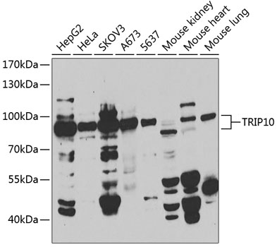
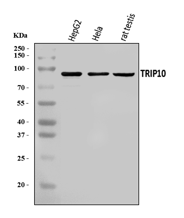
| Gene Name: | TRIP10 |
| Uniprot: | Q15642 |
| Entrez: | 9322 |

| Belongs to: |
|---|
| FNBP1 family |

cdc42-interacting protein 4; CIP4STOT; HSTP; Protein Felic; Salt tolerant protein; salt tolerator; STPTRIP-10; thyroid hormone receptor interactor 10; thyroid receptor interacting protein 10; Thyroid receptor-interacting protein 10; TR-interacting protein 10
Mass (kDA):
68.352 kDA

| Human | |
|---|---|
| Location: | 19p13.3 |
| Sequence: | 19; NC_000019.10 (6739680..6751530) |
Expressed in brain, colon, heart, kidney, liver, lung, megakaryocyte, ovary, pancreas, peripheral blood lymphocytes, placenta, prostate, skeletal muscle, small intestine, spleen, testis, thymus and trachea.
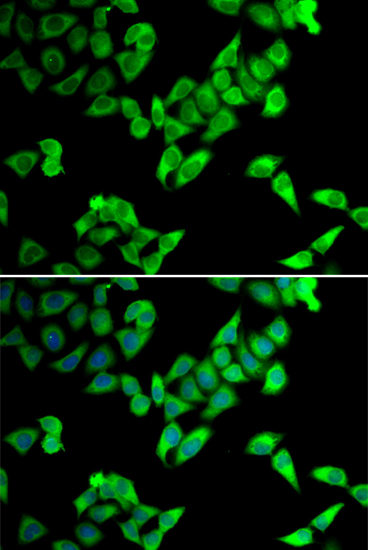
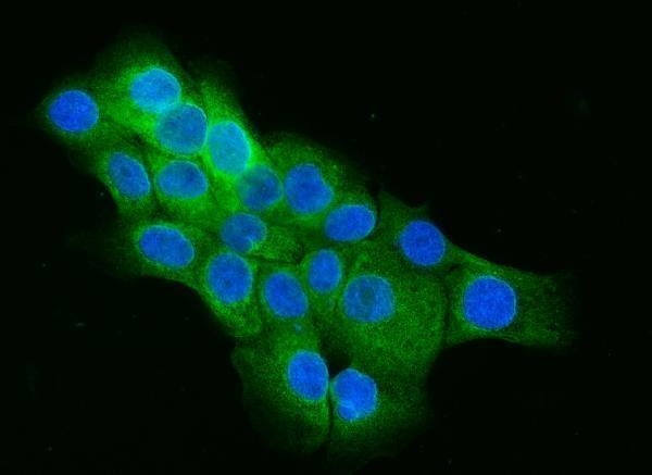
Cytoplasm, cytoskeleton. Cytoplasm, cell cortex. Lysosome. Golgi apparatus. Cell membrane. Cell projection, phagocytic cup. Translocates to the plasma membrane in response to insulin stimulation, and this may require active RHOQ (By similarity). Localizes to cortical regions coincident with F-actin, to lysosomes and to sites of phagocytosis in macrophages. Also localizes to the Golgi, and this requires AKAP9.; [Isoform 5]: Cytoplasm, perinuclear region.


PMID: 9210375 by Aspenstroem P.; A Cdc42 target protein with homology to the non-kinase domain of FER has a potential role in regulating the actin cytoskeleton.
PMID: 12054674 by Wang L., et al. Identification and genetic analysis of human and mouse activated Cdc42 interacting protein-4 isoforms.